27
Cell Communication
| Date post: | 01-Jan-2016 |
| Category: |
Documents |
| Upload: | quincy-landry |
| View: | 21 times |
| Download: | 1 times |

Cell Communication

The Cellular “Internet” Within multicellular organisms, cells
must communicate with one another to coordinate their activities
A signal transduction pathway is a series of steps by which a signal on a cell’s surface is converted into a specific cellular response
Signal transduction pathways are very similar in all organisms, even organisms as different as unicellular yeasts and multicellular mammals

Local (Short-Distance) Signaling Cells may communicate by direct contact
Plasmodesmata in plant cells Gap junctions in animal cells
Animal cells can also use cell-cell recognition Membrane-bound surface molecules can interact and
communicate

Local (Short-Distance) Signaling Messenger molecules can also be secreted by the signaling cell Paracrine signaling:
One cell secretes (releases) molecules that act on nearby “target” cells
Example: growth factors Synaptic Signaling:
Nerve cells release chemical messengers (neurotransmitters) that stimulate the target cell
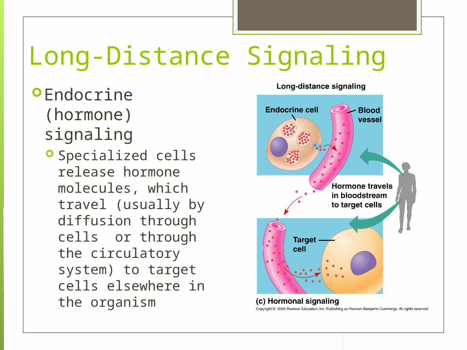
Long-Distance SignalingEndocrine
(hormone) signaling Specialized cells
release hormone molecules, which travel (usually by diffusion through cells or through the circulatory system) to target cells elsewhere in the organism

The Three Stages of Cell Signaling There are 3 stages at the “receiving end”
of a cellular conversation:1. Reception2. Transduction3. Response

Stage 1: Reception The target cell “detects” that there is a signal
molecule coming from outside the cell The signal is detected when it binds to a protein on the
cell’s surface or inside the cell (receptor protein) The signal molecule “searches out” specific receptor
proteins The signal molecule is a ligand
It is a molecule that specifically binds to another one and induces a change in the shape of the receptor protein
Ligands can be hydrophobic or hydrophillic

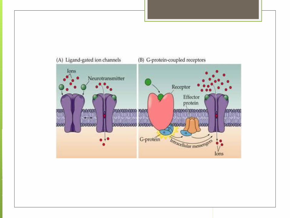
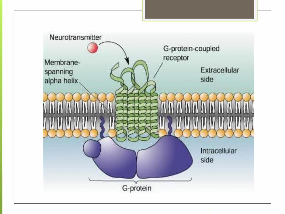
Receptor proteins There are 2 different types of receptor proteins:
Membrane receptors: transmembrane proteins Intracellular receptors: proteins that occur in the
cytoplasm or the nucleus
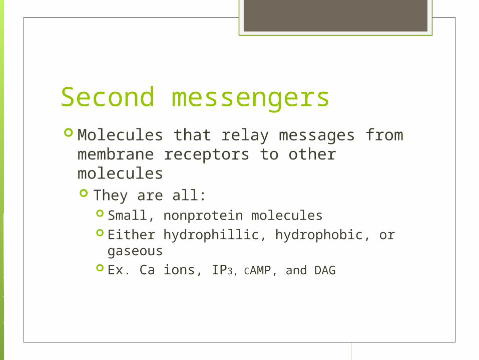
Second messengers Molecules that relay messages from
membrane receptors to other molecules They are all:
Small, nonprotein molecules Either hydrophillic, hydrophobic, or gaseous Ex. Ca ions, IP3, CAMP, and DAG

Stage 2: Transduction This stage converts the signal into a form
that can bring about a specific cellular response One signal-activated receptor activates another
protein, which activates another molecule, etc., etc. - this is called a signaling cascade
These act as relay molecules Often the message is transferred using protein
kinases, which transfer phosphate groups from ATP molecules to proteins
These can be very complicated

Stage 2: Transduction
Advantages of signal transduction Amplification: the effect of the signaling
molecule can be amplified Control: the cell can control the
accuracy of the signaling Multiplicity: a signaling molecule can
start many different processes at once to respond to the signal

Stage 3: Response The signal that was
passed through the signal transduction pathway triggers a specific cellular response Examples: enzyme
action, cytoskeleton rearrangement, activation of genes, etc., etc.
Diagram example: transcription of mRNA
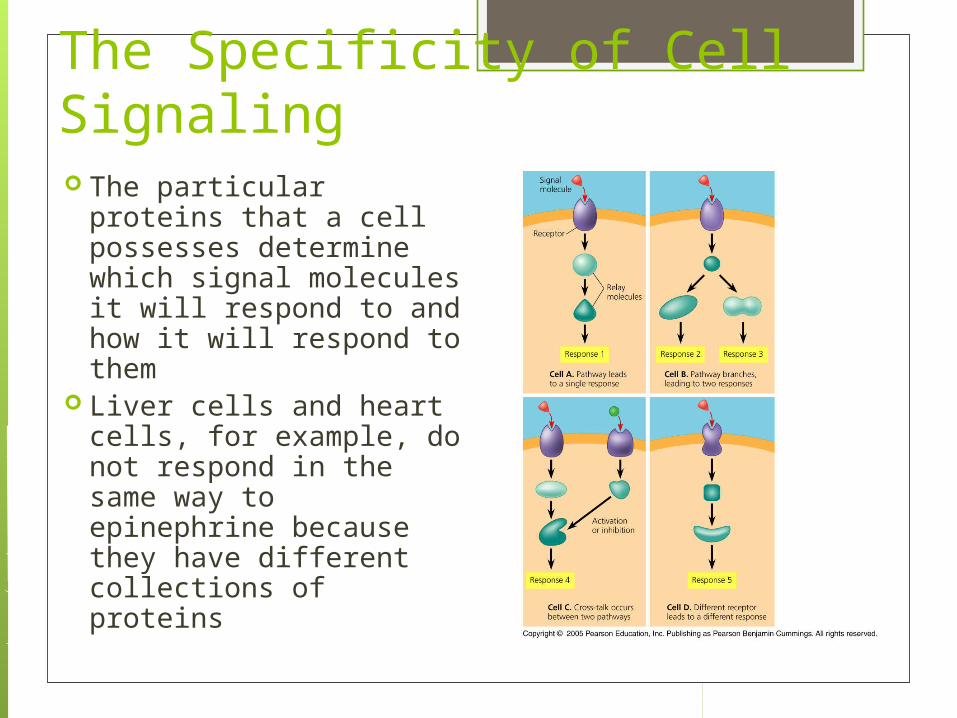
The Specificity of Cell Signaling The particular proteins
that a cell possesses determine which signal molecules it will respond to and how it will respond to them
Liver cells and heart cells, for example, do not respond in the same way to epinephrine because they have different collections of proteins
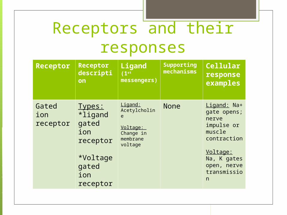
Receptors and their responses
Receptor Receptor description
Ligand(1st messengers)
Supporting mechanisms
Cellular response examples
Gated ion receptor
Types: *ligand gated ion receptor
*Voltage gated ion receptor
Ligand: Acetylcholine
Voltage: Change in membrane voltage
None Ligand: Na+ gate opens; nerve impulse or muscle contraction
Voltage: Na, K gates open, nerve transmission
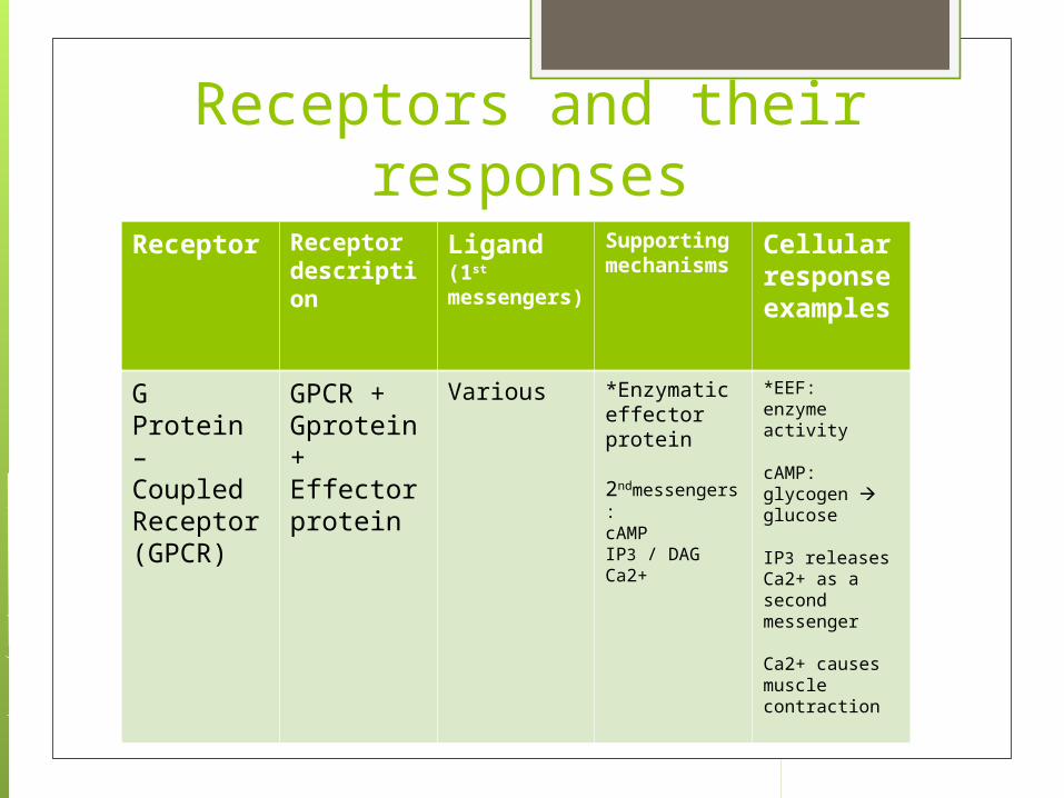
Receptors and their responses
Receptor Receptor description
Ligand(1st messengers)
Supporting mechanisms
Cellular response examples
G Protein – Coupled Receptor (GPCR)
GPCR + Gprotein + Effector protein
Various *Enzymatic effector protein
2ndmessengers:cAMPIP3 / DAGCa2+
*EEF: enzyme activity
cAMP: glycogen glucose
IP3 releases Ca2+ as a second messenger Ca2+ causes muscle contraction
Receptors and their responses
Receptor Receptor description
Ligand(1st messengers)
Supporting mechanisms
Cellular response examples
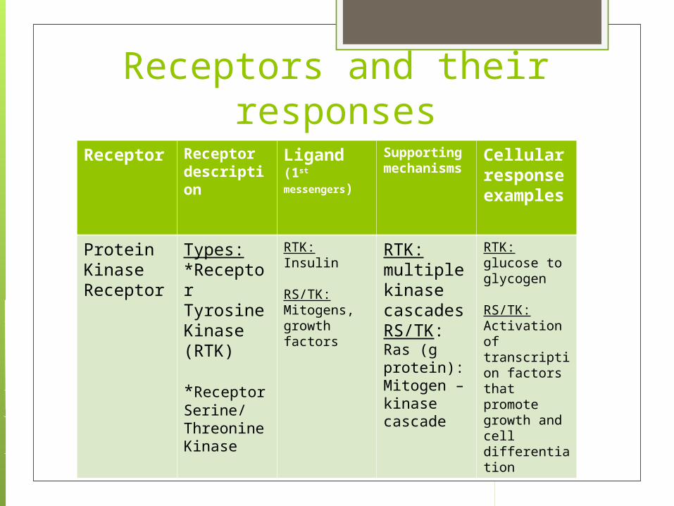
Protein Kinase Receptor
Types: *Receptor Tyrosine Kinase (RTK)
*Receptor Serine/Threonine Kinase
RTK: Insulin
RS/TK: Mitogens, growth factors
RTK: multiple kinase cascadesRS/TK:Ras (g protein):Mitogen – kinasecascade
RTK: glucose to glycogen
RS/TK: Activation of transcription factors that promote growth and cell differentiation

Receptors and their responses
Receptor Receptor description
Ligand(1st messengers)
Supporting mechanisms
Cellular response examples
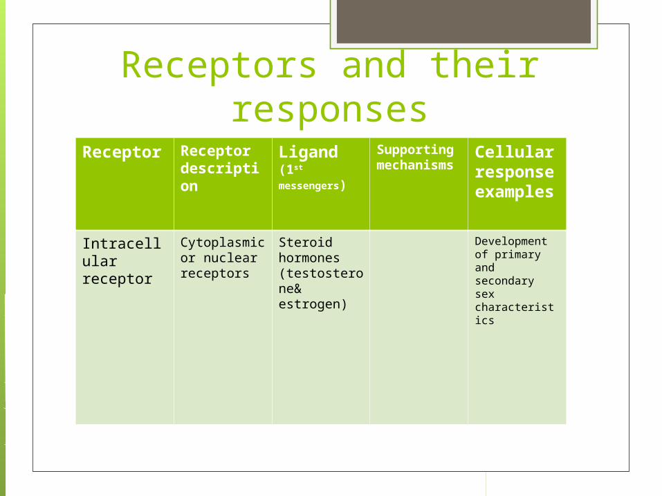
Intracellular receptor
Cytoplasmic or nuclear receptors
Steroid hormones (testosterone& estrogen)
Development of primary and secondary sex characteristics

When cell signaling doesn’t work
Signal transduction pathways can be blocked or defective
Examples: Diabetes Cholera Autoimmune disease Cancer Neurotoxins, poisons, pesticides Drugs (anesthetics, antihistamines, blood
pressure meds)

Cholera
Disease acquired by drinking contaminated water (w/human feces)
Bacteria (Vibrio cholerae) colonizes lining of small intestine and produces toxin
Toxin modifies G-protein involved in regulating salt & water secretion
G protein stuck in active form intestinal cells secrete salts, water
Infected person develops profuse diarrhea and could die from loss of water and salts
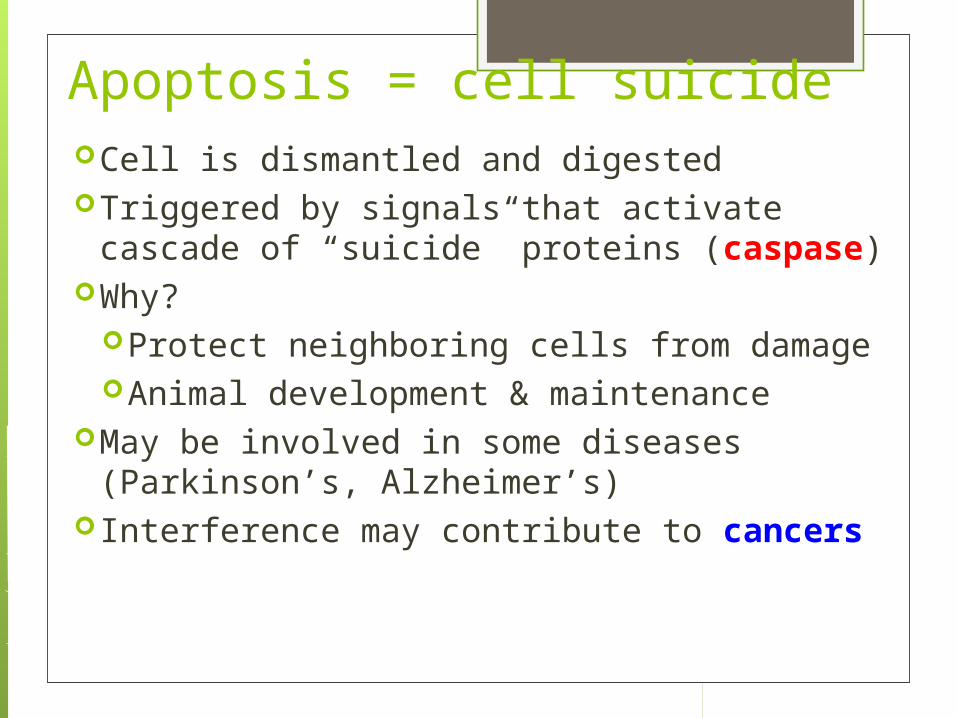
Apoptosis = cell suicideCell is dismantled and digestedTriggered by signals that activate
cascade of “suicide” proteins (caspase)Why?
Protect neighboring cells from damageAnimal development & maintenance
May be involved in some diseases (Parkinson’s, Alzheimer’s)
Interference may contribute to cancers
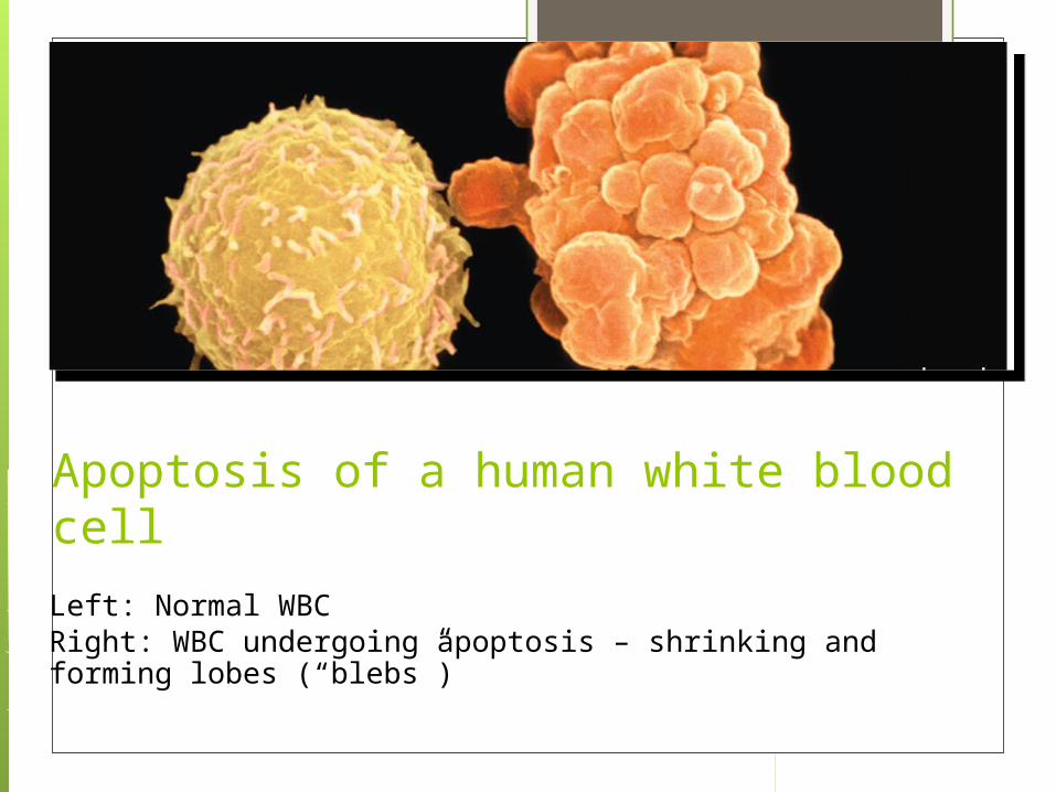
Apoptosis of a human white blood cell
Left: Normal WBCRight: WBC undergoing apoptosis – shrinking and forming lobes (“blebs”)

Effect of apoptosis during paw development in the mouse

Drugs There are drugs that interfere with cell
signaling can either be agonists or antagonists Agonists: acts the same way that a ligand
does Antagonists: blocks the binding site of the
receptor and does not let cell signaling occur Ex. The poison curare blocks the binding
sites for the chemical acetylcholine which will cause muscle paralysis and death


