APPLICATION NOTE 1850 Millrace Drive, Suite 3A Eugene, Oregon 97403 www.mitosciences.com Telephone: 1-800-910-6486 or 541-284-1800 · Fax: 541-284-1801 www.mitosciences.com Customer Service: [email protected]· Tech Support: [email protected]High Throughput Cell Fractionation Rev.0 Monitoring protein movements to and from the mitochondrion in apoptosis, a high throughput quantitative solution. Summary: The movement of pro-apoptotic factors of the Bcl2-family from the cytosol to the mitochondria and the consequent permeabilization of the mitochondrial outer membrane to release cytochrome c and Smac from the mitochondrial intermembrane space into the cytosol or AIF to the nucleus are now well characterized steps in programmed cell death (Figure 1). However, there are concurrent movements of other proteins, including kinases and transcription factors (e.g. p53), to and from the organelle to both signal and modulate apoptosis. The identification and quantification of these protein movements between cellular compartments is necessary to fully understand and to differentiate between different pathways of cell death. Current methodology of monitoring such movements mostly relies on difficult-to-quantify immunocytochemical approaches or on biochemical cell fractionation that involves mechanical disruption of cells followed by a complicated centrifugation scheme. We have developed a simple, rapid cell fractionation method to obtain cytosol-, mitochondria- and nuclei- containing fractions. The method involves sequential and selective extraction of cytosolic and then mitochondrial proteins by detergents from a nucleus containing fraction. This cell fractionation procedure can be performed either on cells in suspension or in a high throughput microplate format on adherent cells (Figure 2). Cell fractionations in a 96 well plate format followed by Western blot analyses were used to monitor translocation of Bax from the cytosol to the mitochondria and cytochrome c and Smac from the mitochondria into the cytosol in HeLa cells induced to undergo apoptosis by Staurosporine treatments. Alternatively, the cell fractionation was followed by a cytochrome c sandwich ELISA assay, thus offering a complete quantitative high throughput approach to measure cytochrome c release from mitochondria in cells undergoing apoptosis. Figure 1. Mitochondrial control of apoptosis.
Transcript
APPLICATION NOTE 1850 Millrace Drive, Suite 3A
Eugene, Oregon 97403 www.mitosciences.com
Telephone: 1-800-910-6486 or 541-284-1800 · Fax: 541-284-1801 www.mitosciences.com
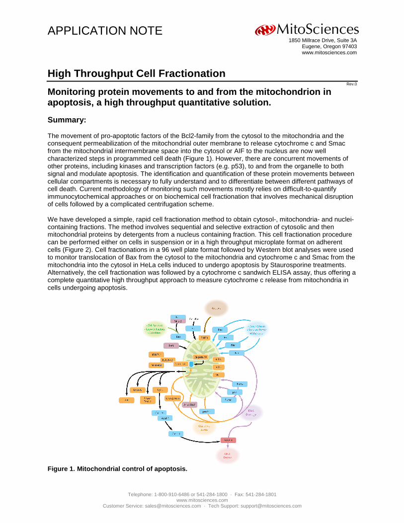
Monitoring protein movements to and from the mitochondrion in apoptosis, a high throughput quantitative solution. Summary: The movement of pro-apoptotic factors of the Bcl2-family from the cytosol to the mitochondria and the consequent permeabilization of the mitochondrial outer membrane to release cytochrome c and Smac from the mitochondrial intermembrane space into the cytosol or AIF to the nucleus are now well characterized steps in programmed cell death (Figure 1). However, there are concurrent movements of other proteins, including kinases and transcription factors (e.g. p53), to and from the organelle to both signal and modulate apoptosis. The identification and quantification of these protein movements between cellular compartments is necessary to fully understand and to differentiate between different pathways of cell death. Current methodology of monitoring such movements mostly relies on difficult-to-quantify immunocytochemical approaches or on biochemical cell fractionation that involves mechanical disruption of cells followed by a complicated centrifugation scheme. We have developed a simple, rapid cell fractionation method to obtain cytosol-, mitochondria- and nuclei-containing fractions. The method involves sequential and selective extraction of cytosolic and then mitochondrial proteins by detergents from a nucleus containing fraction. This cell fractionation procedure can be performed either on cells in suspension or in a high throughput microplate format on adherent cells (Figure 2). Cell fractionations in a 96 well plate format followed by Western blot analyses were used to monitor translocation of Bax from the cytosol to the mitochondria and cytochrome c and Smac from the mitochondria into the cytosol in HeLa cells induced to undergo apoptosis by Staurosporine treatments. Alternatively, the cell fractionation was followed by a cytochrome c sandwich ELISA assay, thus offering a complete quantitative high throughput approach to measure cytochrome c release from mitochondria in cells undergoing apoptosis.
Figure 1. Mitochondrial control of apoptosis.
ICE Application Note
Telephone: 1-800-910-6486 or 541-284-1800 · Fax: 541-284-1801 www.mitosciences.com
Introduction: Biochemical cell fractionation approaches mostly utilize mechanical cell disruption. Mechanical cell disruption methods are often difficult to standardize, they require a relatively large amount of cell sample, and the lack of multiple instruments often limits these methods to process one sample at a time. Thus, they are time consuming and difficult to perform on a large number of samples. They also carry the risk of disrupting the mitochondrial membranes leading, in particular, to artificial release of mitochondrial intermembrane space pro-apoptotic proteins. The mechanical cell disruption is often incomplete and thus a removal of unbroken cells is frequently required. This leads to losses of uncharacterized cell material which is difficult to account for. We have developed a simple and rapid method to fractionate cells in suspension into cytosol-, mitochondria- and nuclei-containing fractions. This fractionation is based on a selective and sequential extraction of cytosolic and mitochondrial proteins thus eliminating the need of mechanical cell disruption and differential centrifugation. This methodology was used to measure protein movements during apoptosis in a variety of trypsin-detached adherent cells including HeLa cells, 143B osteosarcoma cells, SHSY5Y neuroblastoma cells, HepG2 cells, HdFN fibroblasts and cardiomyocytes or suspension-grown Jurkat TIB152 cells and HL60 cells, treated with several apoptosis inducers including Staurosporine and an anti-Fas antibody. Here we present a new generation of this methodology specifically designed to fractionate adherent cells directly, without their detachment, in a high throughput microplate format (Figure 2). We demonstrate the utility of this high throughput fractionation on following the movement of Bax, cytochrome c and Smac in HeLa cells induced to undergo apoptosis by Staurosporine treatment. When coupled to a microplate sandwich ELISA assay, these methodologies present a complete high throughput solution, as shown on monitoring of the cytochrome c release in apoptosis.
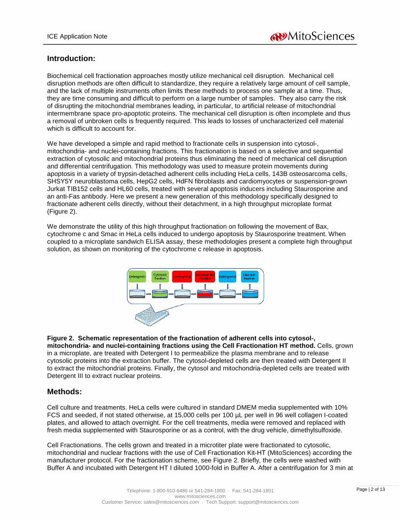
Figure 2. Schematic representation of the fractionation of adherent cells into cytosol-, mitochondria- and nuclei-containing fractions using the Cell Fractionation HT method. Cells, grown in a microplate, are treated with Detergent I to permeabilize the plasma membrane and to release cytosolic proteins into the extraction buffer. The cytosol-depleted cells are then treated with Detergent II to extract the mitochondrial proteins. Finally, the cytosol and mitochondria-depleted cells are treated with Detergent III to extract nuclear proteins. Methods: Cell culture and treatments. HeLa cells were cultured in standard DMEM media supplemented with 10% FCS and seeded, if not stated otherwise, at 15,000 cells per 100 µL per well in 96 well collagen I-coated plates, and allowed to attach overnight. For the cell treatments, media were removed and replaced with fresh media supplemented with Staurosporine or as a control, with the drug vehicle, dimethylsulfoxide. Cell Fractionations. The cells grown and treated in a microtiter plate were fractionated to cytosolic, mitochondrial and nuclear fractions with the use of Cell Fractionation Kit-HT (MitoSciences) according the manufacturer protocol. For the fractionation scheme, see Figure 2. Briefly, the cells were washed with Buffer A and incubated with Detergent HT I diluted 1000-fold in Buffer A. After a centrifugation for 3 min at
ICE Application Note
Telephone: 1-800-910-6486 or 541-284-1800 · Fax: 541-284-1801 www.mitosciences.com
300 x g, the supernatants containing extracted cytosolic proteins were collected. The cytosol-depleted cells were then extracted with Detergent HT II diluted 200-fold in Buffer A. After a centrifugation for 3 min at 300 x g, the supernatants containing extracted mitochondrial proteins were collected. The cell remainders were than extracted with Detergent HT III diluted 10-fold in Buffer A. After a centrifugation for 3 min at 300 x g, the supernatants containing extracted nuclear proteins were collected.
Western Blot Analysis. 20 µL of cytosolic, mitochondrial and nuclear fractions were analyzed by Western blotting according MitoSciences protocol (http://www.mitosciences.com/PDF/western.pdf ) with the use of HRP-conjugated secondary antibodies and chemiluminescence detection (ECL PLUS, GE Healthcare). Primary antibodies, diluted according manufacturers’ instructions, are listed in Table I. The blots were imaged by UltraLum imaging system and the proteins were quantified by UltraQuant software. The distribution of a protein between cytosolic, mitochondrial and nuclear fractions was calculated as a percentage of the protein present in a fraction out of the sum of the protein present in all three fractions. Cytochrome c Sandwich ELISA: The cytochrome c quantity was assayed in cytosolic, mitochondrial and nuclear fractions using Cytochrome c Protein Quantity Microplate Assay Kit (MitoSciences). Protein Name
Second mitochondria-derived activator of caspase (Smac)
Q9NR28
Mitochondrial intermembrane space
Cell Signaling Tech. 2954
Transcription factor Sp1 (SP1)
P08047
Nucleus
Abcam ab13370
Table I. Analyzed proteins and their subcellular locations. This table lists the assayed proteins and the antibodies used in Western blotting to analyze cytosolic, mitochondrial and nuclear fractions prepared with Cell Fractionation Kit HT. All analyzed proteins were found in their correct subcellular location. Results: I. Optimization of separation of cytosolic, mitochondrial and nuclear fractions. Here we describe optimization of cell fractionation method of sequential and selective extraction of cytosolic, mitochondrial and nuclear proteins with proprietary detergents that allow a sequential release of cytosolic, mitochondrial and nuclear proteins to the extracellular buffer. The complete permeabilization of the plasma membrane by Detergent HT I and thus the release of cytosolic proteins from the cells, as well as the complete extraction of mitochondrial proteins by Detergent HT II, nuclear proteins by Detergent HT III, and thus the separation of mitochondrial and nuclear compartments are prerequisite for assaying the redistribution of cytochrome c, and other intermembrane-space localized pro-apoptotic proteins from mitochondrial intermembrane space into the cytosol or nucleus. The cell fractionation was developed on
ICE Application Note
Telephone: 1-800-910-6486 or 541-284-1800 · Fax: 541-284-1801 www.mitosciences.com
HeLa cells and the fractions were analyzed by Western blotting with ApoTrack Apoptosis Cytochrome c WB Antibody Cocktail supplemented with additional antibodies of appropriate subcellular markers. To determine the optimal conditions of plasma membrane permeabilization and thus the release of cytosolic proteins without an extraction of mitochondrial proteins, the amount Detergent HT I used to extract the cells was titrated (Figure 3). When Detergent HT I was diluted 1000-fold, the great majority of glyceraldehyde-3-phosphate dehydrogenase (GAPDH), a cytosolic protein of 38 kDa, and Bax, a cytosolic pro-apoptotic Bcl-2 family protein of 20 kDa, were present in the C fraction, while little or no of their signals were present in the M or N fractions, indicating sufficient plasma membrane permeabilization by Detergent HT I to release cytosolic proteins out of the cells. Under the same conditions, the great majority of cytochrome c, an intermembrane space protein of 13 kDa, remained in the M fraction indicating intactness of mitochondrial outer membrane towards the Detergent HT I. As expected, pyruvate dehydrogenase subunit E1α (PDH E1 α), a mitochondrial matrix protein of 44 kDa, and ATP synthase subunit α (F1-ATPase α), a mitochondrial inner membrane protein of 52 kDa, were present in the M fraction.
Figure 3. Optimization of cell permeabilization to separate cytosolic and mitochondrial fractions. C and M fractions of HeLa cells, seeded at 30,000 per well of a 48-well plate, were prepared using the Cell Fractionation HT method and variable concentrations of Detergent I. Fractions were analyzed by Western blotting using MSA12 ApoTrack™ Cytochrome c Apoptosis WB Antibody Cocktail (MitoSciences), containing antibodies against mitochondrial matrix (pyruvate dehydrogenase subunit E1 α, PDH E1 α), mitochondrial inner membrane (F1-ATPase α), mitochondrial intermembrane space (cytochrome c) and cytosolic (glyceraldehyde-3-phosphate dehydrogenase, GAPDH) markers, and supplemented with antibodies against an additional cytosolic (Bax) marker, followed by appropriate HRP-conjugated goat secondary antibodies and ECL detection. Arrow indicates the dilution of Detergent I for the optimal separation of cytosolic and mitochondrial proteins. To determine the optimal conditions of extraction of mitochondrial proteins without the extraction of nuclear proteins, the amount of Detergent HT II used to extract the cytosol-depleted cells was titrated (Figure 4). When Detergent HT II was diluted 200-fold, the great majority of cytochrome c, PDH E1 α and F1-ATPase α were present in the M fraction while little or no of their signals were present in the N fraction. Under the same conditions, the majority of nuclear markers PARP-1 and transcriptional factor SP1 were found in the N fraction, while little or no signals of these proteins were present in the C and M fractions. The fractions prepared using the optimized detergent dilutions (1000-fold diluted Detergent HT I, 200-fold diluted Detergent HT II and 10-fold diluted Detergent HT III) were further characterized by Western blot analysis (Figure 5). All compartmental markers discussed above localized as expected: the GAPDH was found in C fraction; cytochrome c, PDH E1 α and F1-ATPase α were found in M fraction; and PARP-1 and SP1 mainly in the N fraction. In addition, Hsp70, a mitochondrial matrix protein of 70 kDa, and Smac, a mitochondrial intermembrane space protein of 20 kDa, were found in the M fraction. Based on the correct distribution of all cytosolic, mitochondrial and nuclear markers analyzed, it was concluded that according this method HeLa cells can be fractionated into cytosolic protein-containing (C), mitochondrial protein-containing (M) and nuclear protein-containing (N) fractions. Although the fractionation procedure
ICE Application Note
Telephone: 1-800-910-6486 or 541-284-1800 · Fax: 541-284-1801 www.mitosciences.com
was optimized for 96-well plates seeded at 12,500-18,000 HeLa cells per well and 48-well plates seeded at 29,000-40,000 HeLa cells per well, it can be utilized in a variety of cell culture formats. When scaling up or down, it is important to keep constant the ratio of the cell number to the plate surface so the cells form a monolayer. It is also important to keep constant the ratio of the amount of detergents to the cell number thus the ratio of detergents’ volume to plate surface. When different amounts of cells per the surface area and/or different cell types are seeded, the optimal extraction conditions can be determined as described above.
Figure 4. Optimization of separation of mitochondrial and nuclear fractions. C, M and N fractions of HeLa cells, seeded at 18,000 per well of a 96-well plate, were prepared using the Cell Fractionation HT method and variable concentrations of Detergent II. Fractions were analyzed by Western blotting using MSA12 ApoTrack™ Cytochrome c Apoptosis WB Antibody Cocktail (MitoSciences), containing antibodies against F1-ATPase α, PDH E1 α, GAPDH and cytochrome c, as well as with antibodies against nuclear (poly (ADP-ribose 1) polymerase (PARP1) and SP1 markers, followed by appropriate HRP-conjugated goat secondary antibodies and ECL detection. Arrow indicates the dilution of Detergent II for the optimal separation of mitochondrial and nuclear proteins. Representative blots as well as the quantitative analysis are shown.
Figure 5. Characterization of cytosolic (C), mitochondrial (M) and nuclear (N) fractions of HeLa cells prepared using the Cell Fractionation HT method. Fractions, each derived from one well of a 48-well plate, were analyzed by Western blotting using MSA12 ApoTrack™ Cytochrome c Apoptosis WB Antibody Cocktail (MitoSciences), containing antibodies against mitochondrial matrix (PDH E1 α), mitochondrial inner membrane (F1-ATPase α), mitochondrial intermembrane space (cytochrome c) and cytosolic (GAPDH) markers, and supplemented with antibodies against additional mitochondrial intermembrane space (Smac) and mitochondrial matrix (Hsp70) markers, as well as with antibodies against nuclear (PARP1 and SP1) markers. Representative blots as well as the quantitative analysis are show.
ICE Application Note
Telephone: 1-800-910-6486 or 541-284-1800 · Fax: 541-284-1801 www.mitosciences.com
II. Cell fractionation followed by Western blot analysis to monitor relocalization of Bax, cytochrome c and Smac in HeLa cells induced to undergo apoptosis by Staurosporine treatment.
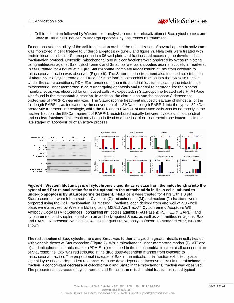
To demonstrate the utility of the cell fractionation method the relocalization of several apoptotic activators was monitored in cells treated to undergo apoptosis (Figure 6 and figure 7). Hela cells were treated with protein kinase c inhibitor Staurosporine in a 96 well plate and fractionated according the developed cell fractionation protocol. Cytosolic, mitochondrial and nuclear fractions were analyzed by Western blotting using antibodies against Bax, cytochrome c and Smac, as well as antibodies against subcellular markers. In cells treated for 4 hours with 1 µM Staurosporine, complete relocalization of Bax from cytosolic to mitochondrial fraction was observed (Figure 6). The Staurosporine treatment also induced redistribution of about 65 % of cytochrome c and 40% of Smac from mitochondrial fraction into the cytosolic fraction. Under the same conditions, PDH E1α remained in the mitochondrial fraction indicating the intactness of mitochondrial inner membrane in cells undergoing apoptosis and treated to permeabilize the plasma membrane, as was observed for uninduced cells. As expected, in Staurosporine treated cells F1-ATPase was found in the mitochondrial fraction. In addition, the distribution and the caspase-3-dependent proteolysis of PARP-1 was analyzed. The Staurosporine treatment induced cleavage of almost all of the full-length PARP-1, as indicated by the conversion of 113 kDa full-length PARP-1 into the typical 89 kDa proteolytic fragment. Interestingly, while the full-length PARP-1 of untreated cells was found mostly in the nuclear fraction, the 89kDa fragment of PARP-1 redistributed equally between cytosolic, mitochondrial and nuclear fractions. This result may be an indication of the lost of nuclear membrane intactness in the late stages of apoptosis or of an active process.
Figure 6. Western blot analysis of cytochrome c and Smac release from the mitochondria into the cytosol and Bax relocalization from the cytosol to the mitochondria in HeLa cells induced to undergo apoptosis by Staurosporine treatment. HeLa cells were treated for 4 hrs with 1.0 µM Staurosporine or were left untreated. Cytosolic (C), mitochondrial (M) and nuclear (N) fractions were prepared using the Cell Fractionation HT method. Fractions, each derived from one well of a 96-well plate, were analyzed by Western blotting using MSA12 ApoTrack™ Cytochrome c Apoptosis WB Antibody Cocktail (MitoSciences), containing antibodies against F1-ATPase α, PDH E1 α, GAPDH and cytochrome c, and supplemented with an antibody against Smac, as well as with antibodies against Bax and PARP. Representative blots as well as the quantitative analysis (mean +/- standard error, n=2) are shown.
The redistribution of Bax, cytochrome c and Smac was further analyzed in greater details in cells treated with variable doses of Staurosporine (Figure 7). While mitochondrial inner membrane marker (F1-ATPase α) and mitochondrial matrix marker (PDH E1 α) remained in the mitochondrial fraction at all concentration of Staurosporine, Bax was redistributed in the drug dose-dependent manner from cytosolic to mitochondrial fraction. The proportional increase of Bax in the mitochondrial fraction exhibited typical sigmoid type of dose-dependent response. With the dose-dependent increase of Bax in the mitochondrial fraction, a concomitant decrease of cytochrome c and Smac in the mitochondrial fraction was observed. The proportional decrease of cytochrome c and Smac in the mitochondrial fraction exhibited typical
ICE Application Note
Telephone: 1-800-910-6486 or 541-284-1800 · Fax: 541-284-1801 www.mitosciences.com
sigmoid type of dose-dependent response. These data show tight coupling of these two reciprocal relocalizations, and support the well established sequence of events in which Bax relocalization to mitochondrial outer membrane is followed by the cytochrome c and Smac release from the mitochondrial outer membrane space into the cytosol [1]. In addition, the full-length PARP-1 was in a dose-dependent manner cleaved to the 89 kDa fragment and, in a later stage, about two-thirds of the 89 kDa fragment were released from the nuclear fraction and re-distributed equally between mitochondrial and cytosolic fractions.
Figure 7. Staurosporine dose-dependent release of cytochrome c and Smac from the mitochondria into the cytosol and Bax relocalization from the cytosol to mitochondria in HeLa cells. Cytosolic (C), mitochondrial (M) and nuclear (N) fractions of HeLa cells treated for 4 hrs with 0.00, 0.02, 0.06, 0.18, 0.54, 1.62, 4.86 and 14.58 µM Staurosporine were prepared using the Cell Fractionation HT method. Fractions, each derived from one well of a 96-well plate, were analyzed as in Figure 6. Representative blots as well as the quantitative analysis are shown. III. Cell fractionation followed by quantitative cytochrome c sandwich ELISA to monitor relocalization of
cytochrome c in HeLa cells induced to undergo apoptosis by Staurosporine treatments. The great advantage of the cell fractionation method described in this paper is the throughput of the method; the 96 samples can be separated into the cytosolic, mitochondrial and nuclear fractions in less than one hour. The fraction analysis by Western blotting is however a low throughput semi-quantitative technique. To demonstrate the full utility of the high throughput fractionation, the cytochrome c was assayed in each fraction by quantitative sandwich ELISA to demonstrate the cytochrome c relocalization into the cytosol in HeLa cells treated with Staurosporine. Cytosolic, mitochondrial and nuclear fractions, all three derived from one well of 96 well plate were prepared in quadruplicates and each of them was
ICE Application Note
Telephone: 1-800-910-6486 or 541-284-1800 · Fax: 541-284-1801 www.mitosciences.com
assayed independently using Cytochrome c Protein Quantity Microplate Assay Kit (MitoSciences) (Figure 8). As observed for the fractions analyzed by Western Blotting the proportional decrease of cytochrome c in the mitochondrial fraction exhibited typical sigmoid type of dose-dependent response. The data analysis revealed that the Staurosporine EC50 of cytochrome c release from mitochondria is 0.42 µM. When parallel-prepared fractions were analyzed by Western blotting, the data analysis revealed Staurosporine EC50 of cytochrome c release from mitochondria equal 0.35 µM. Thus both techniques of cytochrome c quantification yielded nearly identical IC50 values.
Figure 8. Quantitative ELISA analysis of cytochrome c release from the mitochondria into the cytosol in HeLa cells induced to undergo apoptosis by Staurosporine treatment. Cytosolic (C), mitochondrial (M) and nuclear (N) fractions of HeLa cells treated for 4 hrs with 0.00, 0.02, 0.06, 0.18, 0.54, 1.62, 4.86 and 14.58 µM Staurosporine (A and B) or with 0.0 and 1.0 µM Staurosporine (C, D, E) were prepared using the Cell Fractionation HT method. Fractions, each derived from one well of a 96-well plate, were analyzed by ELISA Cytochrome c Protein Quantity Microplate Assay Kit (MitoSciences) (A, C and D). Parallel analyses of fractions prepared independently and thus showing the inter-assay variation of the Cell Fractionation HT method are shown in C and D. Western blot analyses of cytochrome c using MSA12 ApoTrack™ Cytochrome c Apoptosis WB Antibody Cocktail (MitoSciences), described in Figures 6 and 7, are shown for comparison (B and E). Data represent mean +/- standard error of the mean, n=4 (A and C), n=3 (D), n=2 (E), n=1 (B). Conclusions: I. Cell Fractionation HT methodology. The sequential, detergent-based cell fractionation method allows a simple and rapid separation of adherent cells into cytosol-, mitochondria-, and nuclei-containing fractions that exhibit minimum cross-contamination while no cell material is being lost. The HT method is designed for parallel fractionation of a large number of small samples of adherent cells in a microplate format. It allows a preparation of
ICE Application Note
Telephone: 1-800-910-6486 or 541-284-1800 · Fax: 541-284-1801 www.mitosciences.com
cytosolic, mitochondrial and nuclear fractions from 96 cell samples in one hour. The fractions prepared by this method are particularly suitable for high throughput sandwich ELISA microplate assays or dot blot analysis but sufficient also for Western blotting.
II. Validation of the Cell Fractionation HT methodology on the proteins known to translocate between
cytosol and mitochondria in cells undergoing apoptosis.
The fractions generated by this methodology were validated not only by the analysis of appropriate standard subcellular marker but also by following well known relocalization of several activators of apoptosis between cytosol and mitochondria in cells induced to undergo apoptosis by Staurosporine treatments. The fraction analyses demonstrated that staurosporine induced concentration-dependent release of cytochrome c (EC50 = 0.4 µM) and Smac (EC50 = 0.5 µM) from the mitochondria into the cytosol, and concentration-dependent translocation of Bax (EC50 = 0.4 µM) from the cytosol into the mitochondria. In addition, the fraction analysis confirmed mitochondrial localization of Bak in healthy and apoptotic cells and cleavage of cytosol-localized pro-caspase-3 in apoptotic cells (data not shown), as well as cleavage of nuclear-localized full length PARP-1 in apoptotic cells. Our cell fractionation methodologies has been already successfully used to assay the cytochrome c release from mitochondria in Vero cells infected with simian varicella virus [2], and in Cos cells transfected with adipocyte-specific protein FSP27 protein [3]. III. Other possible uses of this methodology
This cell fractionation methodology is well suited to monitor movements of other apoptosis inducers (e.g. AIF, Bad, Bid, Endo G, HtrA2), of kinases and transcription factors (e.g. p53) during apoptosis, and in general to follow other protein movements that involve protein re-localization between cytosol mitochondria and nucleus. The fractionation can be used to separate isoforms of enzymes that have differential distribution, for activity assays. Our cell fractionation methodology was used to demonstrate relocalization of hormone sensitive lipase (HSL) and acute regulatory protein (StAR) in prostate cancer (CaP) tumor cells under specific conditions [4]. References: 1. Tait, S. W. and D. R. Green "Mitochondria and cell death: outer membrane permeabilization and
beyond." Nat Rev Mol Cell Biol 11(9): 621-32. 2. Pugazhenthi, S., D. H. Gilden, et al. (2009). "Simian varicella virus induces apoptosis in monkey
kidney cells by the intrinsic pathway and involves downregulation of bcl-2 expression." J Virol 83(18): 9273-82.
3. Liu, K., S. Zhou, et al. (2009). "Functional analysis of FSP27 protein regions for lipid droplet localization, caspase-dependent apoptosis, and dimerization with CIDEA." Am J Physiol Endocrinol Metab 297(6): E1395-413.
4. Locke, J. A., E. S. Guns, et al. "Arachidonic acid activation of intratumoral steroid synthesis during prostate cancer progression to castration resistance." Prostate 70(3): 239-51.
ICE Application Note
Telephone: 1-800-910-6486 or 541-284-1800 · Fax: 541-284-1801 www.mitosciences.com