Cell Host & Microbe Article Phospholipid Scramblase 1 Mediates Type I Interferon-Induced Protection against Staphylococcal a-Toxin Miroslaw Lizak 1 and Timur O. Yarovinsky 1, * 1 Section of Cardiovascular Medicine, Department of Internal Medicine, Yale University School of Medicine, New Haven, CT, USA *Correspondence: [email protected]DOI 10.1016/j.chom.2011.12.004 SUMMARY The opportunistic gram-positive pathogen Staphylo- coccus aureus is a leading cause of pneumonia and sepsis. Staphylococcal a-toxin, a prototypical pore-forming toxin, is a major virulence factor of S. aureus clinical isolates, and lung epithelial cells are highly sensitive to a-toxin’s cytolytic activity. Type I interferon (IFN) signaling activated in response to S. aureus increases pulmonary cell resistance to a-toxin, but the underlying mechanisms are unchar- acterized. We show that IFNa protects human lung epithelial cells from a-toxin-induced intracellular ATP depletion and cell death by reducing extracel- lular ATP leakage. This effect depends on protein palmitoylation and induction of phospholipid scram- blase 1 (PLSCR1). IFNa-induced PLSCR1 associates with the cytoskeleton after exposure to a-toxin, and cellular depletion of PLSCR1 negates IFN-induced protection from a-toxin. PLSCR1-deficient mice display enhanced sensitivity to inhaled a-toxin and an a-toxin-producing S. aureus strain. These results uncover PLSCR1 activity as part of an innate protec- tive mechanism to a bacterial pore-forming toxin. INTRODUCTION Staphylococcus aureus is an opportunistic gram-positive path- ogen and the leading cause of severe and life-threatening infec- tions, including pneumonia and sepsis (Kallen et al., 2010; Schreiber et al., 2011). Many clinical isolates of S. aureus produce a-toxin, a prototypical pore-forming toxin and a major virulence factor (Bartlett et al., 2008; Menzies and Kernodle, 1996; O’Reilly et al., 1986; Wilke and Bubeck Wardenburg, 2010). Human epithelial cells express ADAM10, a high-affinity protein receptor for a-toxin, which makes them intrinsically sensitive to the cytotoxic effects of a-toxin (Wilke and Bubeck Wardenburg, 2010). Formation of a-toxin pores in the plasma membrane of sensitive cells leads to major changes in the gradients of ions across the membrane, depletion of intracellular ATP, activation of pro-nflammatory cascades, and ultimate cell death (Bhakdi and Tranum-Jensen, 1991; Craven et al., 2009; Pre ´ vost et al., 2001; Ratner et al., 2006; Rose et al., 2002). Excessive inflammation and death of respiratory epithelial cells triggered by a-toxin during pneumonia contribute to acute lung injury and worsen the outcome of infection (Bartlett et al., 2008; Bubeck Wardenburg and Schneewind, 2008; Rose et al., 2002). Neutralization of the cytolytic activity of a-toxin protects animals from S. aureus (Bubeck Wardenburg and Schneewind, 2008; Kennedy et al., 2010; McCormick et al., 2009; Menzies and Kernodle, 1996; Ragle and Bubeck Wardenburg, 2009), suggesting that a-toxin is a promising target for intervention during staphylococcal infections. Cellular defense against a-toxin and other pore-forming toxins relies on constriction of the a-toxin pores (Valeva et al., 2000), activation of mitogen-activated protein kinases (Husmann et al., 2006), induction of lipogenic genes via activation of sterol response element binding proteins (SREBPs) by caspase-1 (Gurcel et al., 2006), and accelerated endocytosis and exocy- tosis (Husmann et al., 2009; Idone et al., 2008). Further charac- terization of host-intrinsic mechanisms of resistance to a-toxin should facilitate development of new therapeutic approaches for staphylococcal infection. We have previously reported that type I interferons (IFNs) increase cell resistance to a-toxin, presumably via induction of interferon-regulated genes involved in lipid metabolism (Yarovin- sky et al., 2008). However, activation of type I IFN signaling by staphylococcal protein A increased inflammation in the lungs (Martin et al., 2009). These opposing effects of I IFNs may be exerted through transcriptional regulation of distinct subsets of IFN-regulated genes. Therefore, it is important to identify which IFN-regulated genes and pathways are necessary for protection from a-toxin. In this study, we focused on human lung epithelial cells since they are highly sensitive to a-toxin and represent the first line of defense against S. aureus during respiratory infections. We demonstrated that IFNa protects human lung epithelial cells from a-toxin-induced cell death by reducing release of cellular ATP into extracellular space. Using an ATP-based cell viability screening assay and bioinformatics analyses, we found that IFNa-induced protection from a-toxin is dependent on protein palmitoylation and induction of phospholipid scramblase 1 (PLSCR1). Increased expression of PLSCR1 has been previously implicated in bidirectional translocation of membrane phospho- lipids across plasma membrane and amplification of transcrip- tional responses to type I IFNs (Dong et al., 2004; Stout et al., 1997; Zhou et al., 2000). Here, we show that IFNa-induced protection from a-toxin correlates with accumulation of PLSCR1 in the cytoskeleton-associated protein fractions. 70 Cell Host & Microbe 11, 70–80, January 19, 2012 ª2012 Elsevier Inc.
Transcript
Cell Host & Microbe
Article
Phospholipid Scramblase 1 MediatesType I Interferon-Induced Protectionagainst Staphylococcal a-ToxinMiroslaw Lizak1 and Timur O. Yarovinsky1,*1Section of Cardiovascular Medicine, Department of Internal Medicine, Yale University School of Medicine, New Haven, CT, USA
The opportunistic gram-positive pathogen Staphylo-coccus aureus is a leading cause of pneumoniaand sepsis. Staphylococcal a-toxin, a prototypicalpore-forming toxin, is a major virulence factor ofS. aureus clinical isolates, and lung epithelial cellsare highly sensitive to a-toxin’s cytolytic activity.Type I interferon (IFN) signaling activated in responseto S. aureus increases pulmonary cell resistance toa-toxin, but the underlying mechanisms are unchar-acterized. We show that IFNa protects human lungepithelial cells from a-toxin-induced intracellularATP depletion and cell death by reducing extracel-lular ATP leakage. This effect depends on proteinpalmitoylation and induction of phospholipid scram-blase 1 (PLSCR1). IFNa-induced PLSCR1 associateswith the cytoskeleton after exposure to a-toxin, andcellular depletion of PLSCR1 negates IFN-inducedprotection from a-toxin. PLSCR1-deficient micedisplay enhanced sensitivity to inhaled a-toxin andan a-toxin-producing S. aureus strain. These resultsuncover PLSCR1 activity as part of an innate protec-tive mechanism to a bacterial pore-forming toxin.
INTRODUCTION
Staphylococcus aureus is an opportunistic gram-positive path-
ogen and the leading cause of severe and life-threatening infec-
tions, including pneumonia and sepsis (Kallen et al., 2010;
Schreiber et al., 2011). Many clinical isolates of S. aureus
produce a-toxin, a prototypical pore-forming toxin and a major
virulence factor (Bartlett et al., 2008; Menzies and Kernodle,
1996; O’Reilly et al., 1986; Wilke and Bubeck Wardenburg,
2010). Human epithelial cells express ADAM10, a high-affinity
protein receptor for a-toxin, which makes them intrinsically
sensitive to the cytotoxic effects of a-toxin (Wilke and Bubeck
Wardenburg, 2010). Formation of a-toxin pores in the plasma
membrane of sensitive cells leads to major changes in the
gradients of ions across the membrane, depletion of intracellular
ATP, activation of pro-nflammatory cascades, and ultimate
cell death (Bhakdi and Tranum-Jensen, 1991; Craven et al.,
2009; Prevost et al., 2001; Ratner et al., 2006; Rose et al.,
70 Cell Host & Microbe 11, 70–80, January 19, 2012 ª2012 Elsevier I
2002). Excessive inflammation and death of respiratory epithelial
cells triggered by a-toxin during pneumonia contribute to acute
lung injury and worsen the outcome of infection (Bartlett et al.,
2008; Bubeck Wardenburg and Schneewind, 2008; Rose et al.,
2002). Neutralization of the cytolytic activity of a-toxin protects
animals from S. aureus (Bubeck Wardenburg and Schneewind,
2008; Kennedy et al., 2010; McCormick et al., 2009; Menzies
and Kernodle, 1996; Ragle and Bubeck Wardenburg, 2009),
suggesting that a-toxin is a promising target for intervention
during staphylococcal infections.
Cellular defense against a-toxin and other pore-forming toxins
relies on constriction of the a-toxin pores (Valeva et al., 2000),
activation of mitogen-activated protein kinases (Husmann
et al., 2006), induction of lipogenic genes via activation of sterol
response element binding proteins (SREBPs) by caspase-1
(Gurcel et al., 2006), and accelerated endocytosis and exocy-
tosis (Husmann et al., 2009; Idone et al., 2008). Further charac-
terization of host-intrinsic mechanisms of resistance to a-toxin
should facilitate development of new therapeutic approaches
for staphylococcal infection.
We have previously reported that type I interferons (IFNs)
increase cell resistance to a-toxin, presumably via induction of
interferon-regulated genes involved in lipidmetabolism (Yarovin-
sky et al., 2008). However, activation of type I IFN signaling by
staphylococcal protein A increased inflammation in the lungs
(Martin et al., 2009). These opposing effects of I IFNs may be
exerted through transcriptional regulation of distinct subsets of
IFN-regulated genes. Therefore, it is important to identify which
IFN-regulated genes and pathways are necessary for protection
from a-toxin.
In this study, we focused on human lung epithelial cells since
they are highly sensitive to a-toxin and represent the first line of
defense against S. aureus during respiratory infections. We
demonstrated that IFNa protects human lung epithelial cells
from a-toxin-induced cell death by reducing release of cellular
ATP into extracellular space. Using an ATP-based cell viability
screening assay and bioinformatics analyses, we found that
IFNa-induced protection from a-toxin is dependent on protein
palmitoylation and induction of phospholipid scramblase 1
(PLSCR1). Increased expression of PLSCR1 has been previously
implicated in bidirectional translocation of membrane phospho-
lipids across plasma membrane and amplification of transcrip-
tional responses to type I IFNs (Dong et al., 2004; Stout et al.,
1997; Zhou et al., 2000). Here, we show that IFNa-induced
protection from a-toxin correlates with accumulation of PLSCR1
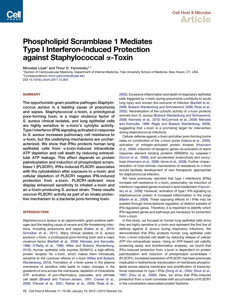
Figure 1. IFNa Protects Lung Epithelial Cells from a-Toxin-Induced Cell Death and Depletion of Intracellular ATPHuman primary SAECs were pretreated with medium or IFNa (1000 U/ml) for 24 hr and treated with a-toxin (0.1 mg/ml) for additional 24 hr.
(A) Representative photomicrographs of cells stained with calcein AM (green, live) and EthD-1 (red, dead). The scale bar represents 100 mm.
(B) Percent dead cells (mean ± SD of five independent experiments, each carried out in quadruplicate wells).
(C) Relative levels of intracellular ATP (perecent remaining, relative to untreated cells at each time point) weremeasured in SAECs at the indicated time points after
a-toxin (mean ± SD of quadruplicate wells; the data are representative of five experiments).
(D) Relative ATP levels in IFNa-pretreated A549 cells at 6 hr after a-toxin (mean ± SD of five independent experiments, each carried out in quadruplicate wells).
(E and F) Relative ATP levels in medium- or IFNa-pretreated A549 cells after incubation with conditioned medium (diluted 1:100) from cultures of Hla+ or Hla–
S. aureus. The data (mean ± SD of quadruplicate wells) are representative of three independent experiments.
Cell Host & Microbe
Scramblase 1 and Staphylococcal Alpha-Toxin
RESULTS
IFNa Reduces a-Toxin-Induced Cell Death and Releaseof Extracellular ATPExposure of human primary small airway epithelial cells (SAECs)
to staphylococcal a-toxin for 24 hr resulted in significant cell
death, which was preceded by rapid depletion of intracellular
ATP (Figure 1). Pretreatment of SAECs with IFNa for 24 hr signif-
icantly inhibited depletion of ATP and reduced cell death trig-
gered by a-toxin. Although higher concentrations of a-toxin
(0.5–2.5 mg/ml) were required to trigger ATP depletion in A549
cells, it was similarly inhibited by IFNa pretreatment (Figure 1D).
Cell H
Pretreatment with IFNa also inhibited ATP depletion from
A549 cells incubated with conditioned medium from a-toxin-
producing strain of S. aureus (Figures 1E and 1F). These data
indicate that type I IFNs protect lung epithelial cells from the
cytotoxic effects of a-toxin.
To determine how IFNa protects cells from a-toxin, we
evaluated the early events after exposure to a-toxin. IFNa
pretreatment did not decrease oligomerization of a-toxin, activa-
tion of p38 mitogen-activated protein (MAP) kinase, and efflux of
potassium (Figure S1 available online). Within the first 30 min
of a-toxin exposure, association of a-toxin with ADAM10
or recently reported a-toxin-induced ADAM10-dependent
ost & Microbe 11, 70–80, January 19, 2012 ª2012 Elsevier Inc. 71
Figure 2. Exposure to a-Toxin Triggers Release of eATP that Is In-
hibited by IFNa Pretreatment
(A and B) A549 cells were treated with the indicated concentrations of a-toxin
(A) or diluted conditioned medium from cultures of Hla+ or Hla– S. aureus (B).
The curves show normalized mean luminescence intensity kinetics measured
Cell Host & Microbe
Scramblase 1 and Staphylococcal Alpha-Toxin
72 Cell Host & Microbe 11, 70–80, January 19, 2012 ª2012 Elsevier I
cleavage of E-cadherin (Inoshima et al., 2011) were not affected
by IFNa pretreatment, albeit they were slightly reduced at 2 hr
(Figure S1). To evaluate whether a-toxin triggers release of
ATP into extracellular space, we treated A549 cells with a-toxin
in the presence of luciferin and luciferase in the medium. After
a short lag period, a-toxin triggered a robust increase in ATP-
dependent luminescence peaking between 20 and 40 min with
maximal luminescence intensity observed at 2.5 mg/ml of a-toxin
(Figure 2A). Althoughmany bacterial products such as endotoxin
and staphylococcal peptidoglycan may trigger extracellular
ATP (eATP) release (De Vuyst et al., 2007; Robertson et al.,
2010), we found that a-toxin was a necessary and sufficient
secreted factor of S. aureus for eATP release from lung epithelial
cells: incubation of A549 cells with conditioned medium from
a-toxin-producing Hla+ strain of S. aureus triggered rapid and
robust eATP release, which was virtually nondetectable after
incubation with conditioned medium from isogenic Hla– a-toxin
mutant strain (Figure 2B). Thus, exposure of lung epithelial cells
to a-toxin leads to eATP release.
We also measured eATP in the conditioned medium collected
from cells at 15 min intervals after exposure to a-toxin, which al-
lowed us to avoid luciferase inactivation by prolonged incubation
at 37�C (Seminario-Vidal et al., 2009). The baseline eATP levels
released by A549 cells within 1 hr of culture under those condi-
tions were below 20 nM and not affected by IFNa treatment.
The concentrations of eATP peaked at 30 min after a-toxin and
reached 1805 ± 248 nM eATP. IFNa pretreatment did not change
the kinetics of eATP release after a-toxin treatment but signifi-
cantly reduced peak eATP concentrations to 1341 ± 170 nM
(p < 0.01; the data are mean ± SEM of seven independent exper-
iments each performed in quadruplicate cultures). Treatment of
human primary SAECs with a-toxin also triggered eATP release,
albeit peak eATP concentrations were much lower than in A549
cells (Figure 2C). Importantly, IFNa pretreatment of SAEC signif-
icantly decreased eATP levels after a-toxin exposure.
The reduction in a-toxin-induced eATP concentrations in
IFNa-pretreated cells could not be explained by changes in
every minute within 1 hr after a-toxin. Data in (A) are mean of quadruplicate
cultures and are representative of two independent experiments. Data in (B)
are mean ± SEM of triplicate cultures and are representative of three inde-
pendent experiments.
(C) SAECs were pretreated with IFNa for 24 hr and exposed to 0.1 mg/ml
a-toxin for 30 min. Cell-free conditioned medium was used to measure eATP.
The data are mean ± SD of quadruplicate cultures and are representative of
three independent experiments.
(D) Exogenous ATP was added to medium- or IFNa-pretreated A549 cells for
30 min. Cell-free conditioned medium was used to measure remaining ATP
(percent input, the data are mean ± SD of quadruplicate cultures and are
representative of three experiments).
(E) A549 cells were pretreated with oxidized ATP for 2 hr prior to exposure to
2.5 mg/ml a-toxin. Cell death at 24 hr after a-toxin is shown. The data are
mean ± SD of quadruplicate cultures and are representative of three inde-
pendent experiments.
(F) C57BL6/ mice (8 weeks old, females) were administered a-toxin or
a-toxin with oxidized ATP diluted in 50 ml sterile PBS via intranasal route.
Body temperature was measured at the indicated time points. The data are
mean ± SD, n = 5. Control mice received 50 ml sterile PBS. Asterisks indicate
the time points when the body temperature in mice treated with a-toxin alone
was significantly lower than in other groups of mice (p < 0.05).
See also Figure S1.
nc.
Cell Host & Microbe
Scramblase 1 and Staphylococcal Alpha-Toxin
eATP hydrolysis. After 30 min incubation with 10–100 nM of
exogenous ATP, the remaining ATP levels were similar between
medium- and IFNa-pretreated cells. IFNa pretreatment actually
slowed degradation of exogenously added ATP at 1000 nM
concentration range (Figure 2D).
Extracellular nucleotides may contribute to cell lysis by
bacterial pore-forming toxins through autocrine or paracrine
activation of P2 receptors (Skals et al., 2009). We found that
pretreatment of A549 cells with 500–1000 mM of oxidized ATP
(a nonselective antagonist of P2 receptors) significantly inhibited
a-toxin-induced cell death (Figure 2E). Moreover, oxidized
ATP protected mice from hypothermia induced by intranasal
administration of sublethal doses of a-toxin (Figure 2F). These
data suggest that a-toxin-induced release of eATP and, probably
other nucleotides, contribute to the cytotoxic effects of a-toxin
via P2 receptors.
Identification of PLSCR1 as a Candidate Gene Involvedin IFNa-Induced Protection from a-ToxinTo characterize signaling pathways involved in IFNa-induced
protection from a-toxin, we carried out a screening assay in
A549 cells treated with IFNa, chemical inhibitors and a-toxin as
depicted in Figure S2. By measuring remaining intracellular
ATP after a-toxin treatment as the readout, we found that
IFNa-induced protection from a-toxin was not affected by inhib-
itors of caspase-1 (Ac-YVAD-FMK, 10 mM), sterol response
element binding proteins (25-OH-cholesterol, 50 mM), ERK
MAP kinase (UO126, 10 mM), and EGFR tyrosine kinase
(AG1478, 15 mM) (Figure S2). Inhibitors of caspases (zVAD-
FMK as a pan-caspase inhibitor, 10 mM), gene transcription
(actinomycin D, 2.5 mg/ml), protein synthesis (cycloheximide,
50 mg/ml), and removal of serum (i.e., treatment of cells in
serum-free medium) showed moderate, but statistically sig-
nificant, protection of both medium- and IFNa-pretreated cells
from a-toxin. However, inhibitors of p38-MAP kinase
(SB203580, 10 mM), PI3 kinase (LY294002, 10 mM), and fatty
acid synthase (cerulenin, 10 mg/ml) significantly affected the
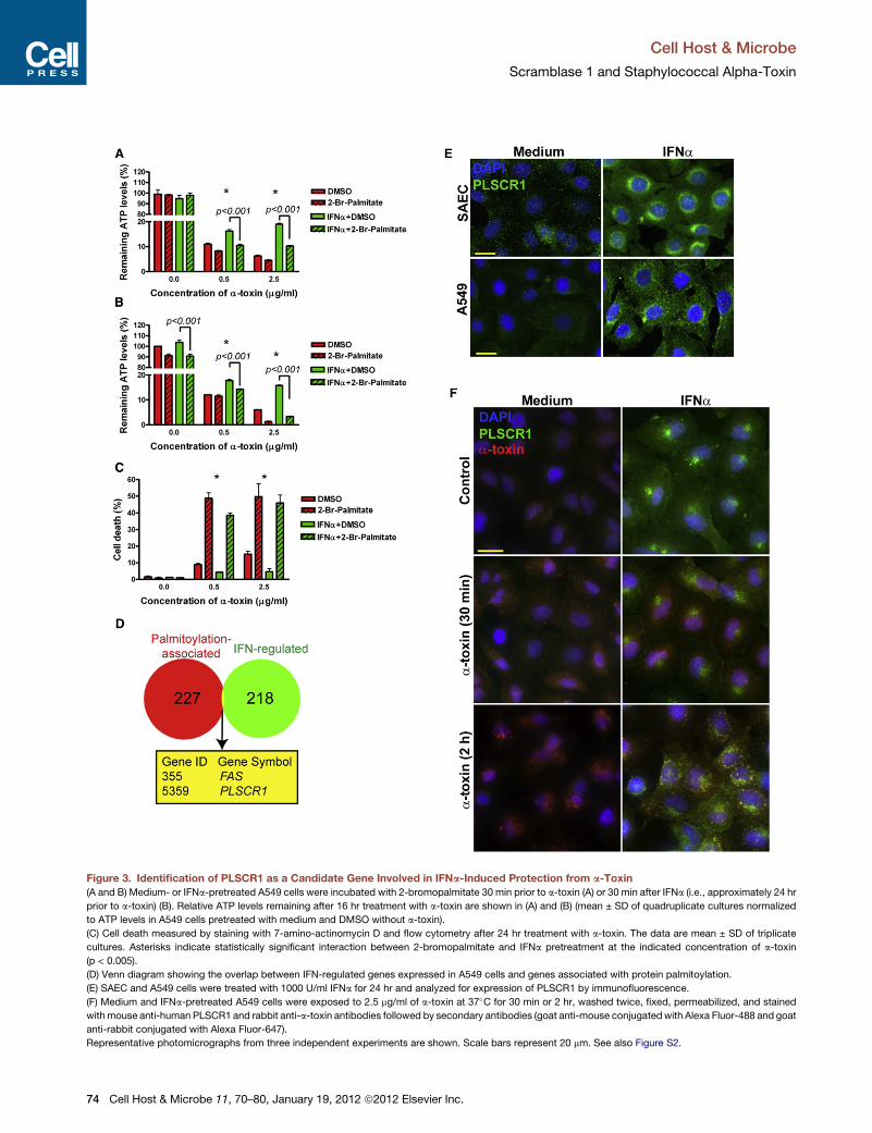
protective effect of IFNa (Figure S2). Furthermore, inhibition of
protein palmitoylation with 2-bromopalmitate (100 mM) effec-
tively wiped out IFNa-induced protection from a-toxin (Fig-
ure 3A). The effects of 2-bromopalmitate were also evident
when it was added to the cells 30 min after IFNa, i.e., approxi-
mately 24 hr prior to a-toxin (Figure 3B). This treatment also
sensitized A549 cells to a-toxin-induced cell death (Figure 3C),
providing additional evidence for involvement of protein palmi-
toylation in protection of lung epithelial cells from a-toxin.
Using palmitoylation and regulation by type I IFNs as selection
criteria, we analyzed the overlap between the 227 human genes
associated with protein palmitoylation (Yang et al., 2010) and
218 IFN-regulated genes expressed in A549 cells (Sanda et al.,
2006) and identified FAS (tumor necrosis factor receptor super-
family, member 6) and PLSCR1 (Figure 3D). We dismissed FAS
since interferons increase its expression, which is more likely
to sensitize cells to FasL-mediated cell death than protect
them from a-toxin (Chawla-Sarkar et al., 2003). Therefore, we
focused our further analyses on PLSCR1 since it is induced by
type I IFNs (Zhou et al., 2000), palmitoylation of PLSCR1 protein
directs it to lipid rafts in plasma membrane (Wiedmer et al.,
2003), where a-toxin preferentially binds (Valeva et al., 2006;
Cell H
Wilke and Bubeck Wardenburg, 2010), and PLSCR1 is abun-
dantly expressed by macrophages and neutrophils (Chen
et al., 2011; Zhou et al., 2002), which are highly resistant to
a-toxin-induced cell death (Valeva et al., 1997).
To determine whether PLSCR1 is induced in lung epithelial
cells, we analyzed PLSCR1 expression in medium- and IFNa-
pretreated human primary SAECs and A549 cells. At the base-
line, PLSCR1 was hardly detectable; however, IFNa treatment
led to robust induction of PLSCR1 protein (Figure 3E). PLSCR1
localized primarily in a perinuclear region, albeit some nuclear
and membrane staining was also evident. Remarkably, after
incubation of IFNa-pretreated cells with a-toxin for 2 hr, most
of the endocytosed a-toxin colocalized with a fraction of
PLSCR1 (Figure 3F). Thus, IFNa-induced PLSCR1 may be in
proximity with a-toxin.
PLSCR1 Is Necessary for IFNa-Induced Protectionfrom a-ToxinTo examine the role of PLSCR1 in IFNa-induced protection from
a-toxin, we suppressed its induction by transfecting cells with
PLSCR1-specific short hairpin RNA (shRNA). A nonsilencing
(NS) control shRNA had no effect on PLSCR1 expression,
whereas PLSCR1-specific shRNA further decreased the base-
line and prevented IFNa-induced expression of PLSCR1 (Figures
4A and 4B). Furthermore, PLSCR1-specific shRNA did not affect
baseline and IFNa-induced expression of STAT1 (Figures 4A and
4C). NS shRNA had no effect on a-toxin-induced ATP depletion
in medium- or IFNa-pretreated A549 cells to a-toxin (Figure 4D).
However, PLSCR1-specific shRNA negated the protective
effects of IFNa on a-toxin-induced depletion of ATP (Figure 4D).
protection from a-toxin-induced cell death (Figure 4E and Fig-
ure S3). These data suggest that induction of PLSCR1 is neces-
sary for IFNa-induced protection from a-toxin.
To explore the role of PLSCR1 in responses to a-toxin in vivo,
we treated PLSCR1 knockout (PLSCR1�/�) mice and littermate
heterozygous (PLSCR1+/�) mice (Zhou et al., 2002) with a-toxin
via intranasal route. PLSCR1 deficiency (Figure 4F) had no effect
on the early a-toxin-induced hypothermia but resulted in
impaired recovery of body temperature (Figure 4G), suggesting
that lack of PLSCR1 increases sensitivity of mice to inhaled
a-toxin. Subsequently, we tested whether PLSCR1 deficiency
alters the outcome of experimental staphylococcal pneumonia
due to infection with a-toxin-producing Hla+ strain of S. aureus
or isogenic a-toxinmutantHla– strain ofS. aureus. When infected
with 2.5 3 108 CFU of Hla+ S. aureus, a larger fraction of
PLSCR1�/� mice developed moribund condition sooner than
PLSCR1+/� littermates (Figure 4H). Infection with Hla+ resulted
in severe hypothermia at 6 hr postinfection (p.i.) in PLSCR1+/�
and PLSCR1�/� mice (Figure 4I). Among the survivors at 16 hr
p.i., three PLSCR1�/� mice and seven PLSCR+/� mice started
recovering their body temperature. These mice eventually
survived the infection with Hla+ S. aureus. Transient and less
severe hypothermia was observed in PLSCR1+/� and
PLSCR1�/� mice infected with the Hla– strain (3.65 3 108 CFU/
mouse). Importantly, all mice recovered their body temperature
by 16 hr and none of the mice were moribund within 72 hr p.i.
with Hla– S. aureus. These data indicate that PLSCR1 deficiency
increases sensitivity of mice to live a-toxin-producing S. aureus.
ost & Microbe 11, 70–80, January 19, 2012 ª2012 Elsevier Inc. 73
Figure 3. Identification of PLSCR1 as a Candidate Gene Involved in IFNa-Induced Protection from a-Toxin
(A and B) Medium- or IFNa-pretreated A549 cells were incubated with 2-bromopalmitate 30 min prior to a-toxin (A) or 30 min after IFNa (i.e., approximately 24 hr
prior to a-toxin) (B). Relative ATP levels remaining after 16 hr treatment with a-toxin are shown in (A) and (B) (mean ± SD of quadruplicate cultures normalized
to ATP levels in A549 cells pretreated with medium and DMSO without a-toxin).
(C) Cell death measured by staining with 7-amino-actinomycin D and flow cytometry after 24 hr treatment with a-toxin. The data are mean ± SD of triplicate
cultures. Asterisks indicate statistically significant interaction between 2-bromopalmitate and IFNa pretreatment at the indicated concentration of a-toxin
(p < 0.005).
(D) Venn diagram showing the overlap between IFN-regulated genes expressed in A549 cells and genes associated with protein palmitoylation.
(E) SAEC and A549 cells were treated with 1000 U/ml IFNa for 24 hr and analyzed for expression of PLSCR1 by immunofluorescence.
(F) Medium and IFNa-pretreated A549 cells were exposed to 2.5 mg/ml of a-toxin at 37�C for 30 min or 2 hr, washed twice, fixed, permeabilized, and stained
withmouse anti-human PLSCR1 and rabbit anti-a-toxin antibodies followed by secondary antibodies (goat anti-mouse conjugatedwith Alexa Fluor-488 and goat
anti-rabbit conjugated with Alexa Fluor-647).
Representative photomicrographs from three independent experiments are shown. Scale bars represent 20 mm. See also Figure S2.
Cell Host & Microbe
Scramblase 1 and Staphylococcal Alpha-Toxin
74 Cell Host & Microbe 11, 70–80, January 19, 2012 ª2012 Elsevier Inc.
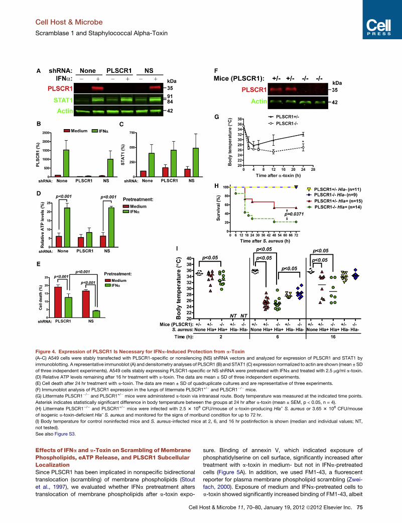
Figure 4. Expression of PLSCR1 Is Necessary for IFNa-Induced Protection from a-Toxin
(A–C) A549 cells were stably transfected with PLSCR1-specific or nonsilencing (NS) shRNA vectors and analyzed for expression of PLSCR1 and STAT1 by
immunoblotting. A representative immunoblot (A) and densitometry analyses of PLSCR1 (B) and STAT1 (C) expression normalized to actin are shown (mean ± SD
of three independent experiments). A549 cells stably expressing PLSCR1-specific or NS shRNA were pretreated with IFNa and treated with 2.5 mg/ml a-toxin.
(D) Relative ATP levels remaining after 16 hr treatment with a-toxin. The data are mean ± SD of three independent experiments.
(E) Cell death after 24 hr treatment with a-toxin. The data are mean ± SD of quadruplicate cultures and are representative of three experiments.
(F) Immunoblot analysis of PLSCR1 expression in the lungs of littermate PLSCR1+/� and PLSCR1�/� mice.
(G) Littermate PLSCR1�/� and PLSCR1+/� mice were administered a-toxin via intranasal route. Body temperature was measured at the indicated time points.
Asterisk indicates statistically significant difference in body temperature between the groups at 24 hr after a-toxin (mean ± SEM, p < 0.05, n = 4).
(H) Littermate PLSCR1�/� and PLSCR1+/� mice were infected with 2.5 3 108 CFU/mouse of a-toxin-producing Hla+ S. aureus or 3.65 3 108 CFU/mouse
of isogenic a-toxin-deficient Hla– S. aureus and monitored for the signs of moribund condition for up to 72 hr.
(I) Body temperature for control noninfected mice and S. aureus-infected mice at 2, 6, and 16 hr postinfection is shown (median and individual values; NT,
not tested).
See also Figure S3.
Cell Host & Microbe
Scramblase 1 and Staphylococcal Alpha-Toxin
Effects of IFNa and a-Toxin on Scrambling of MembranePhospholipids, eATP Release, and PLSCR1 SubcellularLocalizationSince PLSCR1 has been implicated in nonspecific bidirectional
translocation (scrambling) of membrane phospholipids (Stout
et al., 1997), we evaluated whether IFNa pretreatment alters
translocation of membrane phospholipids after a-toxin expo-
Cell H
sure. Binding of annexin V, which indicated exposure of
phosphatidylserine on cell surface, significantly increased after
treatment with a-toxin in medium- but not in IFNa-pretreated
cells (Figure 5A). In addition, we used FM1-43, a fluorescent
reporter for plasma membrane phospholipid scrambling (Zwei-
fach, 2000). Exposure of medium and IFNa-pretreated cells to
a-toxin showed significantly increased binding of FM1-43, albeit
ost & Microbe 11, 70–80, January 19, 2012 ª2012 Elsevier Inc. 75
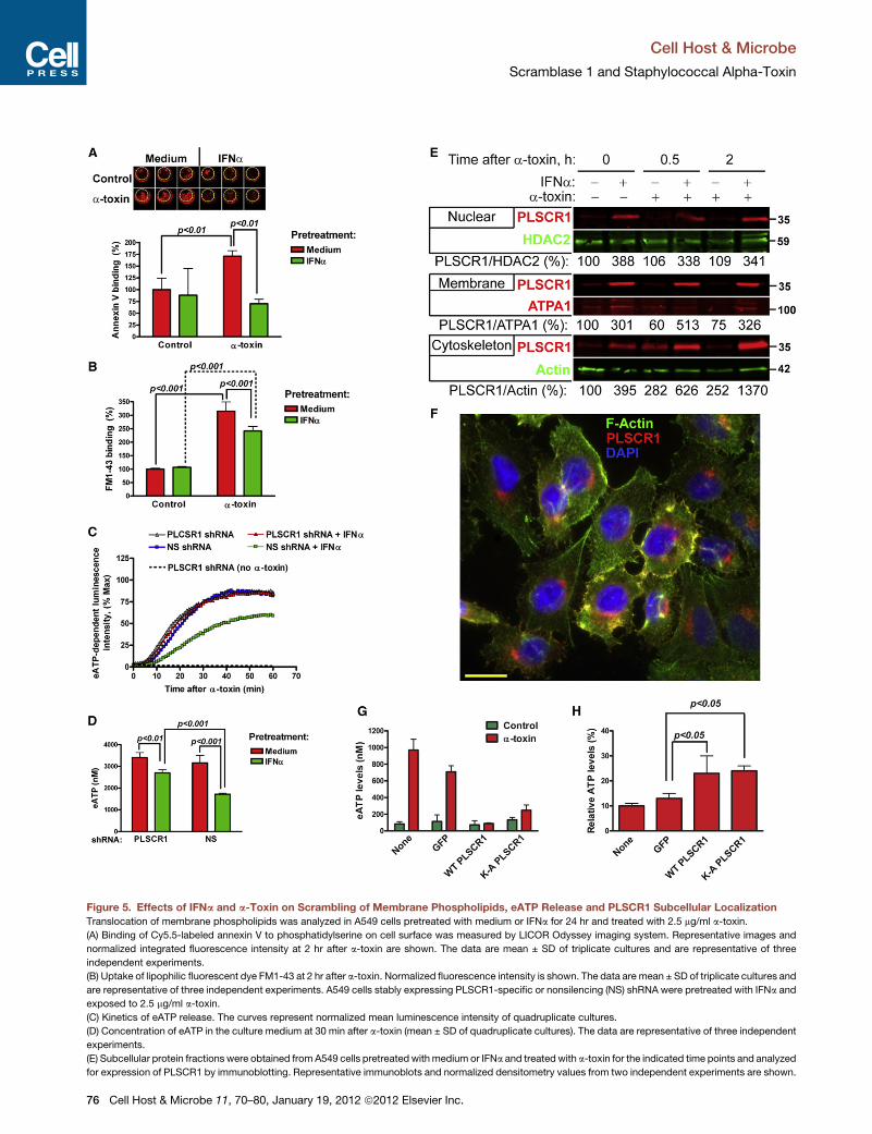
Figure 5. Effects of IFNa and a-Toxin on Scrambling of Membrane Phospholipids, eATP Release and PLSCR1 Subcellular Localization
Translocation of membrane phospholipids was analyzed in A549 cells pretreated with medium or IFNa for 24 hr and treated with 2.5 mg/ml a-toxin.
(A) Binding of Cy5.5-labeled annexin V to phosphatidylserine on cell surface was measured by LICOR Odyssey imaging system. Representative images and
normalized integrated fluorescence intensity at 2 hr after a-toxin are shown. The data are mean ± SD of triplicate cultures and are representative of three
independent experiments.
(B) Uptake of lipophilic fluorescent dye FM1-43 at 2 hr after a-toxin. Normalized fluorescence intensity is shown. The data are mean ± SD of triplicate cultures and
are representative of three independent experiments. A549 cells stably expressing PLSCR1-specific or nonsilencing (NS) shRNA were pretreated with IFNa and
exposed to 2.5 mg/ml a-toxin.
(C) Kinetics of eATP release. The curves represent normalized mean luminescence intensity of quadruplicate cultures.
(D) Concentration of eATP in the culture medium at 30 min after a-toxin (mean ± SD of quadruplicate cultures). The data are representative of three independent
experiments.
(E) Subcellular protein fractions were obtained from A549 cells pretreatedwith medium or IFNa and treated with a-toxin for the indicated time points and analyzed
for expression of PLSCR1 by immunoblotting. Representative immunoblots and normalized densitometry values from two independent experiments are shown.
Cell Host & Microbe
Scramblase 1 and Staphylococcal Alpha-Toxin
76 Cell Host & Microbe 11, 70–80, January 19, 2012 ª2012 Elsevier Inc.
Cell Host & Microbe
Scramblase 1 and Staphylococcal Alpha-Toxin
the increase was less dramatic in IFNa-pretreated cells (Fig-
ure 5B). Thus, two independent assays showed that increased
expression of PLSCR1 in IFNa-pretreated cells does not corre-
late with translocation of membrane phospholipids after a-toxin
exposure.
To define how PLSCR1 mediates IFNa-induced protection
from a-toxin, we measured eATP release from A549 cells stably
expressing NS or PLSCR1-specific shRNA. In the absence of
IFNa pretreatment, A549 cells stably expressing NS or
PLSCR1-specific shRNA released similarly high levels of eATP
in response to a-toxin (Figures 5C and 5D). Pretreatment with
IFNa significantly reduced eATP release from the cells express-
ing NS shRNA. However, the response to IFNa pretreatment
was attenuated in the cells expressing PLSCR1-specific shRNA.
These data indicated that IFNa-induced PLSCR1 is necessary
for reduction of eATP release after exposure to a-toxin.
Nuclear translocation of PLSCR1 augments transcriptional
responses to type IFNs and G-CSF (Chen et al., 2011; Dong
et al., 2004). To provide further insight into how IFNa-induced
PLSCR1may protect cells from a-toxin, we analyzed subcellular
protein fractions for the presence of PLSCR1 after treatment
with IFNa and/or a-toxin. After IFNa pretreatment, PLSCR1
accumulated in the nuclear, membrane, and cytoskeleton-
associated fractions (Figure 5E). Treatment with a-toxin had
no effect on the amounts of PLSCR1 in the nuclear fractions (Fig-
ure 5E, top). After exposure to a-toxin, membrane-associated
PLSCR1 decreased in medium-pretreated cells, but increased
in IFNa-pretreated cells (Figure 5E, middle). Remarkably, treat-
ment with a-toxin led to sustained increases of PLSCR1 in the
cytoskeleton-associated fractions, both in the medium- and