Cell Reproduction Disclaimer: Please remember that none of the information provided is in any way an encouragement for students to “investigate” on their own All “experiments” must be approved by: teacher, parents/guardians, and a possible religious leader of your choice Thank you and enjoy the show
Transcript
Cell Reproduction
Disclaimer:Please remember that none of the information provided is in any way an encouragement for students to “investigate” on their ownAll “experiments” must be approved by: teacher, parents/guardians, and a possible religious leader of your choiceThank you and enjoy the show
Structure of a Chromosome
Structure (cont.)
Rod-shaped made of DNA wrapped around proteins In Prokaryotes, DNA is circular and have
associated proteins In Eukaryotes, the proteins are histones
Maintain shape Aid in tight packing of DNA
Nonhistone proteins Involved in controlling activities of specific regions
of DNA
Structure (cont.)
Made of two chromatids attached at the centromere
These form as DNA copies itself
Chromatin
Between cell divisions, DNA loosens up and (under a microscope in most situations) chromatin forms
Easier to read Information can be used to direct cells
activities
Chromosome NumbersA human adult – 40+ Sex chromosomes are 2
of the 46 -determine the sex of the offspring-carry genes for other characteristics-humans are X or Y
Autosomes
All other chromosomes Every cell of organism produced by sexual
reproduction Autosome from mother + Autosome from
father = Homologous chromosome Carry genes for same trait Same size and shape
46 Human Chromosomes
22 Homologous Pairs of Autosomes 2 Sex Chromosomes
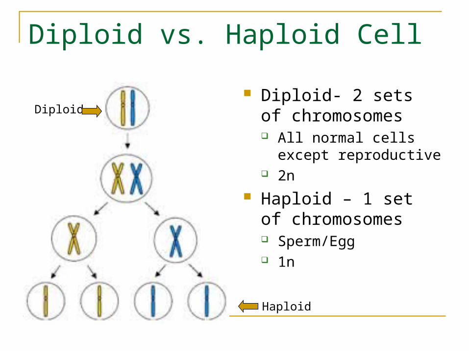
Diploid vs. Haploid Cell
Diploid- 2 sets of chromosomes All normal cells except
reproductive 2n
Haploid – 1 set of chromosomes Sperm/Egg 1n
Diploid
Haploid
Cell Cycle
Interphase – precedes mitosis DNA duplicates Nucleolus may be
visible in this phase, however, the chromosomes are not visible
Cell is involved in metabolic activity
Nucleolus
How do you knowthis is a plant cell?
Nuclear membrane
CellWall
Prophase
This stage is the first stage of cell division Chromosomes become more visible Chromatin fibers are thick enough to be seen
with a light microscope. Sister chromatids joined at the centromere
can be seen under high power. Late in this phase the nuclear envelope
breaks down (looks like dashes) Mitotic spindle (made from microtubles) forms
in the cytoplasm – chromatids attach to microtubles; spindle is football shaped; spindle tugs chromosomes towards middle of cell.
Cell wall
NuclearEnvelope – Begins breakingdown
Metaphase
This stage is very brief Chromosomes gather in the Middle Mitotic spindle is completely formed
and all chromosomes are attached to these microtubules
Centromeres are lined up about halfway between the two poles
Mitotic Spindle
Chromosomes are lined up in center
Poles
Anaphase
This is the third stage Sister chromatids suddenly separate
and are now considered “daughter” chromosomes
Microtubules shorten, pulling the chromosomes closer to the poles
Spindle microtubules that are not attached to the centromeres grow longer and push the poles farther apart.
Poles
“Daughter”chromosomespulled towardspoles
Spindle attachedto chromosomes
Spindle elongatesand stretches polesapart
Telophase and Cytokinesis
This is the final stage of mitosis Chromosomes reach the poles of the
spindle Spindle disappears Two nuclear envelopes form around each
new daughter chromosome Chromosomes uncoil and lengthen Nucleoli reappear Cytokinesis divides the cytoplasm into two
daughter cells, each with a new nucleus – this usually occurs along with telophase
Cytokinesis separates thecytoplasm
Interphase
This stage starts the whole cycle over again.
MeiosisCell division in eukaryotes
Meiosis vs. Mitosis
Yet another comparison…
Meiosis Outcome
The products of meiosis are 4 Haploid daughter cells: Sperm cells Egg cell and polar bodies
Meiosis I and Meiosis II
What are the main differences between Meiosis I and Meiosis II? Look at what is going on inside Write in your notes what is happening….