Cell Stem Cell, volume 14 Supplemental Information Fundamental Differences in Dedifferentiation and Stem Cell Recruitment during Skeletal Muscle Regeneration in Two Salamander Species Tatiana Sandoval-Guzmán, Heng Wang, Shahryar Khattak, Maritta Schuez, Kathleen Roensch, Eugeniu Nacu, Akira Tazaki, Alberto Joven, Elly M. Tanaka, and András Simon
Transcript
Cell Stem Cell, volume 14
Supplemental Information Fundamental Differences in Dedifferentiation and Stem Cell Recruitment during Skeletal Muscle Regeneration in Two Salamander Species Tatiana Sandoval-Guzmán, Heng Wang, Shahryar Khattak, Maritta Schuez, Kathleen Roensch, Eugeniu Nacu, Akira Tazaki, Alberto Joven, Elly M. Tanaka, and András Simon
1
Inventory of Supplemental Information Supplemental Figures Figure S1 Tracing in the regenerating limb after CMV:cre mediated recombination in newt. (relates to Figure 1) Figure S2 In newt, PAX7+ satellite cells are not electroporated during tracing of limb cells. (relates to Figure 1) Figure S3 No evidence for myofiber-derived progeny contributing to cartilage in newt. (relates to Figures 1, 2) Figure S4 The cell fusion-mediated, cre/loxP-based, muscle cell labeling strategy in axolotl; negative controls. (relates to Figure 4). Figure S5 Specificity of tamoxifen-induced Cherry expression in LB Transplant axolotls. (relates to Figure 4) Figure S6 Labeled upper limb muscle via blastema transplant (LB Transplant) does not contribute to regenerated muscle in axolotl. (relates to Figure 4) Figure S7 No evidence for proliferative, myofiber-derived cells in the 10-day axolotl limb blastema. (relates to Figure 4) Supplemental Experimental Procedures Supplemental References
2
Figure S1. Tracing in the regenerating limb after CMV:cre mediated recombination in newt. (relates to Figure 1) (A) Schematic outline of the experimental paradigm.
(B and C) YFP+ nuclei in the distal part of a 60 days old regenerate within and in
close proximity to skeletal muscle.
(D and E) YFP+ nuclei in a 60 days old regenerate are found in epidermis.
(E and F) YFP+ nuclei in a 60 days old regenerate are found in cartilage. Scale bars: (B) (E): 200 m, (C) (D) (F): 20 m.
3
4
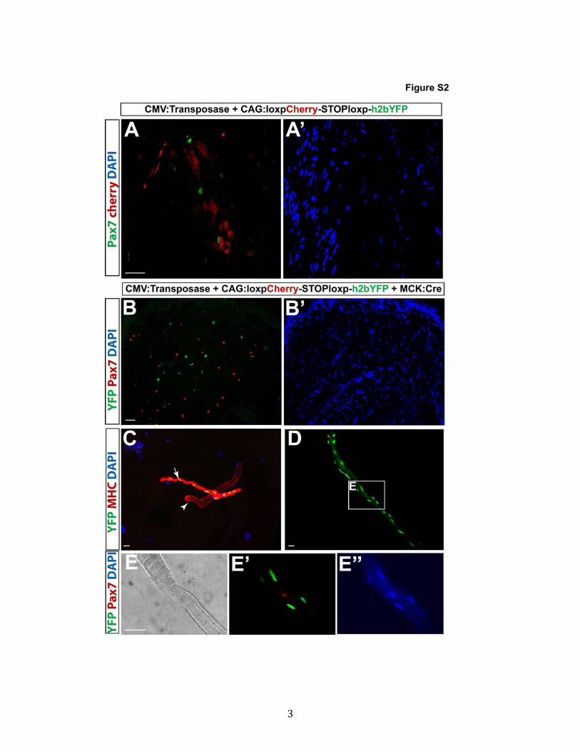
Figure S2 In newt, PAX7+ satellite cells are not electroporated during tracing of
limb cells. (relates to Figure 1)
(A) Lack of Cherry expression in PAX7+ satellite cells without Cre recombinase.
(B) PAX7+ nuclei are YFP- in the newt limb after transfection with Cre recombinase.
(C and D) In vitro culture of dissociated limbs containing single myofibers. Arrow
points to an YFP+ myofiber. Arrowhead points to an YFP- myofiber.
(E) YFP+ nuclei are PAX7- in cultured myofibers.
Scale bars: 20 m.
5
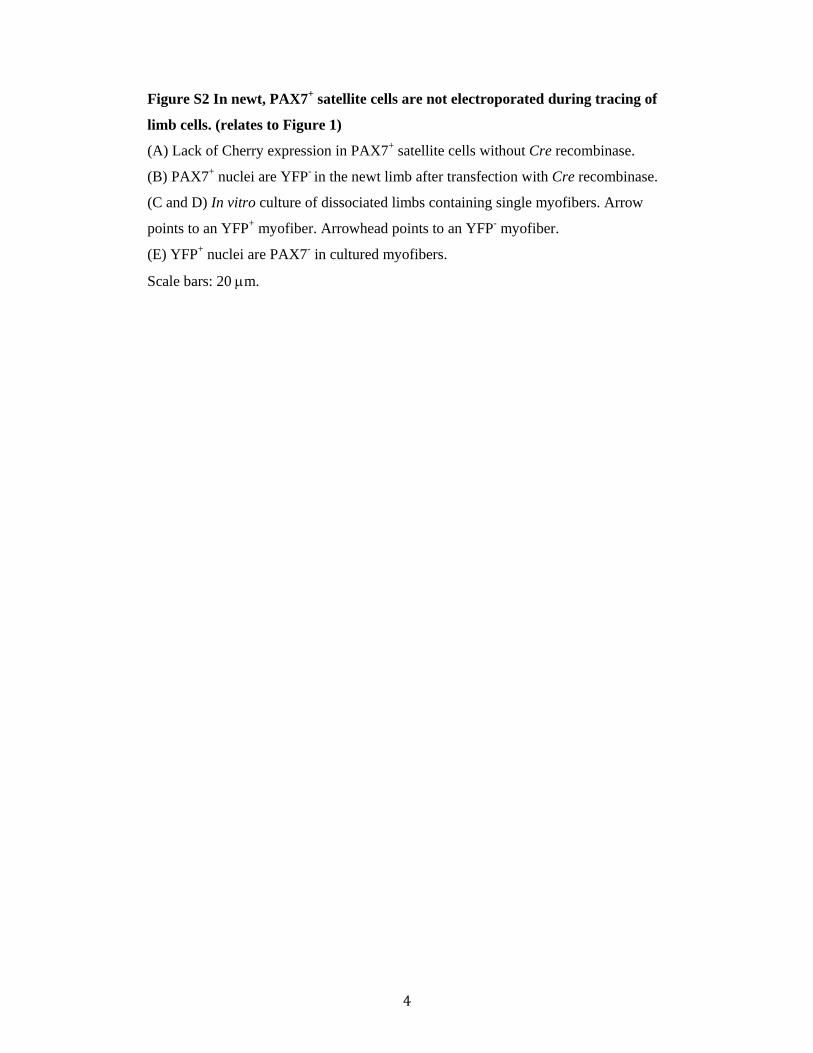
Figure S3 No evidence for myofiber-derived progeny contributing to cartilage in
newt. (relates to Figures 1, 2)
(A) Image showing a palette stage regenerate with the developing humerus, ulna and
radius outlined by collagen-II staining. YFP+ cells are visible both proximally and
distally to the amputation plane, which is indicted by the dashed line.
(B) Close up image illustrating the absence of YFP+ nuclei in cartilage.
Scale bars, (A): 200 m, (B): 20 m.
6
7
Figure S4 The cell fusion-mediated, cre/loxP-based, muscle cell labeling strategy;
negative controls. (relates to Figure 4)
(A) After blastema or presomitic mesoderm transplantation, the animal contains a
mixture of cells of two genotypes, CAGGS:loxpGFP-STOPloxpCherry and
CAGGS:ert2-cre-ert2-T2AnucGFP. During myogenesis the myoblasts of these two
genotypes fuse to form a common cytoplasm. The ERT2-CRE-ERT2 protein is
synthesized in one of the nuclei, and then diffuses into the nucleus of the other
genotype. This results in recombination of the CAGGS:loxpGFP-STOPloxpCherry
transgene in the genome, resulting in Cherry transcription and translation. The Cherry
protein diffuses throughout the cytoplasm of the chimeric cell.
(B) No Cherry fluorescence was observed before tamoxifen injection.
(C) Brightfield image of (B).
(D) No Cherry fluorescence was observed after tamoxifen injection in limbs when the
host is non-transgenic while the donor is CAGGS:ert2-cre-ert2-nucGFP nor when the
host is CAGGS:loxpGFP-STOPloxpCherry while the donor is non-transgenic.
(E and F) Cross-section of a limb from a CAGGS:loxpGFP-STOPloxpCherry host
grafted with a non-transgenic donor after tamoxifen injection. No Cherry+ myofibers
are visible (E) in MHC+ muscle (F, green).
Scale bars, (B, C): 1 mm, (D): 2 mm, (E, F): 200 µm.
8
9
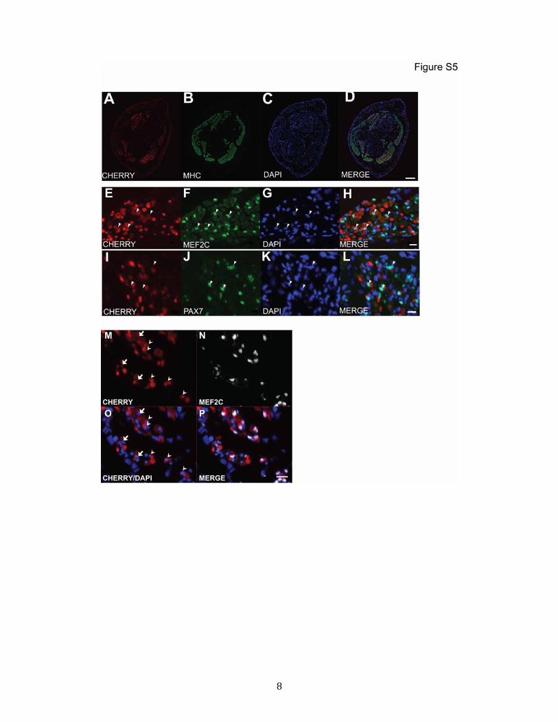
Figure S5 Specificity of tamoxifen-induced Cherry expression in LB Transplant axolotls. (relates to Figure 4) (A-D) Characterization of muscle specificity in LB-transplant axolotl limbs.
Overview, co-localization of Cherry with muscle-specific myosin heavy chain (MHC)
in limb cross-section from chimeric, tamoxifen-injected LB-transplant limb confirms
muscle-restricted expression. Higher magnification examination indicated that all
Cherry+ nuclei counted were surrounded by MHC+ cytoplasm.
(E-H) Co-localization of Cherry+ nuclear signal (E) with MEF2C (F) by
immunofluorescence detection indicates muscle-specific Cherry expression. White
arrowheads denote Cherry, MEF2C double-positive nuclei. 509 out of 513 Cherry+
nuclei were MEF2C+.
(I-L) Cherry+ nuclear signal (I) does not coincide with PAX7+ signal (J), indicating
that satellite cells are not labeled by this method (arrowheads). 2 out of 861 Cherry+
nuclei were PAX7+.
(M-P) Strong versus weak Cherry+ nuclei (MEF2C+) from a mature limb of LB
transplants to detect which nuclei harbored the recombined CAGGS:Cherry gene.
Strong nuclei denoted by arrowhead, weak nuclei by arrows. 43±10% nuclei strongly
expressed Cherry.
Scale bars, (A-D): 400 µm, (E-P): 20 µm.
10
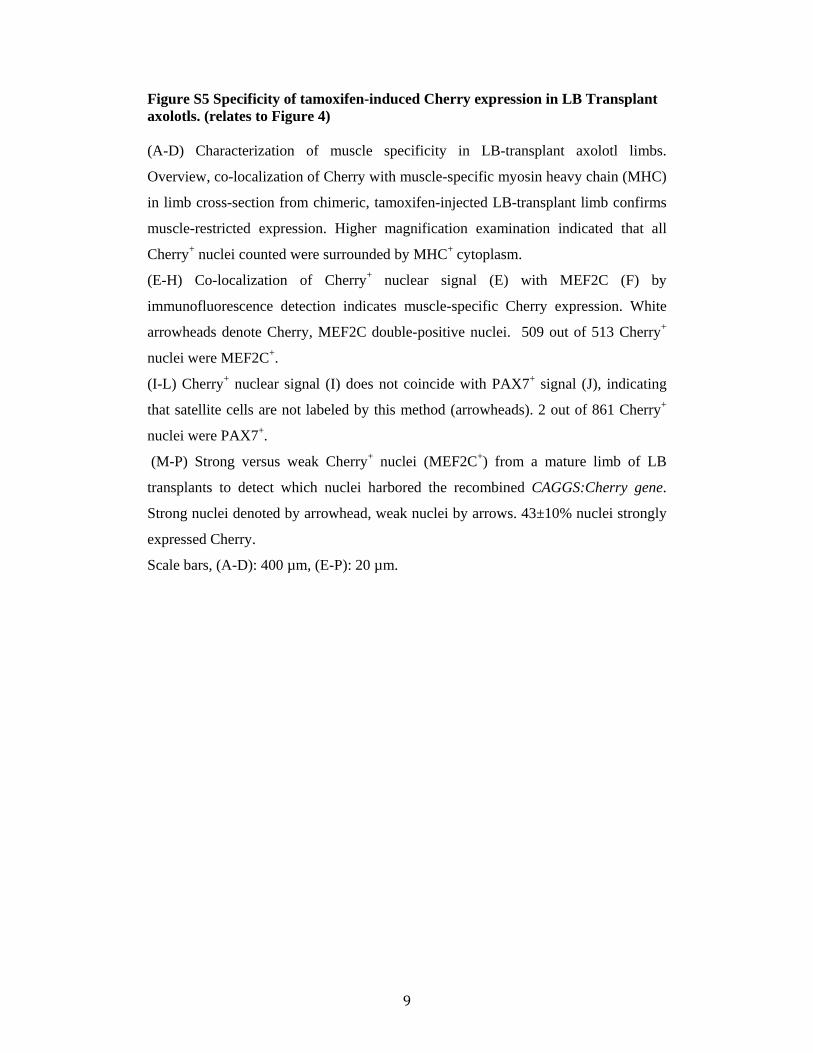
Figure S6 Labeled upper limb muscle via blastema transplant (LB Transplant) does not contribute to regenerated muscle in axolotl. (relates to Figure 4) (A-C) Blastema transplanted limbs with labeled myofibers in the upper arm after
regeneration. No labeled muscle is seen beyond the amputation plane (white line).
(A’-C’) Contralateral non-transplanted control limbs to those shown in (A-C). Scale
bars: 2 mm.
(D) Cherry fluorescence intensity graphs: Left (A, A’), Middle (B, B’), Right (C, C’),
along the proximal-to-distal limb axis of limbs shown in (A-C) and (A’-C’). Red line
is experimental limb (A-C) while blue line represents the control contralateral non-
transplanted limb (A’-C’).
A.U.: arbitrary units.
11
Figure S7 No evidence for proliferative, myofiber-derived cells in the 10-day
axolotl limb blastema. (relates to Figure 4)
(A) Limbs labeled by LB transplant were amputated, and 10-day blastema samples
sectioned. Longitudinal section of a 10-day upper limb blastema immunostained for
PCNA. Solid line indicates amputation plane, and dashed box indicates area of inset
for panel B.
(B) High magnification image of the inset close to the amputation plane. No Cherry+
Hoechst+ PCNA+ cells were detected. Furthermore, no evidence of Cherry+ Hoechst+
cells in the middle or distal tip of the blastema was observed.
(C) Quantitation of Cherry positive structures/cells close to amputation plane (upper
blastema) and in lower/distal tip of blastema. Quantitation of Cherry+ cells that co-
localize with a Hoechst+ nucleus, and those that co-localize with PCNA. No triple
positive (Cherry+, Hoechst+, PCNA+) cells were found, n=2 blastemas.
(D-F) PAX7+ cells in the blastema do not derive from Cherry-labeled myofibers in
LB Transplant limbs. No Cherry+PAX7+ nuclei were found in 10 day LB Transplant
blastemas. Longitudinal section of a 10-day limb blastema showing morphology of
Cherry+ myofibers (D) extending into the blastema with respect to PAX7+ nuclei (E).
Scale bars, (A) 200 µm, (B) 100 µm, (D-F) 50 µm.
12
Supplemental Experimental Procedures
Newt procedures
Animals and procedures
Red-spotted newts, Notophthalmus viridescens were supplied by Charles D. Sullivan
Co. (Nashville, TN, USA) and maintained in a humidified room at 18°C. Animals
were anesthetized by placing them in an aqueous solution of 0.1% ethyl 3-
aminobenzoate methanesulfonate (Sigma) for 15 min. Upper forelimbs were
amputated above the elbow, and the bone and soft tissue were trimmed to produce a
flat amputation surface. Animals were left to recover overnight in an aqueous solution
of 0.5% sulfamerazine (Sigma). At specified time-points, the regenerating limbs were
collected. For EdU-labelling experiments, animals were injected intraperitoneally
with 50-100 µl of 1mg/ml EdU. All experiments were performed according to
European Community and local ethics committee guidelines.
Injections and electroporations
Plasmids were purified using high purity maxiprep system (OriGene) and were
resuspended in 10 mM Tris HCl (pH 8.5) at 10 µg/µl. The three different plasmids
were mixed with a molar ratio of 1:1:1, which gives the most efficient transgene
expression. Plasmid solution (2µl) was injected into the middle of the upper forelimb
with a glass micropipette. A NEPA21 electroporator and a pair of needle electrodes
(CUY611P7-4, NEPA GENE) were used for electroporation according to
manufacturer’s instructions.
Immunohistochemistry
Thin frozen sections were thawed at room temperature and fixed in 4% formaldehyde
for 5 min. Sections were blocked with 10% donkey serum and 0.1% Triton-X for 30
min at room temperature. Sections were incubated with a relevant primary antibody
overnight at 4 °C and with secondary antibodies for 1 h at room temperature.
Antibodies were diluted in blocking buffer and sections were mounted in mounting
medium (DakoCytomation) containing 5µg/ml DAPI (Sigma). Primary antibodies
antibody 4A1025 kindly provided by S. Hughes). After several washes with PBS,
sections were then incubated for 2 hours with secondary antibodies and Hoechst
solution for nuclear staining. EdU was detected using the Click-iT EdU Imaging Kit
(Invitrogen). Images of the stained sections were taken with a 20 × plan-neofluar
objective in a Zeiss Observer microscope (inverted) controlled by an Axiovision
software (Zeiss, Jena, Germany). The Axovision program was also used to stitch
mosaic pictures of the limb sections and for cell quantification. Images in manuscript
often represent one subsection of the stitched image to provide sufficient zoom.
Electroporations
Anesthetized axolotls were injected in lower and upper limbs with MCK:cre,
CAGGS:loxpCHERRY-STOPloxp-H2BYFP, and CMV:Tol2-transposase expression
plasmids. Total of five pulses with a voltage of either 50 or 25 volts with a pulse
length of 50 msec and a pulse interval of 1 sec was applied to each limb with
continuous flow of PBS on the limb. The animals were allowed to recover for two
weeks before amputations.
Whole mount immunostaining
Limbs were harvested from anesthetized axolotl around the shoulder and fixed
overnight at 4°C. Limbs were washed in PBS and then stored in 30 % sucrose at 4°C.
Limbs were washed over night in PBS at 4°C and then washed (PBS/0.3% Triton
X100) over night at 4°C. The limbs were blocked (1x PBS, 0.3 % Triton-X100, 10%
Goat serum, 50mM glycine) overnight at 4°C followed by Anti-MHC (monoclonal
16
antibody 4A1025 kindly provided by S. Hughes) for 60 hours at 4°C. After several
washes, the samples were incubated with secondary antibody (Goat antimouse Cy3
Fab fragment- Jackson Immunoresearch) and Hoechst solution for 60 hours at 4°C.
The limbs were washed for several hours at RT and then overnight 4°C with wash
buffer. The limbs were stored at 4°C in 50% glycerol/PBS. Before imaging, the skin
was manually removed and imaged on a Leica TCS SP5 MP inverse confocal with a
10 X air objective. 3D reconstruction was achieved using Volocity 3D Image
Analysis Software (PerkinElmer). Amputation plane was determined by the visible
change in light refraction of bone at the junction.
Quantitative analysis
For cell and nuclei quantification of the mature limb, images were stitched as mosaics
to assemble the whole cross-section of the limb. Sections from 11 different animals
were taken, including different levels of the limb, lower arm, wrist, hand.
Cherry+ nuclei were counted and each nucleus was confirmed for MEF2C
immunoreactivity.
For MHC quantification, every single myofiber was counted and from those, all
Cherry+ nuclei were counted.
For quantification of the second regenerate, sections from upper limb, proximal lower
limb, distal lower limb and hand from 3 different animals were included. For satellite
cell counting, PAX7+ nuclei in the surroundings of Cherry+ fibers were counted.
For fluorescence intensity measurements of upper arm regenerates (Figure 4F and
S6), FIJI software was used to perform a line scan (width 30) to measure the average
pixel intensity along the line. For each limb segment (UA, LA and Hand), two parallel
lines were drawn to encompass the muscle area within the segment. Regions directly
at the elbow and wrist were not measured. Intensity values from both lines per
segment were averaged. To plot the experimental and control intensity scans on the
same graph, the natural variability in limb lengths had to be taken into account. The
length of the intensity profile of the shorter limb was interpolated to match the longer
limb using R. Profiles were plotted using R.
RT-PCR conditions
About 40 to 100 h2bYFP+ (newt) or GFP+ (axolotl) blastema cells were collected and
stored in lysis buffer of Picopure RNA Isolation kit (Invitrogen). The stump muscle of
the same limb was isolated and homogenized and stored in lysis buffer. The RNA was
extracted with PicoPure RNA Isolation Kit and amplified with RiboAmp HS PLUS
17
RNA Amplification Kit (Invitrogen). In addition, RNA was extracted from the tails
of larva newt and GFP transgenic axolotl. The reverse transcription was performed
with Superscript III (Invitrogen) and the PCR was performed with Platinum Taq
(Invitrogen). The primers are:
Newt:
Mrf4: TGGGACAGGAACAAGACACA and GTCACCACAACACCACAAGG
Myog: GGGGACCACTTGCTAAATGA and GCCCTACAGAGCACCAGTTC
Myf5: CAACCGACAAACTCAGCACT and CCCGAGTCCCTAAAGTCACA
Pax7: CCAAGAACGTGAGCTTGTCA and GTGGAAGGTGTCACACATCG
GFP and YFP: ACGTAAACGGCCACAAGTTC and
AAGTCGTGCTGCTTCATGTG
GAPDH: TGTGGCGTGACGGCAGAGGTG and
TCCAAGCGGCAGGTCAGGTCAAC
Axolotl
Myog: AAGGGGTGTCGAGTGACTGT and TCGACTGTGATGCTTTCGAC
Pax7: TGGAGAAGGCCTTTGAGAGA and TGAAAGCTGCCAGTTGATTG
Mrf4: ACATCGAGAAGCTGCAGGAC and ATCCGAAACATTTTGCCACT
Myf5: AGCAGATTCCTGCGATGTTT and GCACCACATGACAAAACACA
Rp4: TGAAGAACTTGAGGGTCATGG and CTTGGCGTCTGCAGATTTTTT
18
Supplemental References
Khattak, S., Richter, T., and Tanaka, E.M. (2009). Generation of transgenic axolotls (Ambystoma mexicanum). Cold Spring Harb Protoc 2009, pdb prot5264. Kragl, M., Knapp, D., Nacu, E., Khattak, S., Maden, M., Epperlein, H.H., and Tanaka, E.M. (2009). Cells keep a memory of their tissue origin during axolotl limb regeneration. Nature 460, 60-65. Nacu, E., Knapp, D., Tanaka, E.M., and Epperlein, H.H. (2009). Axolotl (Ambystoma mexicanum) embryonic transplantation methods. Cold Spring Harb Protoc 2009, pdb prot5265. Salic, A., and Mitchison, T.J. (2008). A chemical method for fast and sensitive detection of DNA synthesis in vivo. Proc Natl Acad Sci U S A 105, 2415-2420. Sobkow, L., Epperlein, H.H., Herklotz, S., Straube, W.L., and Tanaka, E.M. (2006). A germline GFP transgenic axolotl and its use to track cell fate: dual origin of the fin mesenchyme during development and the fate of blood cells during regeneration. Dev Biol 290, 386-397.