44
Cell Structure and Function Chapter 3
| Date post: | 23-Dec-2015 |
| Category: |
Documents |
| Upload: | elizabeth-gray |
| View: | 215 times |
| Download: | 0 times |
Cell Structure and Function
Chapter 3
The Cell--Considerations• Basic unit of life
• Protection and support
• Movement
• Communication
• Metabolism and energy release
• Inheritance
Cell TheoryAll living things are made up of
cell(s)
Cells are smallest living unit of structure and function for all organisms
All cells arise from preexisting cells
(No spontaneous generation)
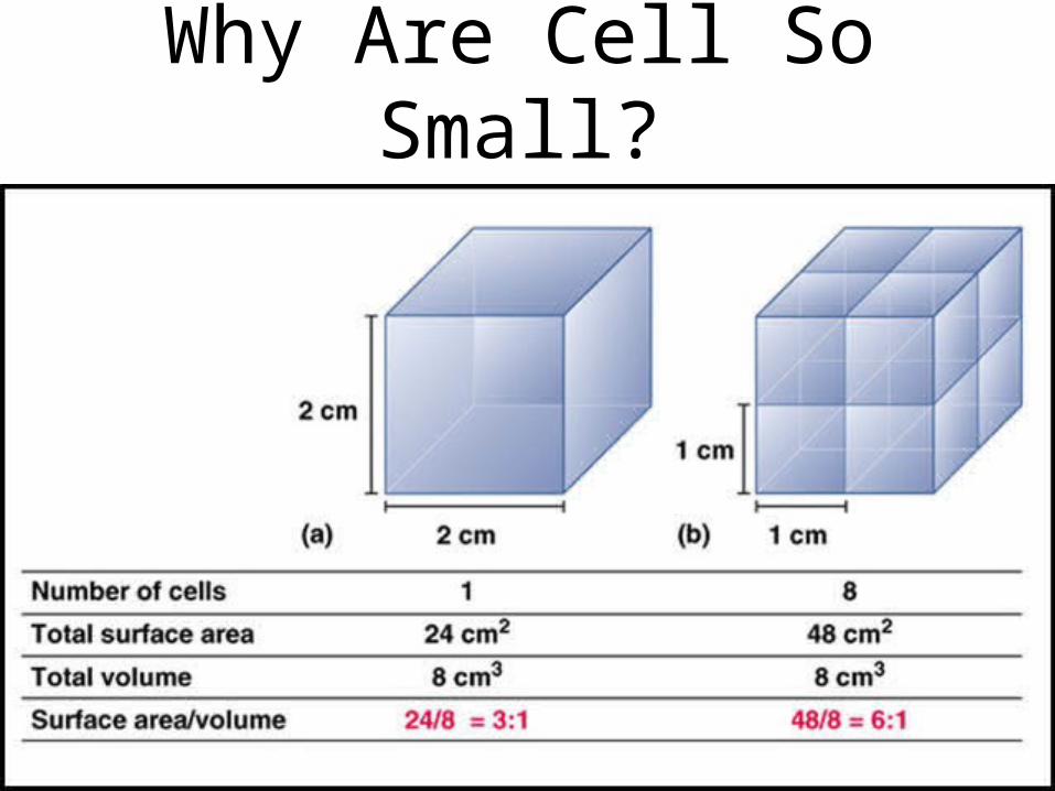
Why Are Cell So Small?

Sizes of living things
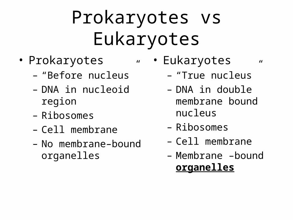
Prokaryotes vs Eukaryotes
• Prokaryotes– “Before nucleus”– DNA in nucleoid region– Ribosomes– Cell membrane– No membrane–bound
organelles
• Eukaryotes– “True nucleus”– DNA in double
membrane bound nucleus
– Ribosomes– Cell membrane– Membrane –bound organelles

Typical Bacterium---A Prokaryote
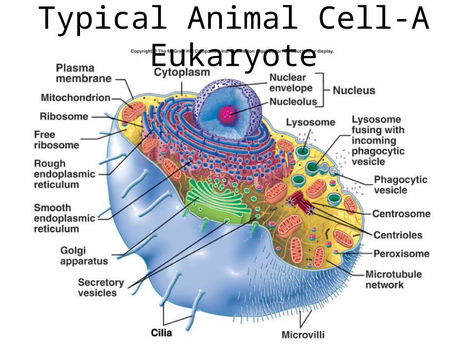
Typical Animal Cell-A Eukaryote

Plasma Membrane

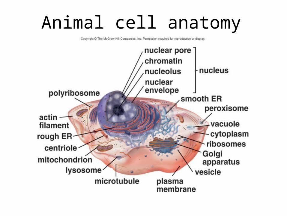
Animal cell anatomy

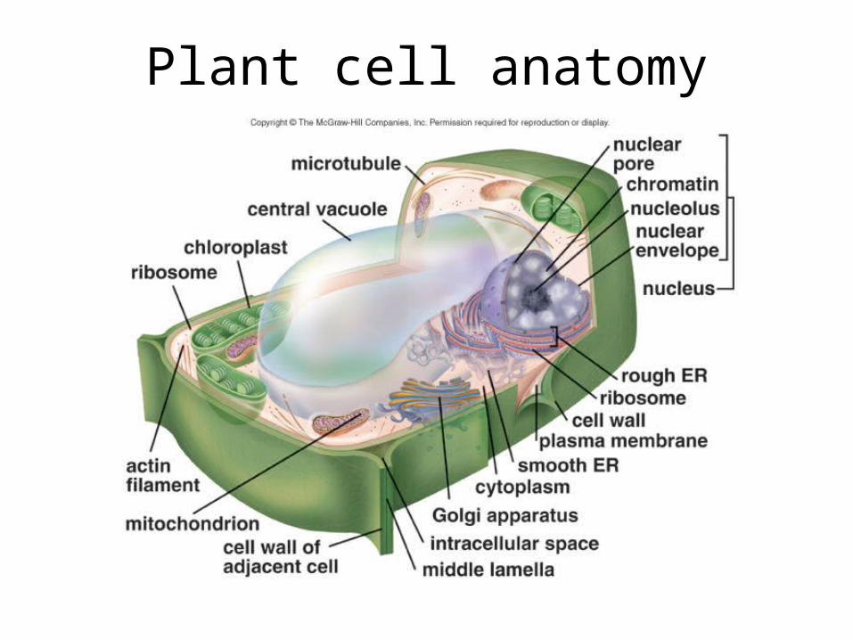
Plant cell anatomy
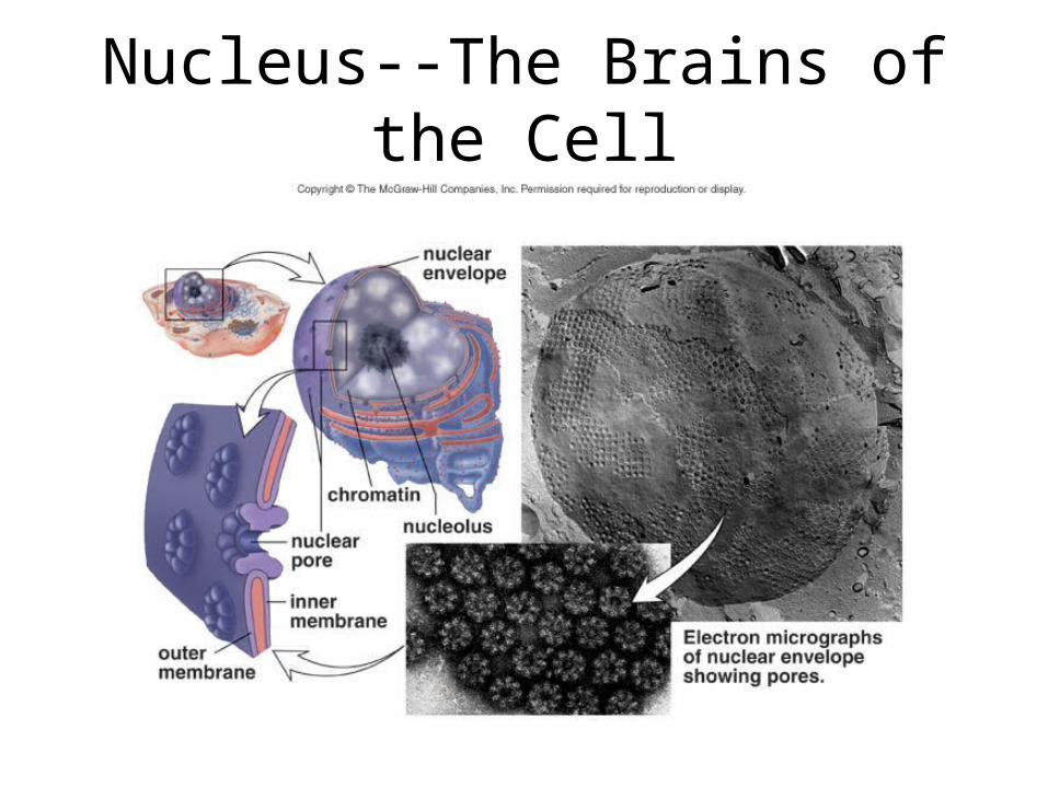
Nucleus--The Brains of the Cell
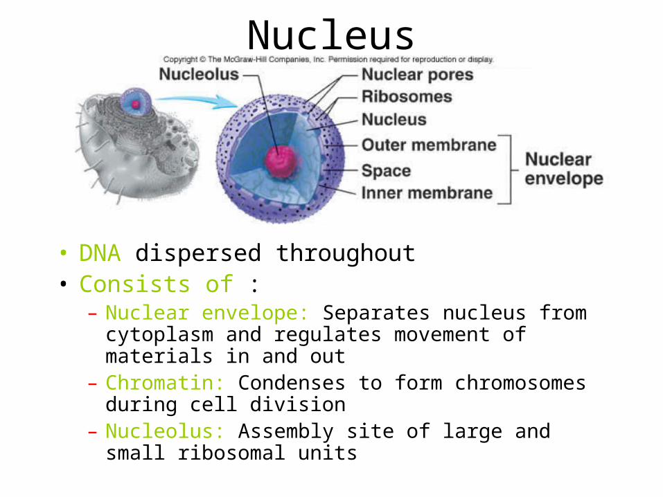
Nucleus
• DNA dispersed throughout• Consists of :
– Nuclear envelope: Separates nucleus from cytoplasm and regulates movement of materials in and out
– Chromatin: Condenses to form chromosomes during cell division
– Nucleolus: Assembly site of large and small ribosomal units

Ribosomes
• Sites of protein synthesis
• Composed of a large and small subunit
• Types– Free– Attached to
endoplasmic reticulum

Composition of eukaryotic and
prokaryotic ribosomes
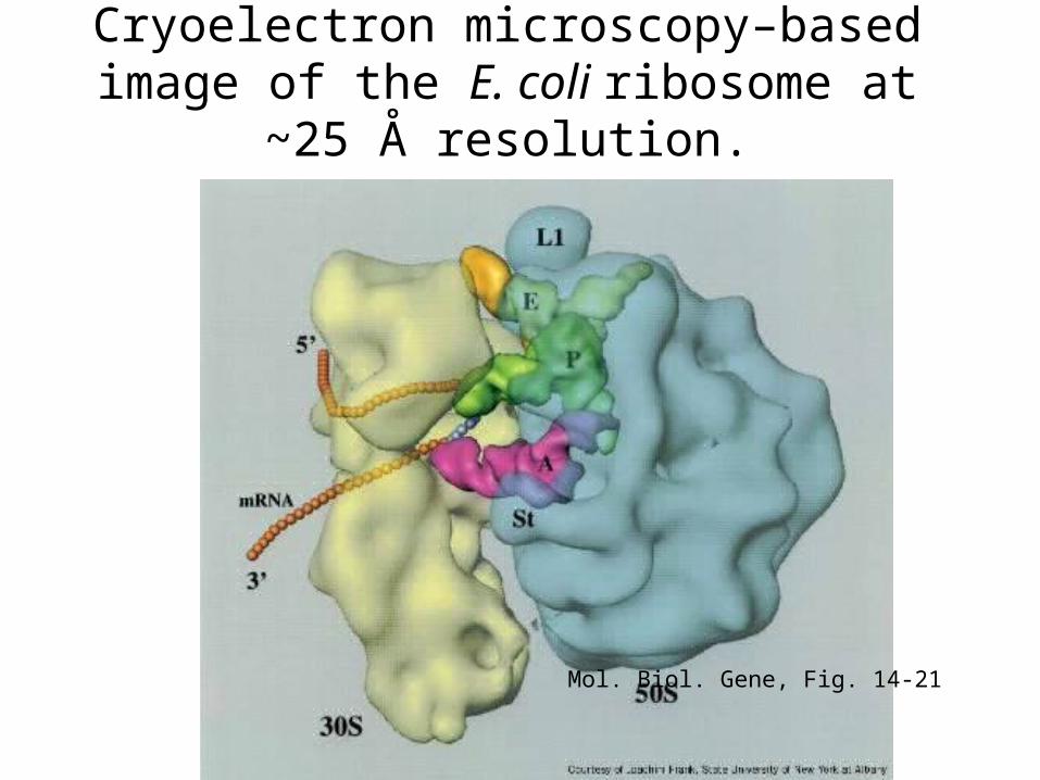
Cryoelectron microscopy–based image of the E. coli ribosome at ~25 Å resolution.
Mol. Biol. Gene, Fig. 14-21

The Endomembrane System
• Nuclear envelope
• Endoplasmic reticulum
• Golgi apparatus
• Vesicles

Endoplasmic Reticulum• Types
– Rough• Attached ribosomes• Proteins produced
and modified– Smooth
• No attached ribosomes
• Manufacture lipids• Cisternae or Lumen:
Interior spaces isolated from rest of cytoplasm

Golgi Apparatus—Traffic Cop
• Modification, packaging, distribution of proteins and lipids for secretion or internal use
• Flattened membrane sacs stacked on each other
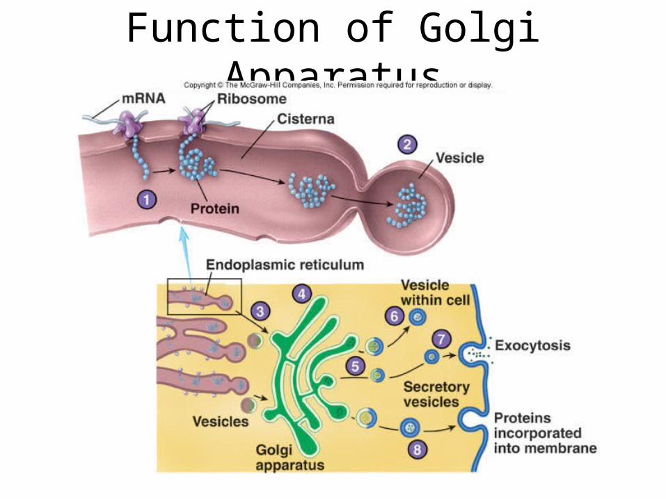
Function of Golgi Apparatus

Action of Lysosomes

Peroxisomes and Proteasomes
• Peroxisomes– Smaller than lysosomes– Contain enzymes to break down fatty and
amino acids– Hydrogen peroxide is a by-product of
breakdown
• Proteasomes– Consist of large protein complexes– Include several enzymes that break down
and recycle proteins in cell
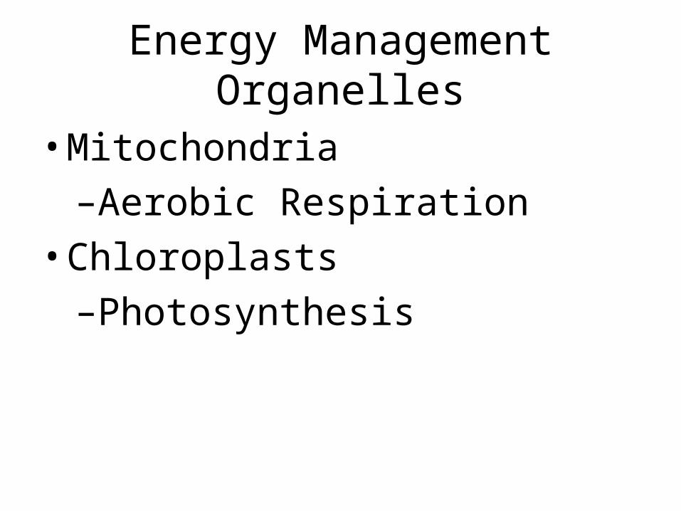
Energy Management Organelles
• Mitochondria
–Aerobic Respiration
• Chloroplasts
–Photosynthesis

Circle of Life
Carbon compounds, O2
CO2, H2O
Respiration
Photosynthesis
Energy
Energy
Mitochondria
Chloroplasts
Complex>>Simple
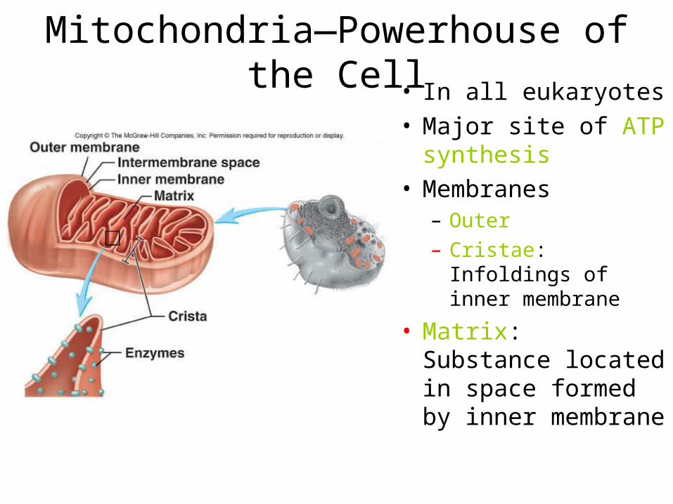
Mitochondria—Powerhouse of the Cell• In all eukaryotes• Major site of ATP
synthesis• Membranes
– Outer– Cristae: Infoldings of
inner membrane
• Matrix: Substance located in space formed by inner membrane

Chloroplast
Animal cell anatomy
Plant cell anatomy
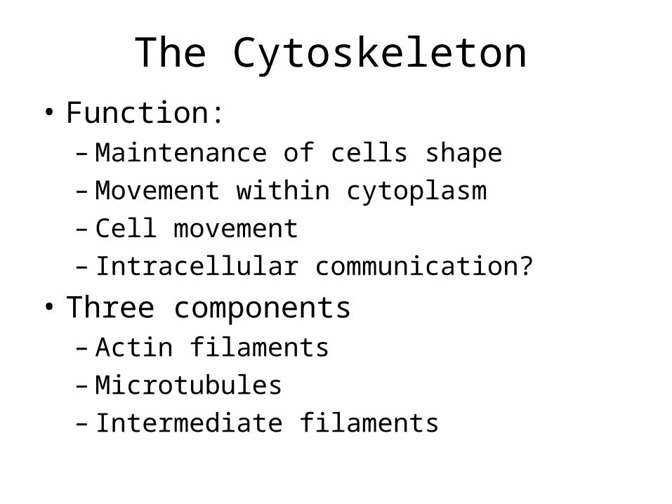
The Cytoskeleton• Function:
– Maintenance of cells shape– Movement within cytoplasm– Cell movement – Intracellular communication?
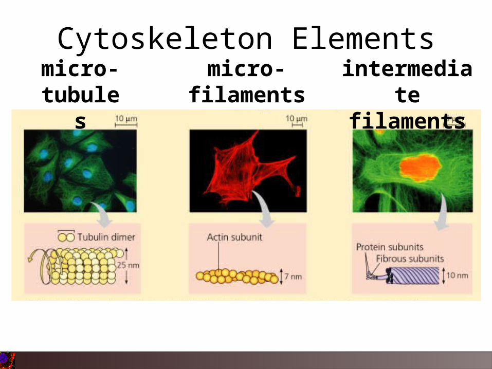
• Three components – Actin filaments– Microtubules– Intermediate filaments
FUNCTION:
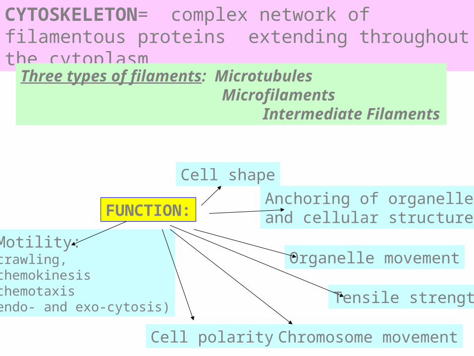
CYTOSKELETON= complex network of filamentous proteins extending throughout the cytoplasm
Three types of filaments: Microtubules Microfilaments Intermediate Filaments
Cell shape
Motility:crawling,chemokinesis chemotaxisendo- and exo-cytosis)
Anchoring of organelles and cellular structures
Organelle movement
Cell polarity
Tensile strength
Chromosome movement

helical structure, diameter ~ 7 nm EX. intestinal microvilli
ACTIN FILAMENTS

MICROFILAMENTSACTIN STRUCTURES IN CELLS:
MICROVILLI STRESS FIBRESFOCAL ADHESIONS
LAMELLIPODIAFILOPODIA(or MICROSPIKES
CONTRACTILERING(cell division)
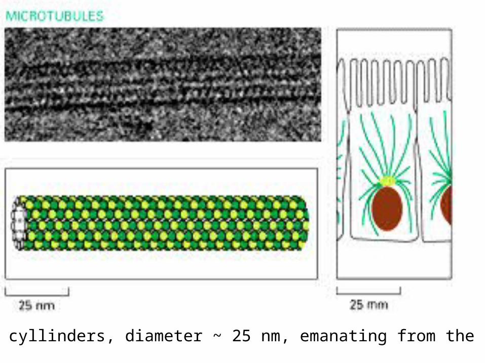
hollow cyllinders, diameter ~ 25 nm, emanating from the MTOC
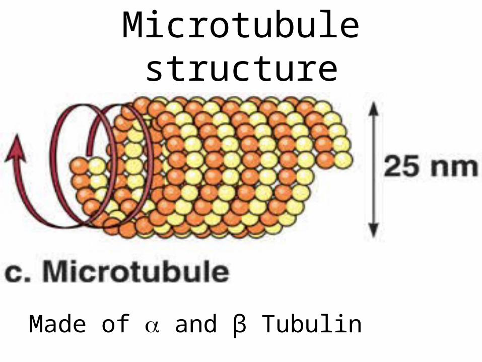
Microtubule structure
Made of and β Tubulin

rope-like fibres, diameter ~ 10 nm, nuclear, cytoplasmic, connecting cell-cell junctions
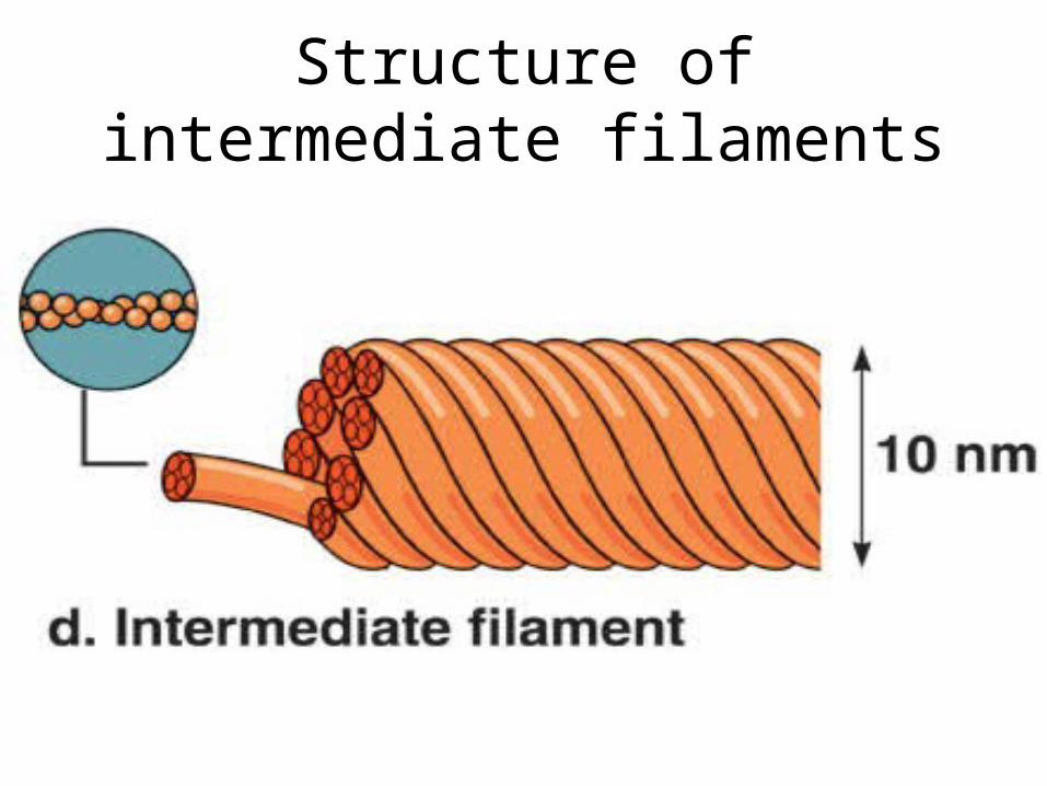
Structure of intermediate filaments
micro-tubule
s
micro-filaments
intermediate
filaments
Cytoskeleton Elements
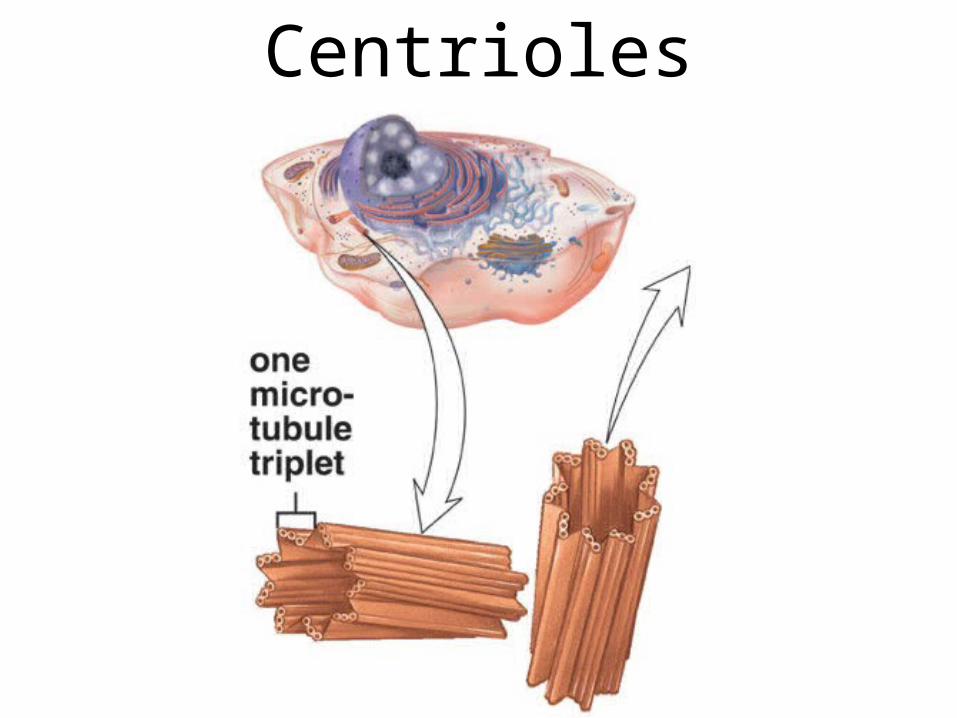
Centrioles
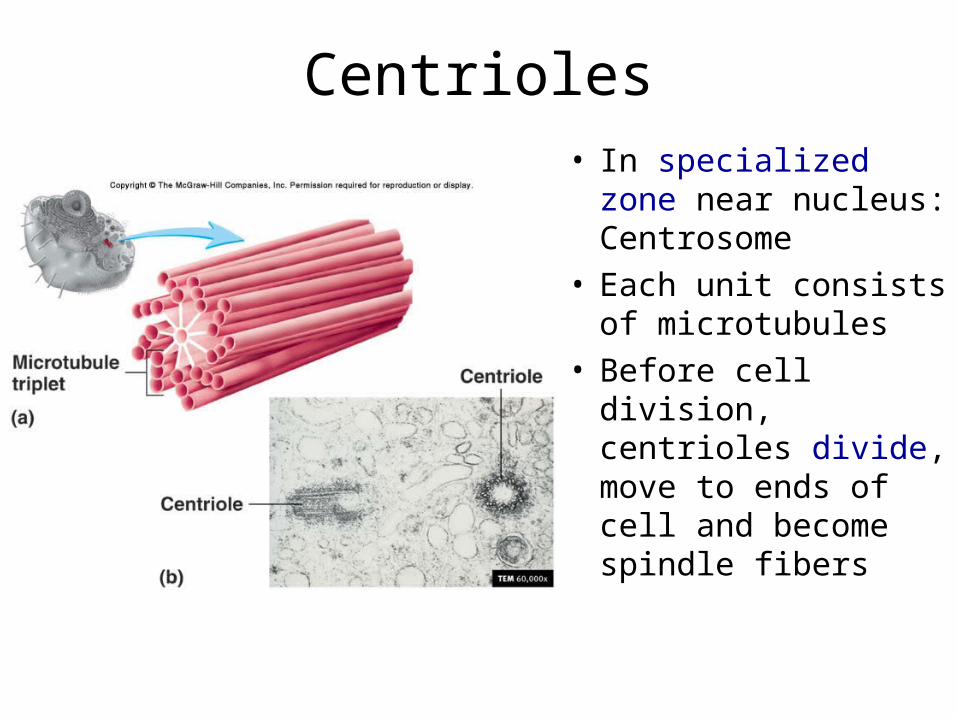
Centrioles• In specialized zone
near nucleus: Centrosome
• Each unit consists of microtubules
• Before cell division, centrioles divide, move to ends of cell and become spindle fibers
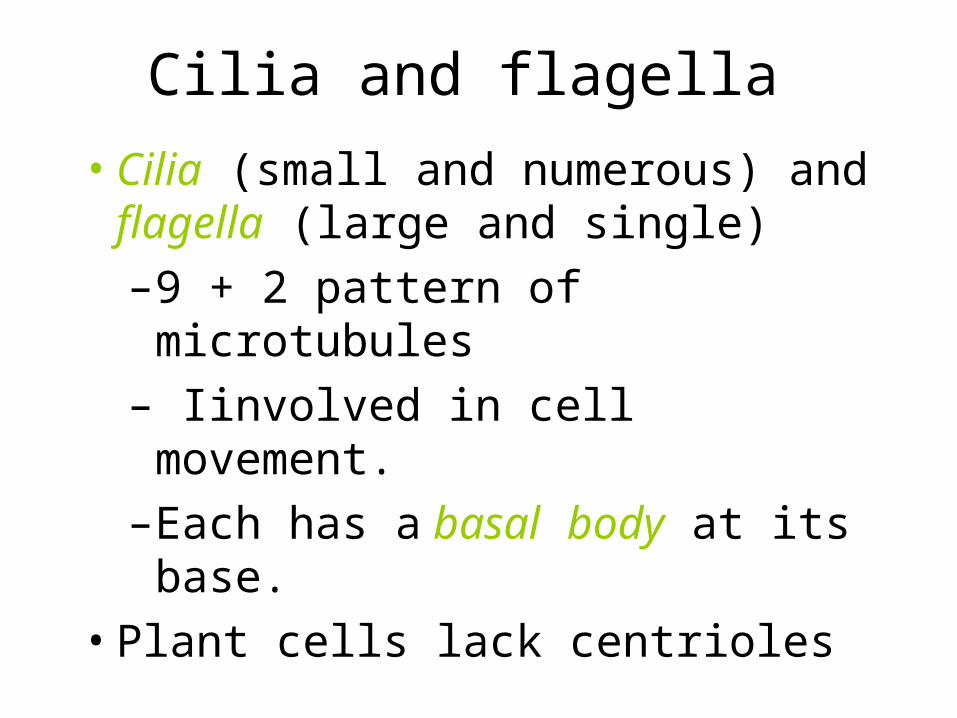
Cilia and flagella
• Cilia (small and numerous) and flagella (large and single)
–9 + 2 pattern of microtubules
– Iinvolved in cell movement.
–Each has a basal body at its base.
• Plant cells lack centrioles
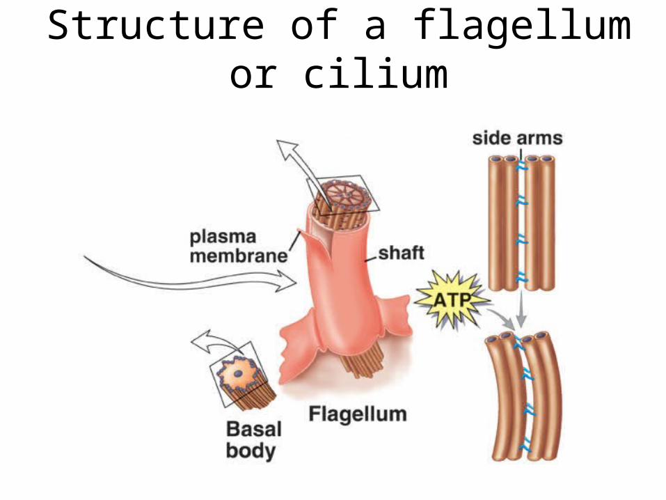
Structure of a flagellum or cilium

Prokaryotes vs Eukaryotes
• Prokaryotes– “Before nucleus”– DNA in nucleoid region– Ribosomes– Cell membrane– No membrane–bound
organelles
• Eukaryotes– “True nucleus”– DNA in double
membrane bound nucleus
– Ribosomes– Cell membrane– Membrane –bound organelles
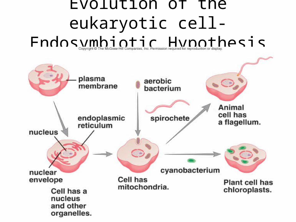
Evolution of the eukaryotic cell- Endosymbiotic Hypothesis
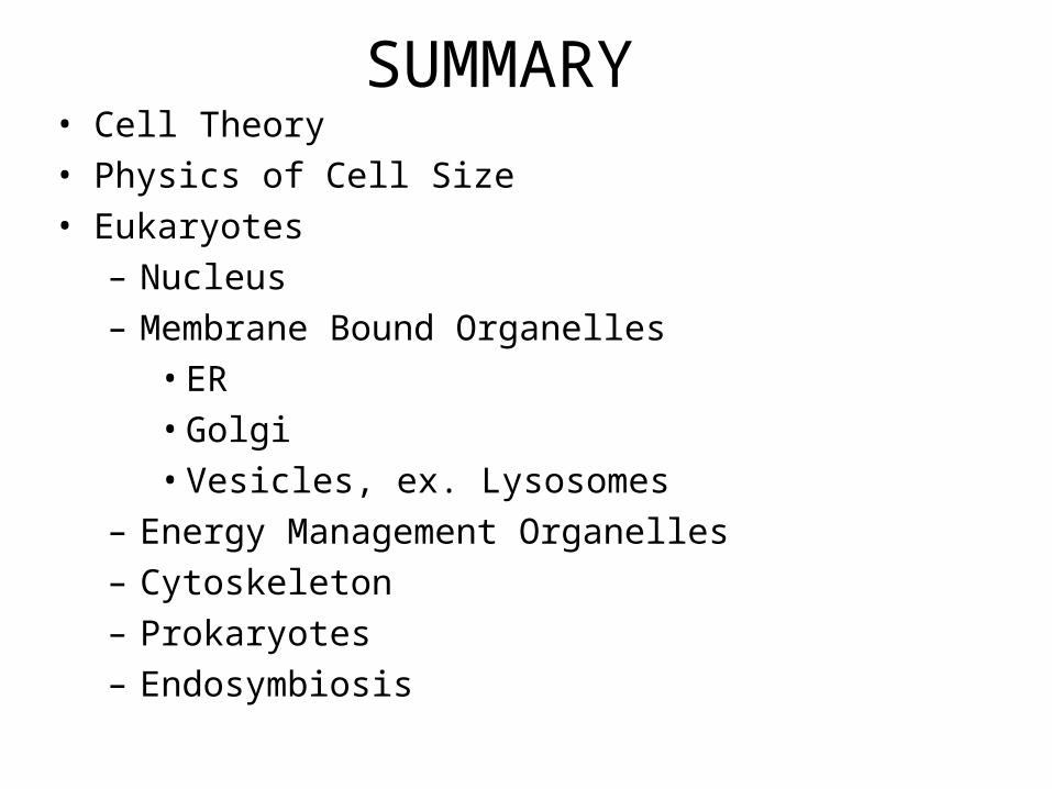
SUMMARY• Cell Theory• Physics of Cell Size• Eukaryotes
– Nucleus– Membrane Bound Organelles
• ER• Golgi• Vesicles, ex. Lysosomes
– Energy Management Organelles– Cytoskeleton– Prokaryotes– Endosymbiosis


