Introduction to Biological Science - BIOL1361 Recitation A.O. Cheek & D. Pattison, Department of Biology & Biochemistry, University of Houston Rev. 1/3/18 Attribution-NonCommercial-ShareAlike 4.0 International (CC BY-NC-SA 4.0) 1 Cell Structure and Protein Secretion
Transcript
Introduction to Biological Science - BIOL1361 Recitation
A.O. Cheek & D. Pattison, Department of Biology & Biochemistry, University of Houston Rev. 1/3/18 Attribution-NonCommercial-ShareAlike 4.0 International (CC BY-NC-SA 4.0)
1
Cell Structure and Protein Secretion
Introduction to Biological Science - BIOL1361 Recitation
A.O. Cheek & D. Pattison, Department of Biology & Biochemistry, University of Houston Rev. 1/3/18 Attribution-NonCommercial-ShareAlike 4.0 International (CC BY-NC-SA 4.0)
2
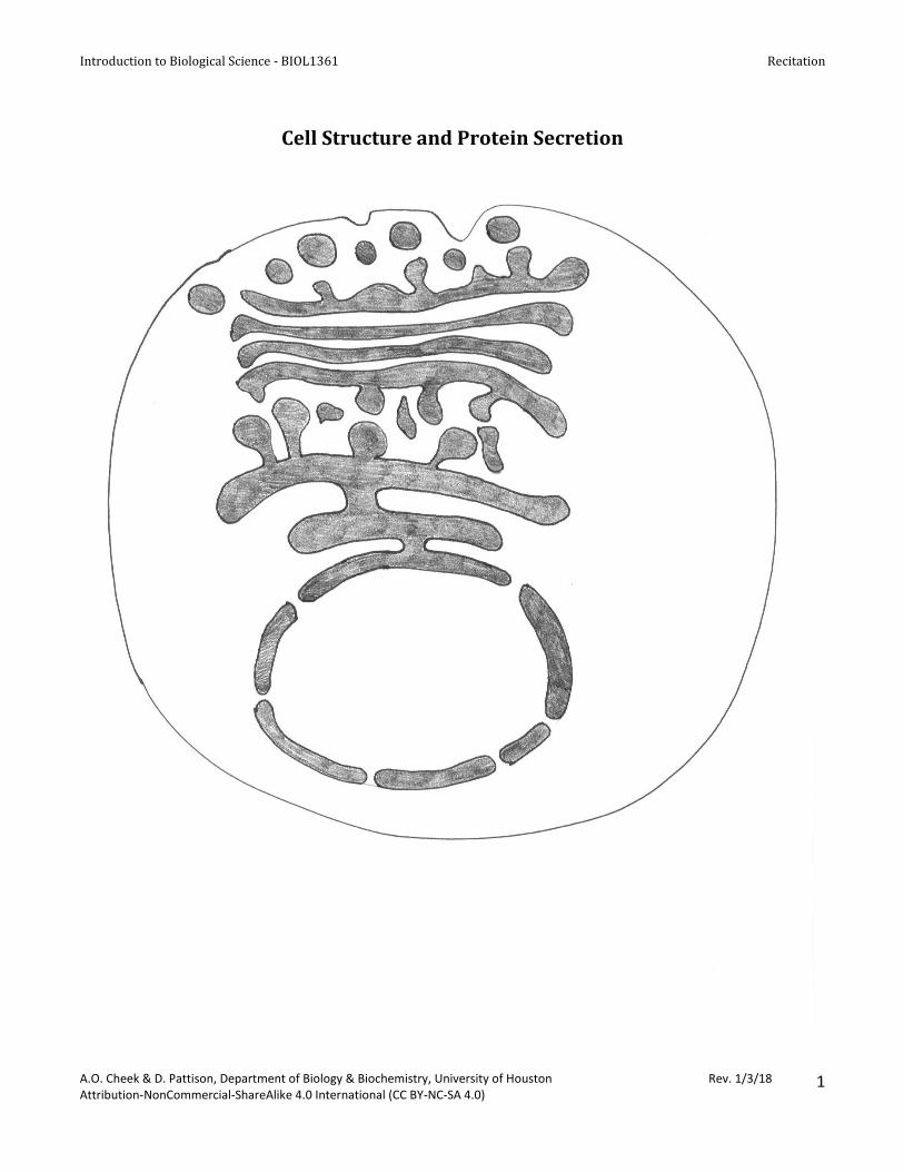
I. Tracing the Intracellular Pathway of Protein Secretion A. Label the nucleus, nuclear envelope, endoplasmic reticulum, Golgi apparatus, secretory vesicles, and extracellular space on the cell diagram on page 1. Use colored beads and the large diagram of a cell to reproduce a key experiment performed by James Jamieson and George Palade in the 1960s. Palade was awarded the Nobel Prize in Physiology or Medicine in 1974 for his work on the roles of ribosomes, endoplasmic reticulum, and the Golgi apparatus in protein secretion. http://www.nobelprize.org/nobel_prizes/medicine/laureates/1974/ B. Move beads through cellular compartments according to instruction below and fill in the data table. Times indicate when Jamieson & Palade took pictures to see where the radioactive leucine was during the actual experiment. You don’t need to wait between steps. 1. Time 0: Put 10 beads in the extracellular space. The beads represent radioactively labeled leucine, an
amino acid used to build proteins. 2. 3-minute incubation: Move 9 beads into the endoplasmic reticulum and 1 into the nucleus. 3. 7 minutes later: Distribute beads as follows: Nucleus – 1 bead; ER – 4 beads; Golgi apparatus – 4
Introduction to Biological Science - BIOL1361 Recitation
A.O. Cheek & D. Pattison, Department of Biology & Biochemistry, University of Houston Rev. 1/3/18 Attribution-NonCommercial-ShareAlike 4.0 International (CC BY-NC-SA 4.0)
3
II. Organelles with their own circular DNA
Mitochondria in cross-section.
1. Start at the dot on each mitochondrion and trace the line clockwise until you return to the dot. What do
you notice about this line? What structure does this line represent? What can you conclude about this
structure based on the way it is drawn?
2. Label the mitochondria above using the following terms: outer membrane, inner membrane, inter-
membrane space, matrix
3. What are the cristae?
4. What is in the intermembrane space?
5. Describe the structural difference between the inner membrane and the intermembrane space.
Introduction to Biological Science - BIOL1361 Recitation
A.O. Cheek & D. Pattison, Department of Biology & Biochemistry, University of Houston Rev. 1/3/18 Attribution-NonCommercial-ShareAlike 4.0 International (CC BY-NC-SA 4.0)
4
The Chloroplast
1. Label the chloroplast below using the following terms: outer membrane, inner membrane, thylakoid, stroma, granum.
2. What is the difference between a thylakoid and a granum? 3. What is the stroma? 4. Does a plant have both mitochondria and chloroplasts?
Introduction to Biological Science - BIOL1361 Recitation
A.O. Cheek & D. Pattison, Department of Biology & Biochemistry, University of Houston Rev. 1/3/18 Attribution-NonCommercial-ShareAlike 4.0 International (CC BY-NC-SA 4.0)
5
Cell Structure Plant/Animal/Both Function
Cell wall
Plasma membrane
Nucleus
Nucelolus
Ribosomes
Smooth endoplasmic reticulum (ER)
Rough endoplasmic reticulum (ER)
Golgi Complex
Vacuoles
Lysosomes
Peroxisome
Mitochondria
Chloroplast
Cilia
Flagella
Centrosome
Centriole
Microtubules
Intermediate filaments
Microfilaments
Microvilli
Introduction to Biological Science - BIOL1361 Recitation
A.O. Cheek & D. Pattison, Department of Biology & Biochemistry, University of Houston Rev. 1/3/18 Attribution-NonCommercial-ShareAlike 4.0 International (CC BY-NC-SA 4.0)
6
Chromosome
Chromatin
Plasmodesmata
Central vacuole
Introduction to Biological Science - BIOL1361 Recitation
A.O. Cheek & D. Pattison, Department of Biology & Biochemistry, University of Houston Rev. 1/3/18 Attribution-NonCommercial-ShareAlike 4.0 International (CC BY-NC-SA 4.0)
7
Teaching Tips for Peer Leaders This week emphasize the relationship between cell structure and function. Students usually can label the parts of the cell. Explaining the functions of each organelle, especially the process of protein secretion is more difficult. Part I. A) Have students work in pairs or threes. Each group needs an 8 x 10 cell diagram (page 1) and 10 colored
beads. Make sure students label the nucleus, endoplasmic reticulum, Golgi apparatus, and secretory vesicles. The shaded areas represent the space between the nuclear membranes and the cisternae (or lumen) of the ER, Golgi apparatus, and vesicles. The colored beads represent radioactively labeled leucine, an amino acid. Jamieson & Palade incubated slices of guinea pig pancreas in 14C leucine for 3 minutes. At the end of 3 minutes, the 14C leucine solution was removed and replaced with non-radioactive solution. During that time, pancreatic acinar cells (the cells that produce trypsinogen, chymotrypsinogen and all the other pancreatic digestive enzymes) transported the leucine across the cell membrane and into the cytoplasm. At the end of the 3 minute incubation, the tissue slice was fixed and prepared for autoradiography, a technique where film is placed on top of the tissue sections, allowing the radioactivity to expose the film. The film is developed to see where the radioactivity is within the cell or tissue. Jamieson & Palade found 90% of radioactivity in the endoplasmic reticulum and 10% in the nucleus. Students mimic this in step 2 by moving 9 beads into the endoplasmic reticulum and 1 bead into the nucleus.
Jamieson & Palade fixed and analyzed additional tissue slices 7 minutes, 37 minutes, and 117 minutes after the 3-min incubation with radioactive leucine ended. At each time point, they photographed the location of the 14C leucine. Students move the beads to different organelles to represent what the researchers saw.
B) Students need to move the beads around in the cell and fill in the table with data on where the beads
were at each time. C) Emphasize that the colored beads represent radioactive leucine, a tracking device for protein location
within the cell. Wherever the tracking devices are, that’s where the proteins containing them are. The point of this experiment is to help students understand the sequential roles played by rough ER, Golgi apparatus, secretory vesicles and exocytosis in protein synthesis and secretion. After pairs or groups have completed A – D, send someone to the board to explain the process of protein secretion. Ask the audience to correct any mistakes as you notice them. You can start with a volunteer who is willing to draw the nucleus, ER, Golgi, and vesicles.
D) The protein is secreted by exocytosis (review course textbook if necessary). Part II. Emphasize to students that they must understand the structure of mitochondria and chloroplasts to understand cellular respiration and photosynthesis. Point out that the line drawings of mitochondria are cross-sections. Mitochondria look different in cross-section, depending on how they are sliced – transverse (the round section) or longitudinally (the oval section). Use a sliced cucumber as an analogy: The cut surface is an oval if you cut it lengthways, but the cut surface is a circle if you cut across it.
Introduction to Biological Science - BIOL1361 Recitation
A.O. Cheek & D. Pattison, Department of Biology & Biochemistry, University of Houston Rev. 1/3/18 Attribution-NonCommercial-ShareAlike 4.0 International (CC BY-NC-SA 4.0)
8
Practicing Vocabulary. This vocabulary exercise is intended for individual study outside of recitation. You can recommend they turn this exercise into matching cards and see if they can match terms and definitions without looking at their notes or the book. Point out that nucleus & nucleolus and chromatin & chromosome can be confusing terms. Help them come up with a way to remember the difference.
Notes to Faculty
Supplies needed in addition to the hand-out: Enough plastic beads to give 10 beads to each pair or group of