61
| Date post: | 30-Nov-2015 |
| Category: |
Documents |
| Upload: | prasanth-chitturi |
| View: | 4 times |
| Download: | 0 times |

Physiological Systems of the Human Body
Functioning of the human body is a coordinated action of various systems.
1) skeletal system2) circulatory system3) respiratory system4) Digestive system5) excretory system6) regulatory system7) reproductive system8) muscular system

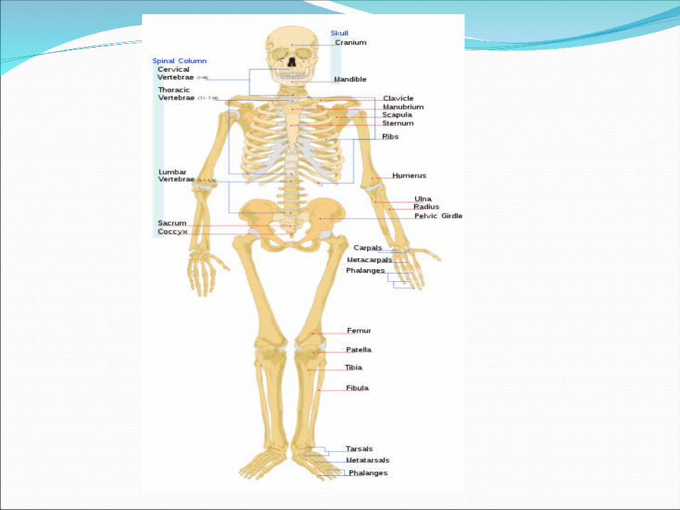
Skeletal systemIt is a frame work of the body.Provides mechanical stability for the body to protect
the delicate organs and serves as an anchorage for the muscles in order to make possible through liver action.
Also serves as a reservoir for calcium and phosphorus and contains the bone marrow in which blood cells are formed.
It consists of 206 bones and enclosed by a membrane called “periosteum” from which a new bone is formed in the healing of fractures.
Top-cranium skullMiddle-pelvisBottom-foot bonesArticular cartilage (capsule)
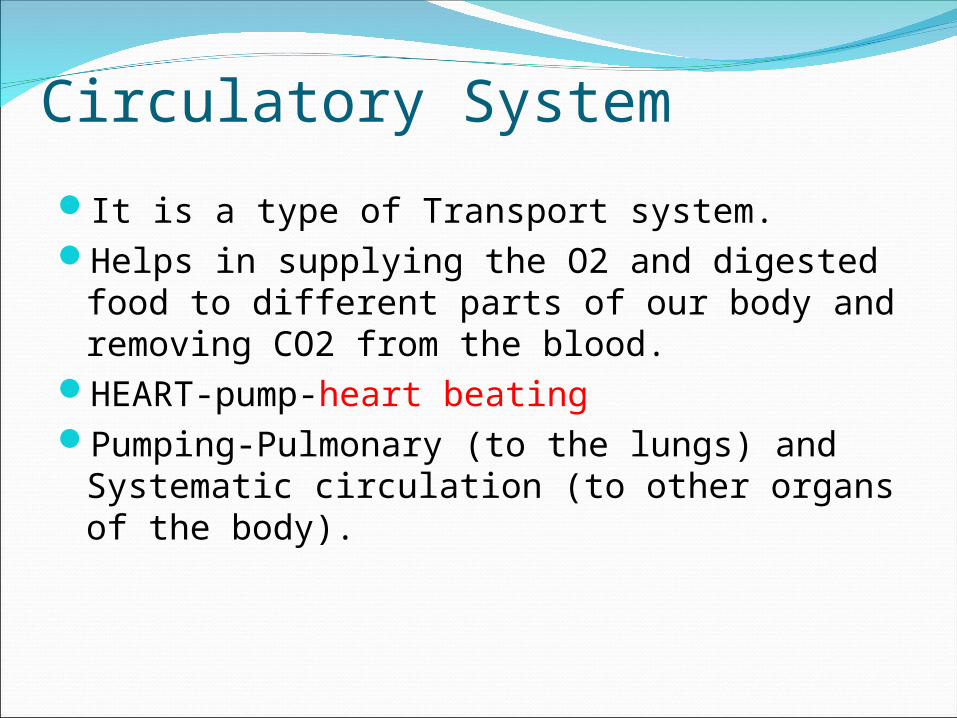
Circulatory System
It is a type of Transport system.Helps in supplying the O2 and digested food
to different parts of our body and removing CO2 from the blood.
HEART-pump-heart beatingPumping-Pulmonary (to the lungs) and
Systematic circulation (to other organs of the body).

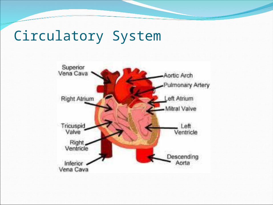
Circulatory System
Arteries-blood vessels which carry pure blood from the heart to various parts
Veins-blood vessels through which impure blood returns to the heart
Superior vena cavaInferior vena cavaPulmonary veins-carry pure blood from lungs
to the heartPulmonary artery-carry impure blood heart to
lungs
Circulatory System

Respiratory System
It is concerned with Breathing and Respiration.
NOSE(starting point)-LUNGS(ending point)Alveoli-gas exchange take placeBreathing 1) Inspiration 2) Expiration

Respiratory System
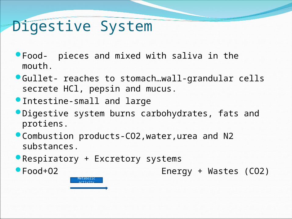
Digestive System
Food- pieces and mixed with saliva in the mouth.Gullet- reaches to stomach…wall-grandular cells
secrete HCl, pepsin and mucus.Intestine-small and largeDigestive system burns carbohydrates, fats and
protiens.Combustion products-CO2,water,urea and N2
substances.Respiratory + Excretory systemsFood+O2 Energy + Wastes (CO2)Metabolic
activity

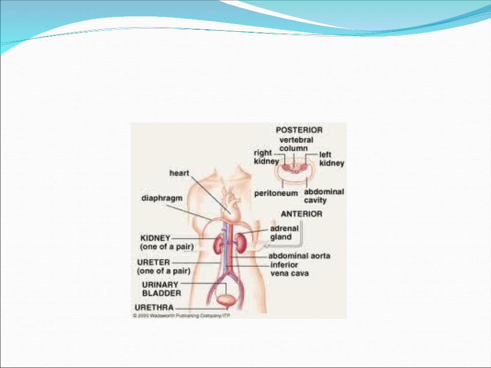
Excretory system
Lungs, Kidneys, Skin and large intestine.Removes waste products formed during
combustion of the food from our body.Lungs-CO2(volatile).Kidneys-nitrogen breakdown products
interms of Urine (non volatile).Sweat glands- water, salt and other products.Liver-via bile remove certain waste.

Regulatory systemNervous system-for regulation of rapid eventsEndocrine system- for regulation of slower metabolic
processes.Central Nervous System Brain- cerebrum, cerebellum and the brain stem. Spinal cord- through the back bone and also acts as
a communicator b/w various parts and brain.Peripheral Nervous System Sensory (inward path) and Motory nerves (outward
path)---communication wires.Neurons Endocrine system Works by using hormones which are carried
through circulatory system.

Reproductive system
A fetus develops through repeated cell division by means of reproductive system.
Fertilization –in fallopian tube.

Muscular systemMovements of various parts are caused by
muscles.1) voluntary muscles : arm muscles2) involuntary muscles: food canal3) cardiac muscles: heartMuscle contraction is regulated via nerves.A bundle of muscle fibers in a muscle supplied by
a single motor nerve fiber is called a “Motor Unit” because all the muscle fibers contract simultaneously when the nerve fiber is stimulated.
Sensors—muscles spindles sends the signals to the central nervous system so that feedback and control is obtained.


SOME FACTORS ABOUT CELLBasic building block of Human body.100 TrillionFluid in charactersimilar cells forms---Tissues, Muscle
nerves, blood, bones etc.Two or more tissues of dissimilar
physiology forms Heart, Lungs, kidney, Skin, Blood vessels etc.
EX: Digestive system---Mouth, trachea, stomach, intestines, liver,
rectum.

SOME FACTORS ABOUT CELLThe cells of the tissues are held by the
product called “Intercellular cement”.The fluid which lies inside the cell is known
as “ intracellular fluid”.To sustain the cells a fluid called “Interstitial
or Extra cellular fluid” is able to flow to each cell to bring nourishment to it, to remove waste products from it and to respond to electrochemical impulses.
CHRACTERISTICS OF LIVING ORGANISMS—(virus, cell, fish, man)
a) Metabolism: Living organism ingests substances containing energy—liberates energy– uses to maintain activities and excrete waste products.
An organism is able to sustain itself for a period of time by this process.

CHRACTERISTICS OF LIVING ORGANISMS
b) Motility: move on its own with out the assistance from the outside.
Tasks : warm itself, secure food, avoid threatening situations.
c) Irritability: ability of an organism to respond to outside stimulus.
Ex: harmful bacteria influence, temperature or current.
Results in contraction , expansion, discharge, chemical or physical change.

CHRACTERISTICS OF LIVING ORGANISMS
d) Growth: ability to grow.Ex: size, energy, form, function and chemical
structure.
e) Reproducibility: to reproduce one’s own kind of species by repeated cell division.
LIVING

General characteristics of a human cell
1) Nearly Spherical in Shape.2) diameter ≈ 20 μ3) semi permeable outer membrane with a
T=100Á4) has inner fluid substance “cytoplasm” (organells+inclusions).5) Inner control unit “nucleus” filled with
nucleoplasm has the nuclear membrane.Nucleus— nuleoli contains RNA & DNA.

Average cell structure
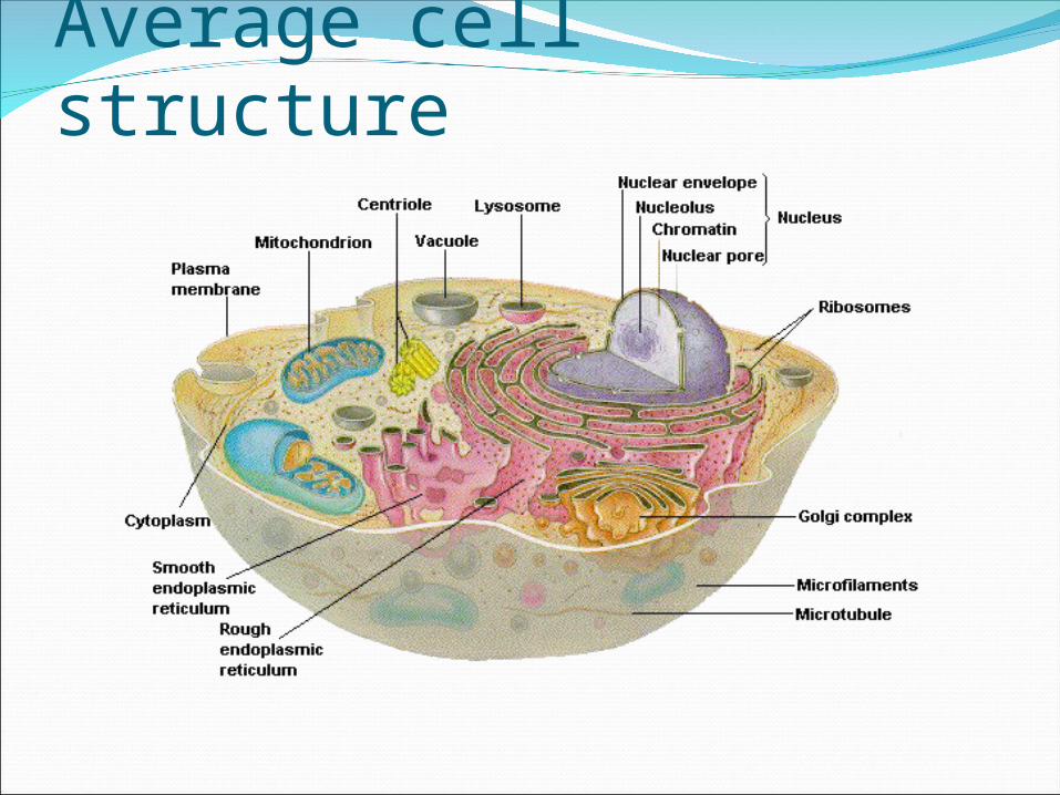
Average cell structure

Basic Processes in Cells
a) Diffusion: takes place as a result of difference of concentration of substances inside & outside of the cell.
Transportation is through intermolecular spaces in the membrane or in combination with a carrier protein.
Factors affecting the rate of diffusion:1) Permeability of the membrane, which the cell
can regulate.2) The relative concentration of the substance
inside & outside of the cell.3) Potential inside and outside of the cell.4) Pressure difference inside & outside of the
cell.
b) Active transport: When a cell moves molecules or ions uphill against a concentration gradient.
By which the cell substance can cross the cell membrane.
In this a carrier can be used.By this substances in a lower
concentration of liquid are moved in to higher levels of concentration.
Creates an balance of charges & energy.

c) Pinocytosis: carrier is not used.
Certain substances approach & rest on the cell membrane the cell can engulf them & forms some sort of capsule.
This capsule then travels through the membrane in to the cytoplasm.
Cell Potential Genesis: Resting stateThe concentration of the ions inside and
outside the cell are markedly different.i) In the interstitial (outside) fluid, the
concentration of Na+ and Cl- ions are much higher than in the intercellular (inside) fluid.
ii) In the intercellular fluid, the concentration of K + ions is much higher than in the interstitial fluid.
iii) There is an electrical potential difference between the inside and outside fluids.
(In skeletal muscles-90 mv approx)


Resting potentialThe membrane of a cell which is a semi
permeable plays an important role in the maintenance of the above differences.
When the membrane is not stimulated i.e; in the resting state, it is highly permeable to K+ but only slightly permeable to Na + .
Because of its steep concentration gradient, K+ tends to leak out at a high rate.
As a result the inside of the cell becomes electrically negative wrto the outside.
Resting voltageIs depending upon :a) the relative difference in concentration
(chemical gradient)b) any external sources resulting from the
transport of ions across the membrane (electrical gradient)
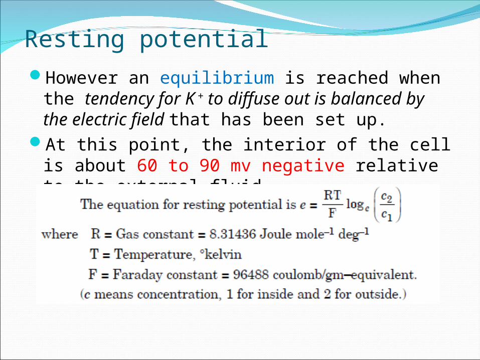
Resting potentialHowever an equilibrium is reached when the
tendency for K + to diffuse out is balanced by the electric field that has been set up.
At this point, the interior of the cell is about 60 to 90 mv negative relative to the external fluid.
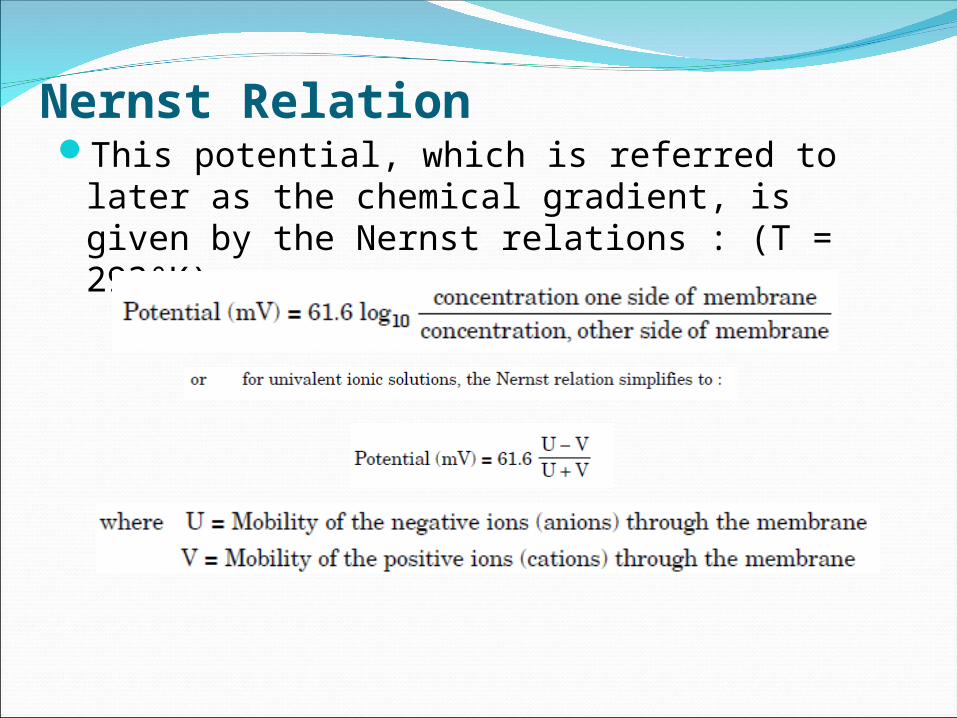
Nernst RelationThis potential, which is referred to later as
the chemical gradient, is given by the Nernst relations : (T = 293°K)

Electrical characteristics of the Human CellNet Gradient: A chemical gradient is formed due to a
difference in concentration (10 to 1) producing a potential gradient as given by the Nernst relation.
An electrical gradient is formed as a result of a potential (90mV)that may exist across the membrane due to some other source.
The net result is that the sodium and potassium currents are equal; the sodium current balances the potassium current with a resultant current of zero.
Since the net current though the membrane is zero, the cell’s internal potential will not change and will remain at its – 90 mV resting level.

Electrical characteristics of the Human CellCell in Resting State or polarized state :
In this state potassium ions can pass fairly readily through the membrane as the membrane offers medium resistance.
This membrane is, however, almost impermeable to sodium ions and, thus, offers a high resistance to the passage of these ions.
A large net gradient affects the movement of sodium ions into the cell.

Resting potential
Cell Condition after a Stimulus
When a cell receives a stimulation above a certain “threshold” value, this balance is upset and the cell will go through a cycle known as an “ Action Potential”.
The membrane permeability to potassium ions is unaltered but the permeability to sodium ions is increased thus flows in to the cell.
A much lower resistance is offered to the flow of sodium ions, thus increasing the sodium ionic current.

This increased sodium ionic current causes more positive ions to pass into the cell than are passing out of the cell, causing the internal cell potential to drop from – 90 mV in an attempt to achieve sodium current and potassium current balance.

Cell DepolarizationAs this potential decreases, the net sodium
gradient across the membrane decreases and the net potassium gradient across the membrane increases, causing the currents to decrease and increase, respectively.
This process continues until current balance is again obtained, at which time the internal cell potential is + 20 mV.
The cell is then referred to as being in a depolarized state.


Cell Repolarization
By the time the cell has fully depolarized the characteristics of the membrane have begun to revert back to their pre-stimulus state.
This causes the sodium ionic current to be considerably lower than the potassium ionic current ; the internal cell potential thus begins to go negative with the process continuing until the – 90 mV resting potential of the cell is once again obtained.

Action Potential from a CellSuppose a stimulus is applied to the cell, the output
of the microelectrode would appear as shown below.This waveform is known as the “Cell action
potential”.Bioelectric currents are due to positive and negative
ion movement within a conductive fluid.As these ions possess finite mass and encounter
resistance to movement within the fluid their speeds are limited.
The cell action potential, thus, shows a finite “rise time” and “fall time”.
Time scale : 1 ms for nerve cells 150-300 ms in heart muscles.

Sodium Potassium Pump Phenomenon
The ionic concentration gradient across the cell membrane is maintained by virtue of metabolic energy expended by the cell in “Pumping” ions against the ionic gradient formed by the differing ionic concentration between the inside and outside of the cell.
This action has been referred to as the “Sodium – potassium pump”.

Threshold of Stimulus Causing Action PotentialA cell may be stimulated, or caused to depolarize
and then repolarize, by subjecting the cell membrane to an ionic current.
Note: This current may be produced by other cells, it may be produced by ionic currents existing as nerve impulses, or it may be artificially produced by some external current stimulus.
A cell will be stimulated when sufficient positive ions are added to the inside of the cell to cause its resting potential to be decreased from its – 90 mV level to approximately – 60 mV.
Once this threshold level is reached, the cell depolarizes without requiring the addition of any further positive ions to the inside of the cell from the stimulus source.

All-or-nothing law
Regardless of the method of excitation of cells or the intensity of the stimulus, which is assumed to be greater than the threshold of stimulus, the action potential is always same for any given cell.
Refractory periodsFollowing the generation of an action
potential , there is a brief period of time during which the cell can not respond to any new stimulus.
This period is called the “ absolute refractory
period”. (1 ms for nerve cells)Following the ARP , there occurs a “relative
refractory period”, during which another action potential can be triggered, but a much stronger stimulation is required. (several ms)

Refractory periods
Propagation rate
The rate at which an action potential moves down a fiber of a nerve cell or is propagated from cell to cell is called the “propagation rate or conduction velocity”.
This value is depending on the type and diameter of the nerve fiber and is from 20-140 mm/s.
But in heart muscle, it is very slower ranging from 0.2-0.4 mm/s.

Characteristics of Action Potential a) once the “threshold” has been reached, the sequence
can’t be turned off or changed, regardless of how long the stimulus is continued or if it is removed.
b) a second stimulus will not have any effect while the cell is in the depolarizing phase.
c) when a cell is repolarizing, a grater than normal stimulus is required in order to initiate another action potential.
d) The threshold level can be increased or decreased by altering the permeability of the membrane, by changing the concentration levels of sodium, potassium, calcium, etc.; by introducing drugs in to the fluid surrounding the cell.
e) Once the threshold is reached, the action potential will take about 10 ms to depolarize the repolarized cells.
Synchronous Depolarization
Consider a group of cells in close proximity to one another…….
under certain conditions of stimulation, these cells may all depolarize at the same time (synchronous depolarization) ; however, the repolarization process is random.
Repolarization of the individual cells will occur at different times.

Asynchronous DepolarizationUnder other conditions of stimulation, the group
of cells described previously will not all depolarize at the same time (asynchronous depolarization).
The stimulation may result in one cell depolarizing; the action of this cell depolarizing will then act as a stimulus on its adjacent
cell causing it to depolarize also. This chain reaction would proceed until all cells in a particular
area have depolarized.
In practice, combinations of synchronous and asynchronous depolarization occur in a group of cells.

Stimulation is not synchronous and the potential externally seen

Electrical Analog of the Cell
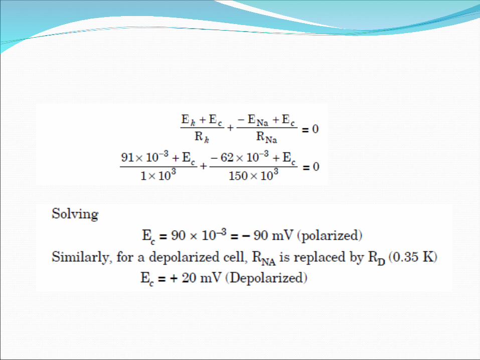
After assigning resistance values inversely proportional to the relative permeability of the membrane
and assuming potassium and sodium concentration ratios, then the intracellular potential for both a polarized cell and a depolarized cell can be determined.
The values assumed are analogous to actual values four in a cell.

Cm = membrane capacitance,ENa Ek are the Sodium potassium Nernst potentials, RK,RNa, the permeability of membrane to K and Na ions flow through membrane, Rd is the permeability of membrane to Na ion flow in depolarizing condition.


Bioelectric potentials----origin
The depolarization and repolarization undergone by a cell from time to time, give rise to voltage waveforms, which are of interest to clinicians and biomedical engineers.
Thus, each cell in the human body is a minute voltage generator, and is the source of all bioelectric potentials.
These are actually ionic voltages produced by the co-ordinated electrochemical activity of large group of cells.
In this type of synchronized action of many cells, the charges tend to migrate through the body fluids towards the still unexcited cell areas.

Bioelectric potentials----origin
Such charge migration constitutes an electrical current and hence, sets up potential differences between various portions of the body including its outer surface.
These PD can be conveniently picked up by placing conducting plates at any two points on the surface of the body and measured with the help of a sensitive instrument.
The bio potentials, so monitored, are highly significant for diagnosis and therapy.

Characteristics of the various Bioelectric signals Bioelectric signal
Spectrum (Hz)
Potential range (µv)
Sensing devices used
Signal origin
ECG 0.05-100 10-5000 covers fetal range
Surface ElectrodesNeedle Electrodes
Heart muscles
EEGElectro encephalogram
0.1-100 2-200 SE &NE Neuronal activity of brain
Cerebral potentials
Pulse duration 0.6 ms-0.1 s
10-100000 Deep needle Cerebrum of the brain
EMG 5-2000 20-5000 SE or NE Skin muscles
EGG 0.05-0.2 10-350 SE Peristaltic movement of the gastro intestinal tract
ERG 0.01-200 0.5-1000 Corneal electrodes
Retina of the eye
EOG DC-100 10-3500 Miniature SE Corneal-retinal potential variations


