54
Cells (Chapter 3) Developed by Dave Werner OCC BIOL-114 Spring 2014
| Date post: | 30-Dec-2015 |
| Category: |
Documents |
| Upload: | dortha-chase |
| View: | 219 times |
| Download: | 0 times |
Cells (Chapter 3)
Developed by Dave WernerOCC BIOL-114Spring 2014

OBJECTIVES:
Outline the discoveries that led to the development of the Cell Theory.
State the cell theory. Describe the relationship between
cell shape & cell function. Distinguish between prokaryotes
and eukaryotes.

3.1 History 1. In 1660, the English Scientist Robert Hooke used a
microscope to examine a thin slice of cork and described it as consisting of "a great many little boxes". Named “cells”.
2. 1673, Antony Von Leeuwenhoek – improved lenses and advanced cell biology by viewing red blood cells and sperm.
3. 1838, German Botanist Matthias Schleiden - PLANT cells
4. 1839, German Zoologist Theodor Schwann –ANIMAL cells
5. In 1855, German Physician Rudolf Virchow induced that THAT CELLS ONLY COME FROM OTHER CELLS".
6. His statement contradicted the idea that life could arise from Nonliving Matter. "Theory of Spontaneous Generation" The process by which life begins when ethers enter nonliving things.
CELL THEORY
A. All living things are composed of one or more cells.
B. Cells are the basic units of structure & function in an organism.
C. Cells come only from reproduction of existing cells.
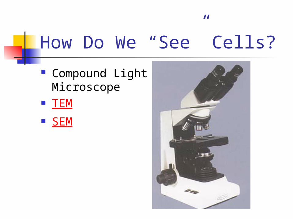
How Do We “See” Cells? Compound Light
Microscope TEM SEM

So What is “A Cell”? The CELL is the smallest unit of matter
that CAN Carry on ALL the PROCESSES OF LIFE.
Both Living and Nonliving Things are composed of molecules made from chemical elements such as carbon, hydrogen, oxygen, and nitrogen.
The organization of these molecules into Cells is one feature that distinguishes Living Things from all other matter.
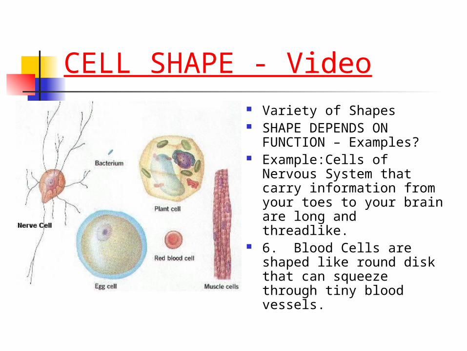
CELL SHAPE - Video Variety of Shapes SHAPE DEPENDS ON
FUNCTION – Examples? Example:Cells of Nervous
System that carry information from your toes to your brain are long and threadlike.
6. Blood Cells are shaped like round disk that can squeeze through tiny blood vessels.
INTERNAL ORGANIZATION 1. Cells contain a variety of Internal
Structures called ORGANELLES. 2. An organelle PERFORMS SPECIFIC
FUNCTIONS FOR THE CELL. 3. The entire cell is Surrounded by A THIN
MEMBRANE, called the CELL MEMBRANE 4. A Large Organelle near the Center of
the Cell is the NUCLEUS. IT CONTAINS THE CELL'S GENETIC INFORMATION AND CONTROLS THE ACTIVITIES OF THE CELL.
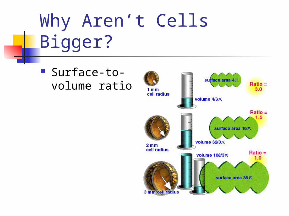
Why Aren’t Cells Bigger? Surface-to-volume
ratio
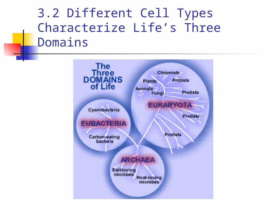
3.2 Different Cell Types Characterize Life’s Three Domains
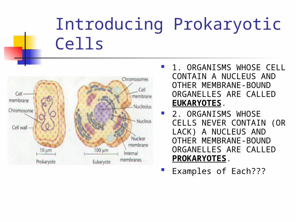
Introducing Prokaryotic Cells
1. ORGANISMS WHOSE CELL CONTAIN A NUCLEUS AND OTHER MEMBRANE-BOUND ORGANELLES ARE CALLED EUKARYOTES.
2. ORGANISMS WHOSE CELLS NEVER CONTAIN (OR LACK) A NUCLEUS AND OTHER MEMBRANE-BOUND ORGANELLES ARE CALLED PROKARYOTES.
Examples of Each???
Differences between
UNICELLULAR ORGANISMS such as bacteria and their relatives are Prokaryotes.
Prokaryotes are placed in Two Kingdoms, Separate from Eukaryotes.
All other organisms are Eukaryotes; plants, fish, mammals, insects and humans.

4.2 Prokaryotic Cells (fig.4.3)
Believed to be the first cells to evolve.
Lack a membrane bound nucleus and organelles.
Genetic material is naked in the cytoplasm
Ribosomes are only organelle.
Http.micro.magnet.fsu.edu/cells.html
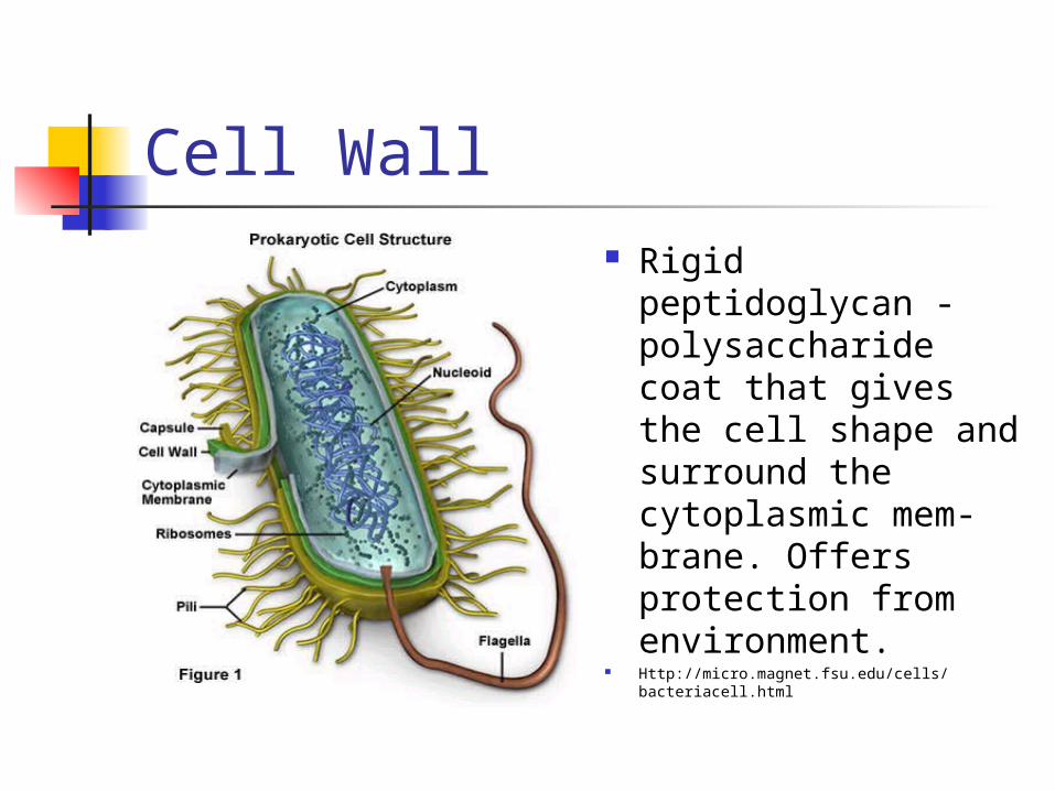
Cell Wall Rigid peptidoglycan
- polysaccharide coat that gives the cell shape and surround the cytoplasmic mem-brane. Offers protection from environment.
Http://micro.magnet.fsu.edu/cells/bacteriacell.html

Plasma Membrane Layer of phospho-
lipids and proteins that separates cytoplasm from external environment.
Regulates flow of material in and out of cell.
Http://micro.magnet.fsu.edu/cells/bacteriacell.html

Cytoplasm Also known as proto-
plasm is location of growth, metabolism, and replication. Is a gel-like matrix of water, enzymes, nutrients, wastes, and gases and contains cell structures.
Http://micro.magnet.fsu.edu/cells/bacteriacell.html
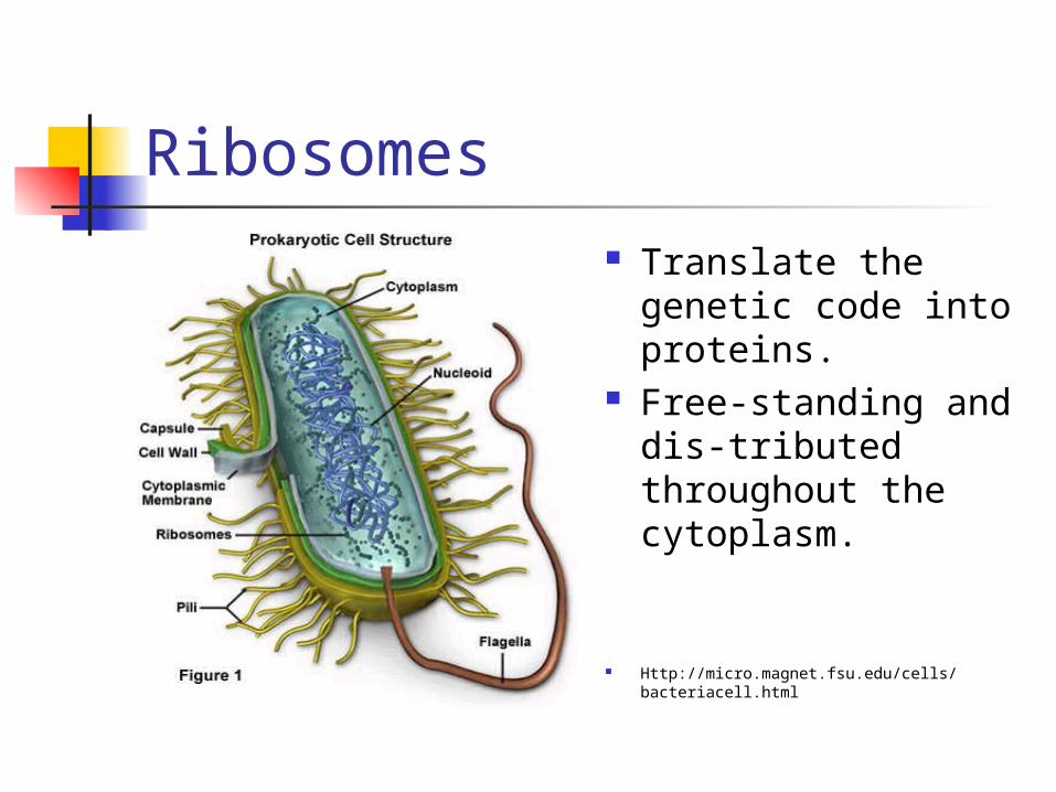
Ribosomes Translate the
genetic code into proteins.
Free-standing and dis-tributed throughout the cytoplasm.
Http://micro.magnet.fsu.edu/cells/bacteriacell.html

Nucleoid Region of the
cytoplasm where chromosomal DNA is located. Usually a singular, circular chromosome. Smaller circles of DNA called plasmids are also located in cytoplasm.
Http://micro.magnet.fsu.edu/cells/bacteriacell.html

Mesosome Infolding of cell
membrane. Possible role in cell
division. Increases surface
area. Photosynthetic
pigments or respira-tory chains here.
Http://www.med.sc.edu:85/fox/protobact.jpg

Prokaryotic vs. Eukaryotic
Http://micro.magnet.fsu.edu/cells.html
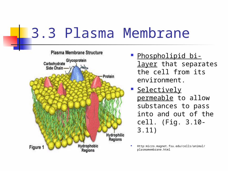
3.3 Plasma Membrane Phospholipid bi-layer
that separates the cell from its environment.
Selectively permeable to allow substances to pass into and out of the cell. (Fig. 3.10-3.11)
Http:micro.magnet.fsu.edu/cells/animal/plasmamembrane.html

Proteins (Fig 3.12)
Transport Proteins Enzymes Recognition Proteins Adhesion Proteins Receptor Proteins
3.4 Eukaryotic Organelles Divide Labor
OBJECTIVES: Describe the structures, composition, &
function of the cell membrane. Name the major organelles found in a
Eukaryotic cell, and describe their function.
Describe the structure and function of the nucleus.
Describe three structures characteristic of plant cells.
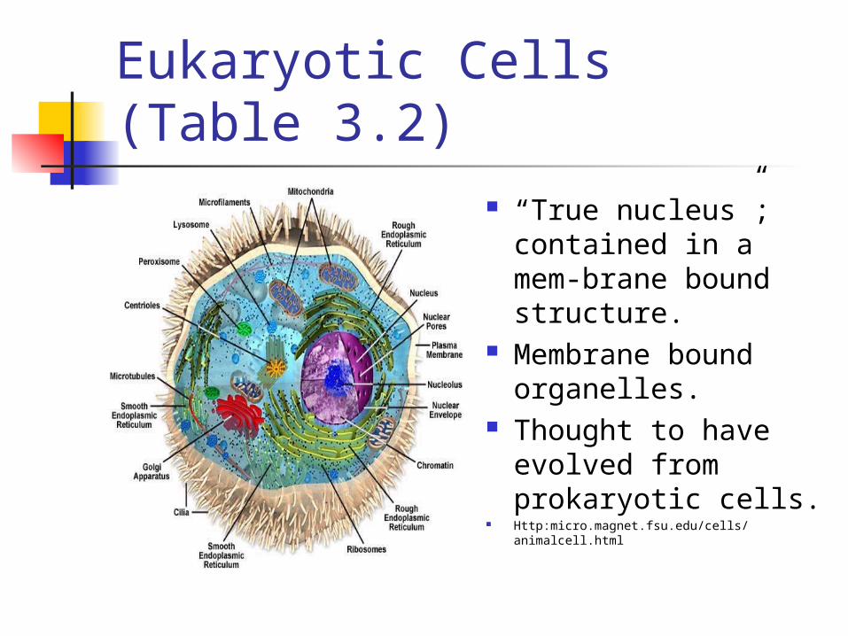
Eukaryotic Cells (Table 3.2)
“True nucleus”; contained in a mem-brane bound structure.
Membrane bound organelles.
Thought to have evolved from prokaryotic cells.
Http:micro.magnet.fsu.edu/cells/animalcell.html
CYTOPLASM EVERYTHING BETWEEN THE CELL MEMBRANE AND THE
NUCLEUS = CYTOPLASM.
Consists of TWO MAIN COMPONENTS: CYTOSOL and ORGANELLES.
CYTOSOL = jellylike mixture that consists MOSTLY OF WATER, along with PROTEINS, CARBOHYDRATES, SALTS, MINERALS and ORGANIC MOLECULES.
Suspended in the Cytosol are tiny ORGANELLES (ORGANS).
ORGANELLES ARE STRUCTURES THAT WORK LIKE MINIATURE ORGANS, THEY CARRY OUT SPECIFIC FUNCTIONS IN THE CELL.
Any analogies???
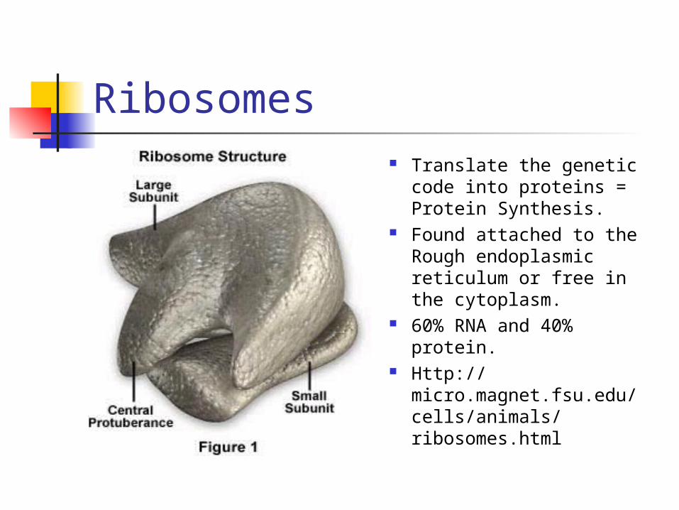
Ribosomes Translate the genetic
code into proteins = Protein Synthesis.
Found attached to the Rough endoplasmic reticulum or free in the cytoplasm.
60% RNA and 40% protein.
Http://micro.magnet.fsu.edu/cells/animals/ribosomes.html

Ribosome
Http://cellbio.utmb.edu/cellbio/ribosome.htm

Rough Endoplasmic Reticulum
Network of continuous sacs, studded with ribosomes.
Manufactures, pro-cesses, and transports proteins for export from cell.
Continuous with nuclear envelope.
Http://micro.magnet.fsu.edu/cels/animal/endoplasmicreticulum.html

Endoplasmic Reticulum (Fig 3.15)
Http://cellbio.utmb.edu/cellbio/ribosome.htm

Smooth Endoplasmic Reticulum
Similar in appearance to rough ER, but without the ribosomes.
Involved in the production of lipids, carbohydrate metabolism, and detoxification of drugs and poisons.
Metabolizes calcium. Http://micro.magnet.fsu.edu/cells/animals/endoplasmicreticulum.html
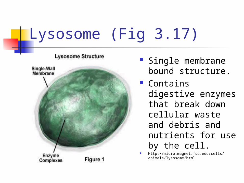
Lysosome (Fig 3.17)
Single membrane bound structure.
Contains digestive enzymes that break down cellular waste and debris and nutrients for use by the cell.
Http://micro.magnet.fsu.edu/cells/animals/lysosome/html

Lysosome
Http://anatomy.med.unsw.edu.au/teach/phph1004/1998/WWWlect3/sld005.htm
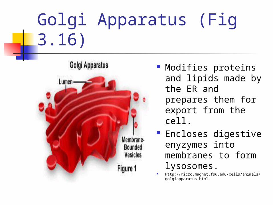
Golgi Apparatus (Fig 3.16) Modifies proteins
and lipids made by the ER and prepares them for export from the cell.
Encloses digestive enyzymes into membranes to form lysosomes.
Http://micro.magnet.fsu.edu/cells/animals/golgiapparatus.html

Golgi Apparatus
Http://cellbio.utmb.edu/cellbio/golgi.htm
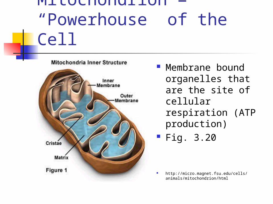
Mitochondrion = “Powerhouse” of the Cell
Membrane bound organelles that are the site of cellular respiration (ATP production)
Fig. 3.20
http://micro.magnet.fsu.edu/cells/animals/mitochondrion/html

Mitochondrion
Http://anatomy.med.unsw.edu.au/teach/phph1004/1998/WWWlect3/sld005.htm
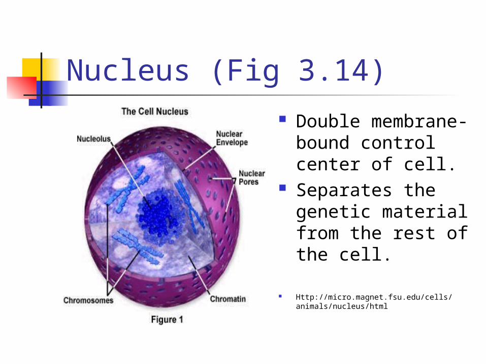
Nucleus (Fig 3.14) Double membrane-
bound control center of cell.
Separates the genetic material from the rest of the cell.
Http://micro.magnet.fsu.edu/cells/animals/nucleus/html
Nucleus
Http://cellbio.utmb.edu/cellbio/nucleus.htm
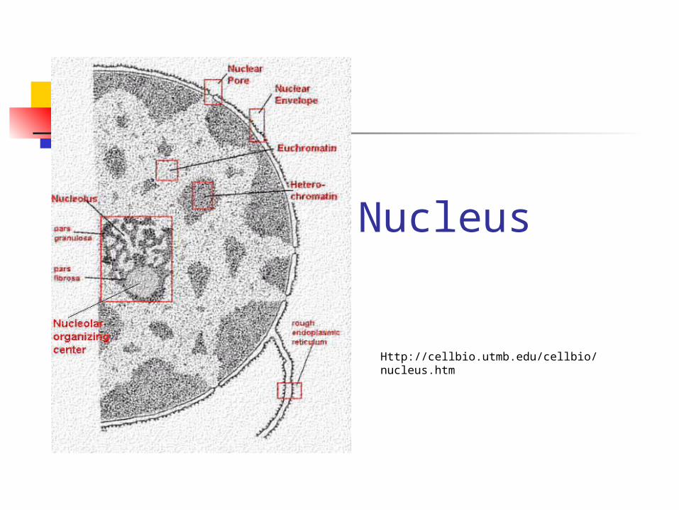
Parts of the nucleus:
Chromatin - genetic material of cell in its non-dividing state.
Nucleolus - dark-staining structure in the nucleus that plays a role in making ribosomes
Nuclear envelope - double membrane structure that separates nucleus from cytoplasm.
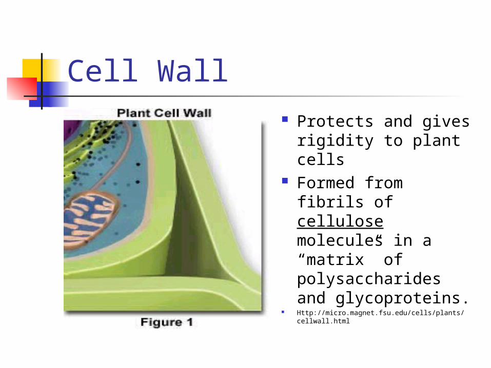
Cell Wall Protects and gives
rigidity to plant cells
Formed from fibrils of cellulose molecules in a “matrix” of polysaccharides and glycoproteins.
Http://micro.magnet.fsu.edu/cells/plants/cellwall.html

PLANT CELLS
2 Additional Structures Not found in animals cells
1. CELL WALLS2. PLASTIDS
PLASTIDS MAKE OR STORE FOOD CHLOROPLAST, (figure 4-17) an organelle that
converts SUNLIGHT, CARBON DIOXIDE, AND WATER INTO SUGARS. This process is called PHOTOSYNTHESIS
Each Chloroplast encloses a system of Flattened, Membranous Sacs called THYLAKOIDS. It is in the Thylakoids that Photosynthesis occurs
Chloroplasts are GREEN because they contain CHLOROPHYLL, a PIGMENT that ABSORBS ENERGY IN SUNLIGHT. THEY ARE FOUND ONLY IN ALGAE, SUCH AS SEAWEED, AND IN GREEN PLANTS.
Other PLASTIDS store reddish-orange pigments that color fruits, vegetables, flowers, and autumn leaves
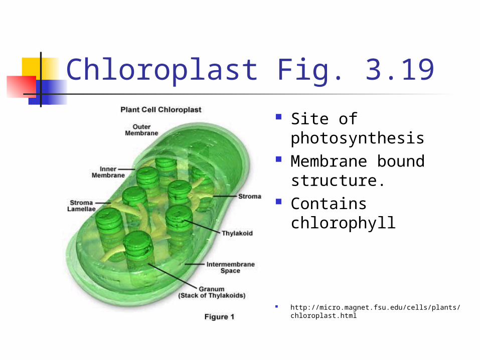
Chloroplast Fig. 3.19 Site of
photosynthesis Membrane bound
structure. Contains chlorophyll
http://micro.magnet.fsu.edu/cells/plants/chloroplast.html
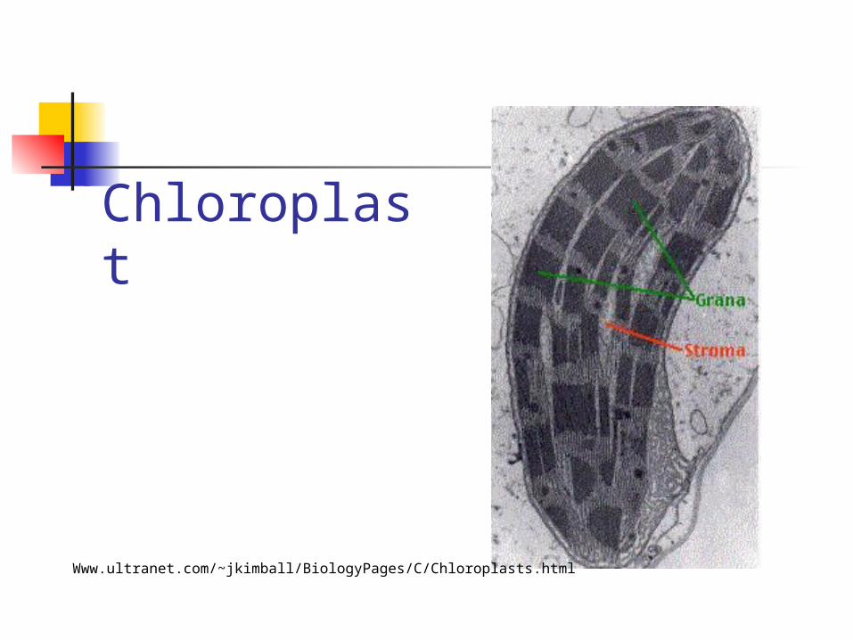
Chloroplast
Www.ultranet.com/~jkimball/BiologyPages/C/Chloroplasts.html
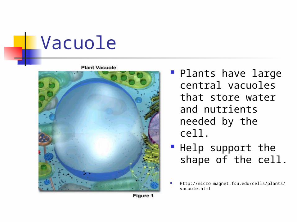
Vacuole Plants have large
central vacuoles that store water and nutrients needed by the cell.
Help support the shape of the cell.
Http://micro.magnet.fsu.edu/cells/plants/vacuole.html
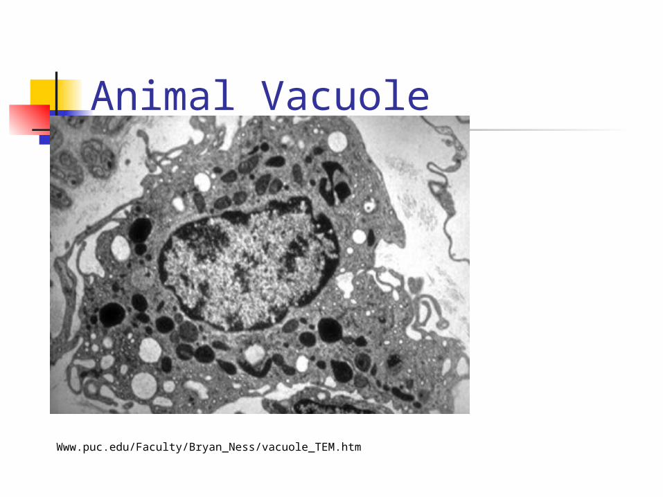
Animal Vacuole
Www.puc.edu/Faculty/Bryan_Ness/vacuole_TEM.htm
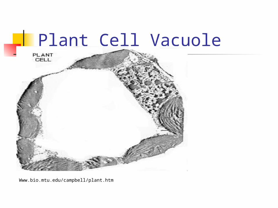
Plant Cell Vacuole
Www.bio.mtu.edu/campbell/plant.htm

Animal Cell vs. Plant Cell
Http://:micro.magnet.fsu.edu/cells/html

3.5 The Cytoskeleton Supports Eukaryotic Cells
In Animal Cells, an internal framework called CYTOSKELETON maintains the Shape of the Cell (Fig. 3.23)
TWO Types of structures: MICROFILAMENTS MICROTUBULES

MICROFILAMENTS
NOT HOLLOW and have a structure that resembles ROPE made of TWO TWISTED CHAINS OF PROTEIN called ACTIN.
CONTRACT, causing movement. Muscle Cells

MICROTUBULES (Fig. 3.24) HOLLOW TUBES like plumbing pipes. They are
the Largest Strands of the Cytoskeleton. Made of a PROTEIN called TUBULIN. THREE FUNCTIONS: A. To maintain the shape of cell. B. To serve as tracks for organelles to move
along within the cell. C. When the Cell is about to divide, bundles of
Microtubules known as SPINDLE FIBERS come together and extend across the cell to assist in the movement of Chromosomes during Cell Division

Microfilaments Solid rods of
globular proteins. Important
component of cytoskeleton which offers support to cell structure.
Http://micro.magnet.fsu.edu/cells/animals/microfilaments.html
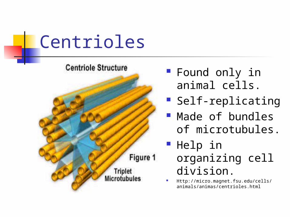
Centrioles Found only in
animal cells. Self-replicating Made of bundles
of microtubules. Help in organizing
cell division. Http://micro.magnet.fsu.edu/cells/
animals/animas/centrioles.html
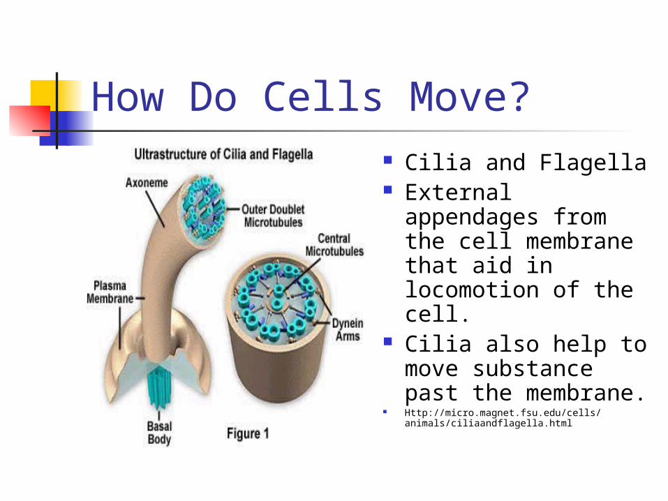
How Do Cells Move? Cilia and Flagella External
appendages from the cell membrane that aid in locomotion of the cell.
Cilia also help to move substance past the membrane.
Http://micro.magnet.fsu.edu/cells/animals/ciliaandflagella.html


