• Cells must communicate to coordinate their activities. • Cells may receive a variety of signals, chemical signals, electromagnetic signals, and mechanical signals. Introduction • The process by which a signal on a cell’s surface is converted into a specific cellular response is a signal-transduction pathway.
Transcript
• Cells must communicate to coordinate their activities.
• Cells may receive a variety of signals, chemical signals, electromagnetic signals, and mechanical signals.
Introduction
• The process by which a signal on a cell’s surface is converted into a specific cellular response is a signal-transduction pathway.
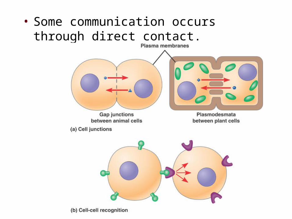
• Some communication occurs through direct contact.
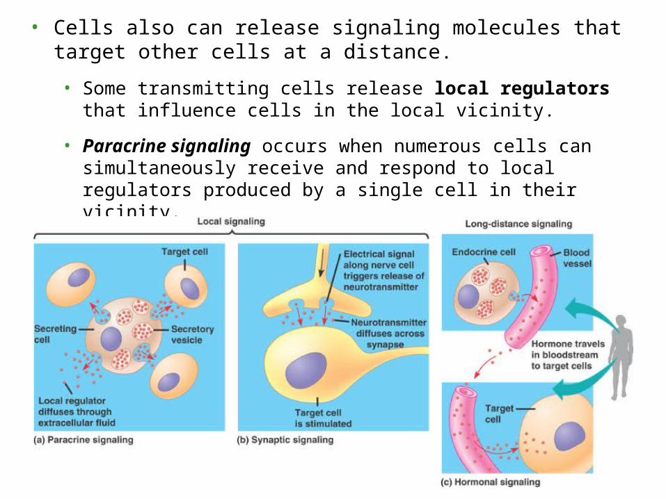
• Cells also can release signaling molecules that target other cells at a distance.
• Some transmitting cells release local regulators that influence cells in the local vicinity.
• Paracrine signaling occurs when numerous cells can simultaneously receive and respond to local regulators produced by a single cell in their vicinity.
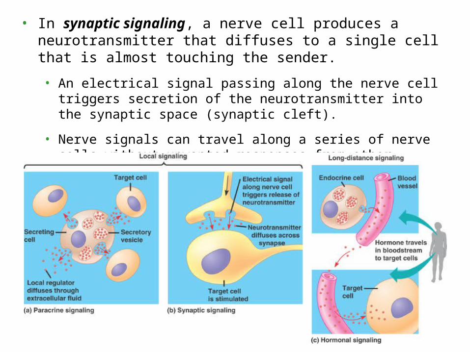
• In synaptic signaling, a nerve cell produces a neurotransmitter that diffuses to a single cell that is almost touching the sender.
• An electrical signal passing along the nerve cell triggers secretion of the neurotransmitter into the synaptic space (synaptic cleft).
• Nerve signals can travel along a series of nerve cells without unwanted responses from other cells.
• Plants and animals use hormones to signal at greater distances.
• In animals, specialized endocrine cells release hormones into the circulatory system and target cells in other parts of the body.
• In plants, hormones may travel in vessels, but more often travel from cell to cell or by diffusion in air.
• Hormones and local regulators range widely in size and type.
• The plant hormone ethylene (C2H4), is
a small gas molecule that promotes fruit ripening and regulates growth.
• Insulin, which regulates sugar levels in the blood of mammals, is a protein with thousands of atoms.
• The origins of our understanding of cell signaling were pioneered by E.W. Sutherland and colleagues.
• Their work investigated how the animal hormone epinephrine stimulates breakdown of glycogen in liver and skeletal muscle.
• Breakdown of glycogen releases glucose derivatives that can be used for fuel in glycolysis.
• Sutherland’s research team discovered that epinephrine activated a cytosolic enzyme, glycogen phosphorylase.
• However, epinephrine did not activate the phosphorylase directly but could only act onintact cells.
• Therefore, there must be an intermediate step or steps occurring inside the cell.
• Also, the plasma membrane must be involved in transmitting the epinephrine signal.
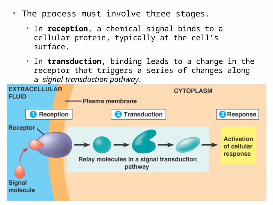
• The process must involve three stages.
• In reception, a chemical signal binds to a cellular protein, typically at the cell’s surface.
• In transduction, binding leads to a change in the receptor that triggers a series of changes along a signal-transduction pathway.
• In response, the transduced signal triggers a specific cellular activity.
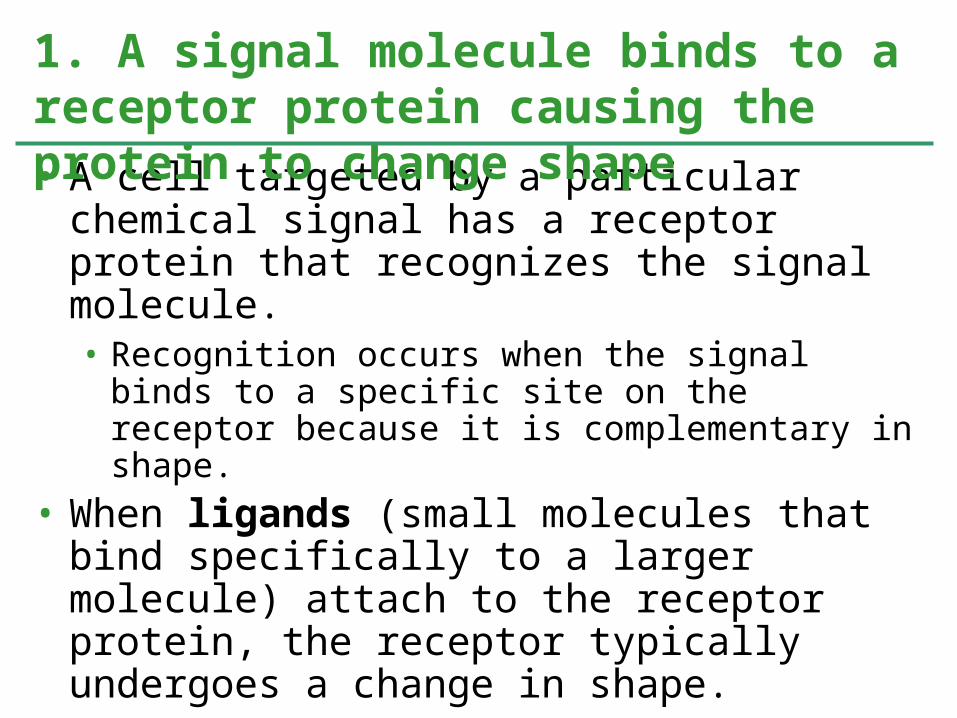
• A cell targeted by a particular chemical signal has a receptor protein that recognizes the signal molecule.• Recognition occurs when the signal binds to a specific site on the receptor because it is complementary in shape.
• When ligands (small molecules that bind specifically to a larger molecule) attach to the receptor protein, the receptor typically undergoes a change in shape.• This may activate the receptor so that it can interact with other molecules inside the cell.
• For other receptors this leads to aggregation of receptors.
1. A signal molecule binds to a receptor protein causing the protein to change shape
• Most signal molecules are water-soluble and too large to pass through the plasma membrane.
• They influence cell activities by binding to receptor proteins on the plasma membrane.
• Three major types of receptors are G-protein-linked receptors, tyrosine-kinase receptors, and ion-channel receptors.
2. Most signal receptors are plasma membrane proteins
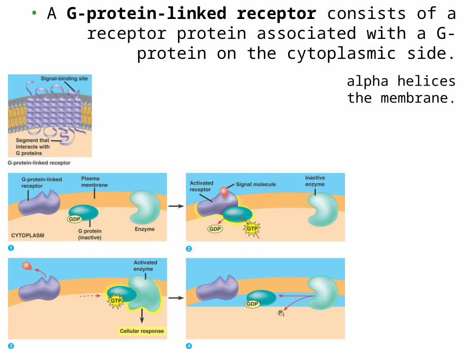
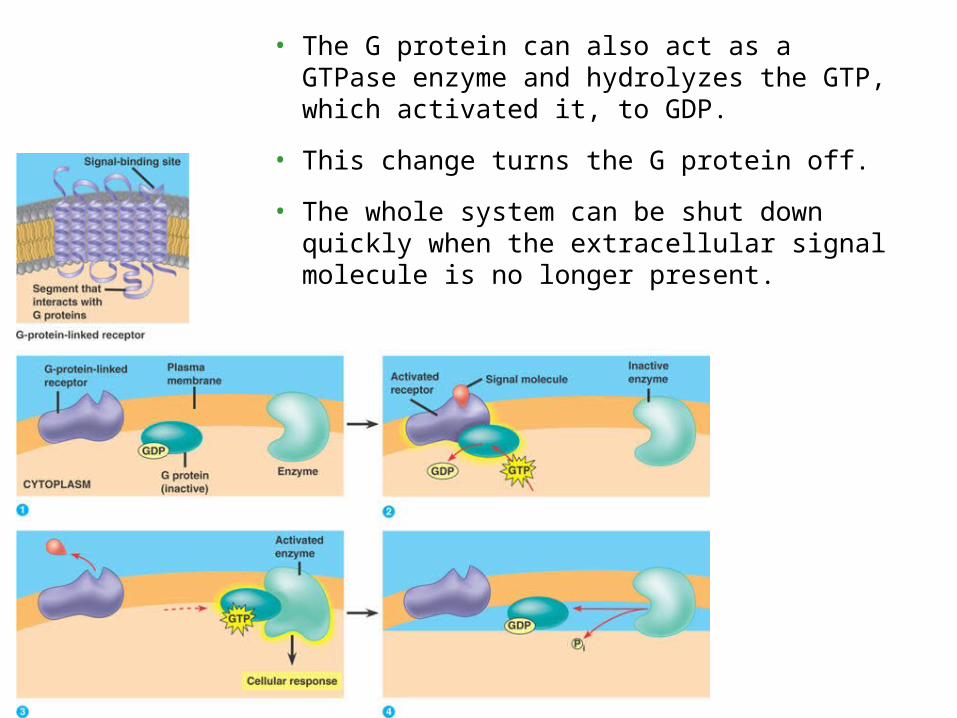
• A G-protein-linked receptor consists of a receptor protein associated with a G-
protein on the cytoplasmic side.
The receptor consists of seven alpha helices spanning the membrane.
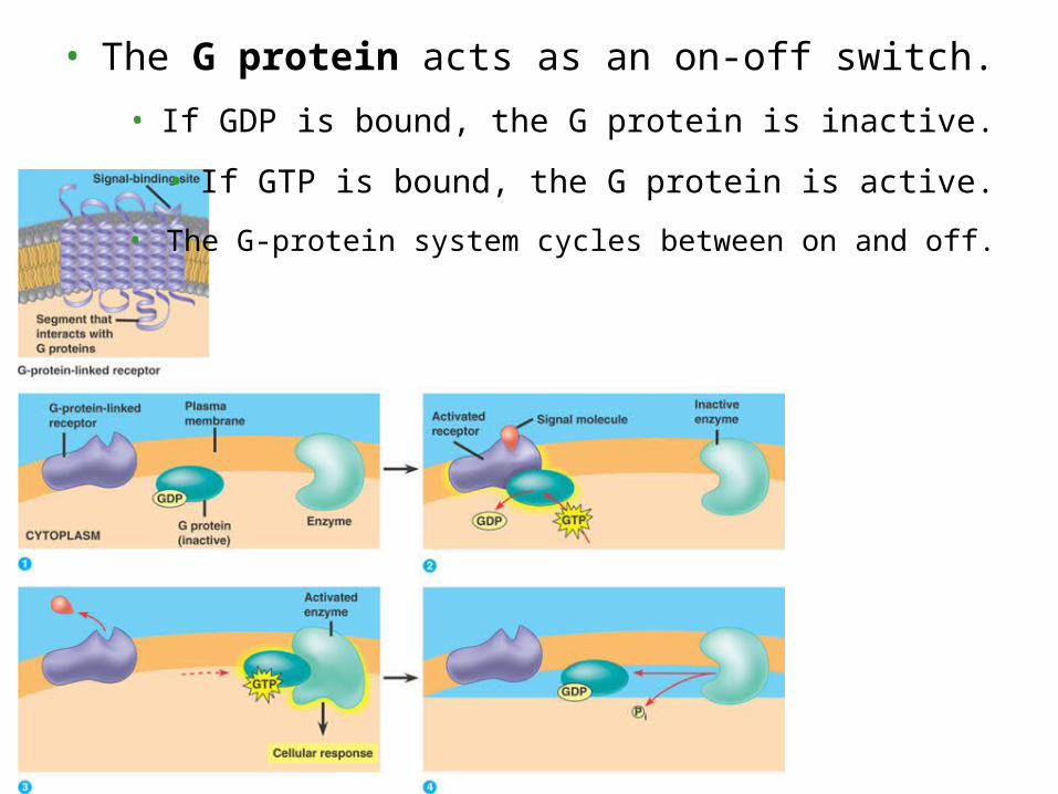
• The G protein acts as an on-off switch.
• If GDP is bound, the G protein is inactive.
• If GTP is bound, the G protein is active.
• The G-protein system cycles between on and off.
• The G protein can also act as a GTPase enzyme and hydrolyzes the GTP, which activated it, to GDP.
• This change turns the G protein off.
• The whole system can be shut down quickly when the extracellular signal molecule is no longer present.
• G-protein receptor systems are extremely widespread and diverse in their functions.
• embryonic development
• sensory systems.
• Several human diseases are the results of activities, including bacterial infections, that interfere with G-protein function.
• Cholera
• Botulism
• The tyrosine-kinase receptor system is especially effective when the cell needs to regulate and coordinate a variety of activities and trigger several signal pathways at once.
• Extracellular growth factors often bind to tyrosine-kinase receptors.
• The cytoplasmic side of these receptors function as a tyrosine kinase, transferring a phosphate group from ATP to tyrosine on a substrate protein.
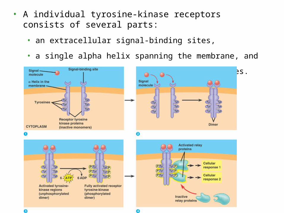
• A individual tyrosine-kinase receptors consists of several parts:
• an extracellular signal-binding sites,
• a single alpha helix spanning the membrane, and
• an intracellular tail with several tyrosines.
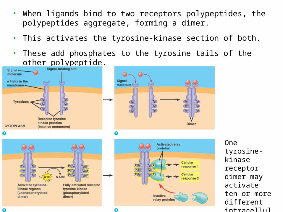
• When ligands bind to two receptors polypeptides, the polypeptides aggregate, forming a dimer.
• This activates the tyrosine-kinase section of both.
• These add phosphates to the tyrosine tails of the other polypeptide.
One tyrosine-kinase receptor dimer may activate ten or more different intracellular proteins simultaneously.
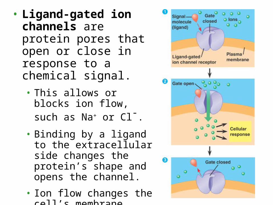
• Ligand-gated ion channels are protein pores that open or close in response to a chemical signal.• This allows or blocks ion flow, such as Na+ or Cl-.
• Binding by a ligand to the extracellular side changes the protein’s shape and opens the channel.
• Ion flow changes the cell’s membrane potential.
• When the ligand dissociates, the channel closes.
• Ligand-gated ion channels are very important in the nervous system.
• Similar gated ion channels respond to electrical signals (voltage-gated ion channels).
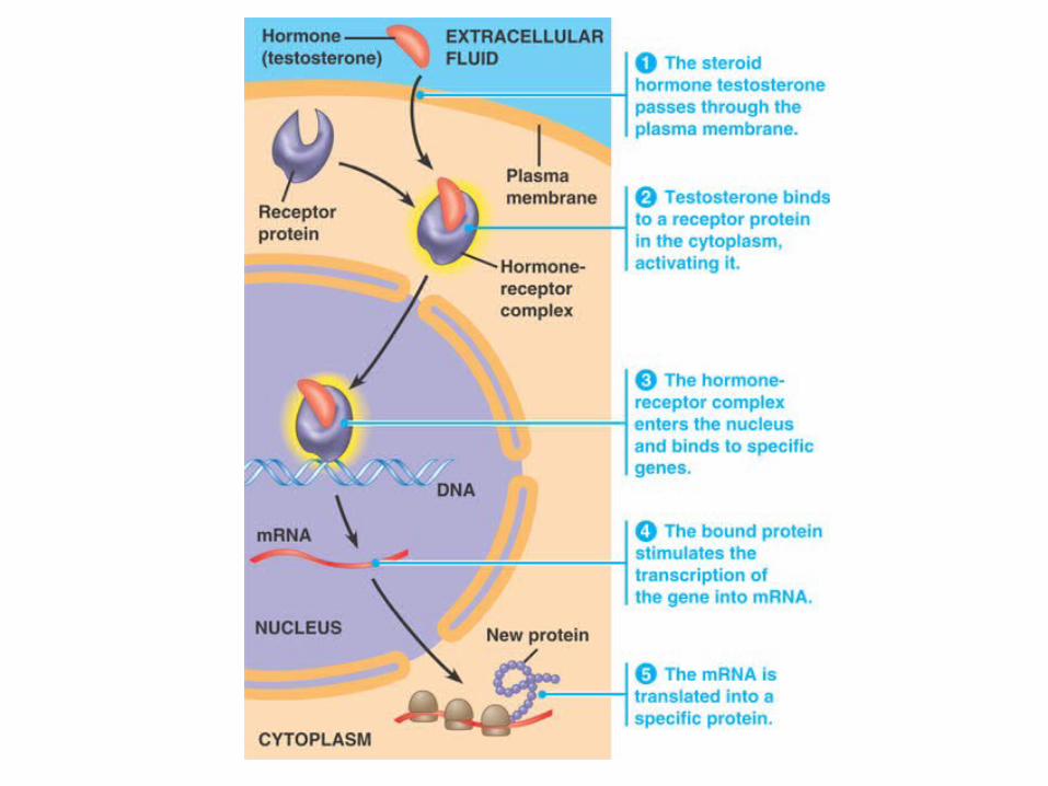
• Other signal receptors are dissolved in the cytosol or nucleus of target cells.
• The signals pass through the plasma membrane.
• These chemical messengers include the hydrophobic steroid and thyroid hormones of animals.
• Also in this group is nitric oxide (NO), a gas whose small size allows it to slide between membrane phospholipids.
• These activated proteins act as transcription factors.
• Transcription factors control which genes are turned on - that is, which genes are transcribed into messenger RNA (mRNA) and translated into protein by ribosomes.
• Other intracellular receptors are already in the nucleus and bind to the signal molecules there (e.g., estrogen receptors).
• The transduction stage of signaling is usually a multistep pathway.
• These pathways often greatly amplify the signal.
• A small number of signal molecules can produce a large cellular response.
• Also, multistep pathways provide more opportunities for coordination and regulation than do simpler systems.
Signal Transduction Pathways
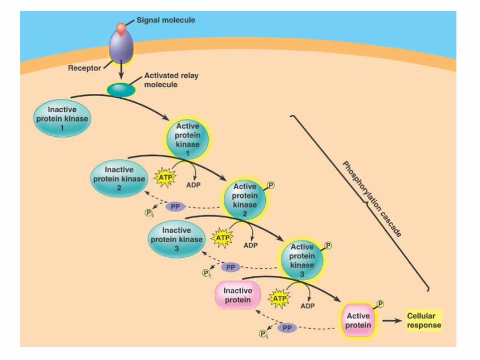
• The phosphorylation of proteins by a specific enzyme (a protein kinase) is a widespread cellular mechanism for regulating protein activity.
• Protein kinases can lead to a “phosphorylation cascade”.
• Each protein phosphorylation leads to a shape change because of the interaction between the phosphate group and charged or polar amino acids.
• Phosphorylation of a protein typically converts it from an inactive form to an active form.
• The reverse (inactivation) is possible too for some proteins.
• A single cell may have hundreds of different protein kinases, each specific for a different substrate protein.
• The responsibility for turning off a signal-transduction pathway belongs to protein phosphatases.
• These enzymes rapidly remove phosphate groups from proteins.
• The activity of a protein regulated by phosphorylation depends on the balance of active kinase molecules and active phosphatase molecules.
• When an extracellular signal molecule is absent, active phosphatase molecules predominate, and the signaling pathway and cellular response are shut down.
• Many signaling pathways involve small, nonprotein, water-soluble molecules or ions, called second messengers.
• These molecules rapidly diffuse throughout the cell.
• Second messengers participate in pathways initiated by both G-protein-linked receptors, tyrosine-kinase receptors, and some ion channels.
• Two of the most important are cyclic AMP and Ca2+.
• Once Sutherland knew that epinephrine caused glycogen breakdown without entering the cell, he looked for a second messenger inside the cell.
• Binding by epinephrine leads to increases in the concentration of cyclic AMP or cAMP.
• This occurs because the receptor activates adenylyl cyclase which converts ATP to cAMP.
• cAMP is short-lived as phosphodiesterase converts it to AMP.
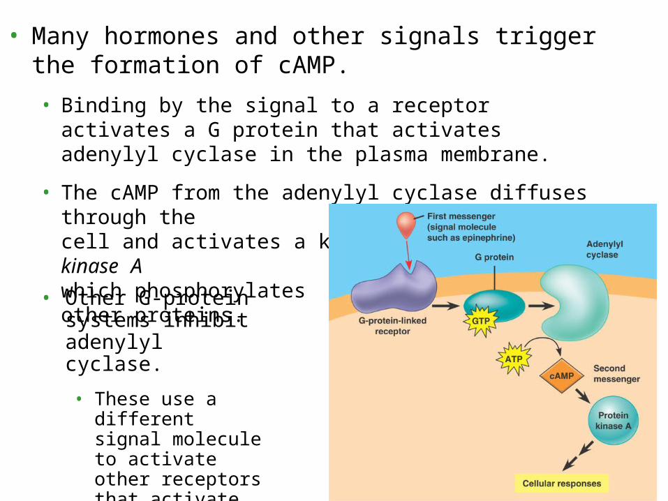
• Many hormones and other signals trigger the formation of cAMP.
• Binding by the signal to a receptor activates a G protein that activates adenylyl cyclase in the plasma membrane.
• The cAMP from the adenylyl cyclase diffuses through the cell and activates a kinase, called protein kinase A which phosphorylates other proteins.
• Other G-protein systems inhibit adenylyl cyclase.
• These use a different signal molecule to activate other receptors that activate inhibitory G proteins.
• Many signal molecules in animals induce responses in their target cells via signal-transduction pathways that increase the cytosolic concentration of Ca2+.
• In animal cells, increases in Ca2+ may cause contraction of muscle cells, secretion of some substances, and cell division.
• Cells use Ca2+ as a second messenger in G-protein pathways, tyrosine-kinase pathways, and for some ion channels.
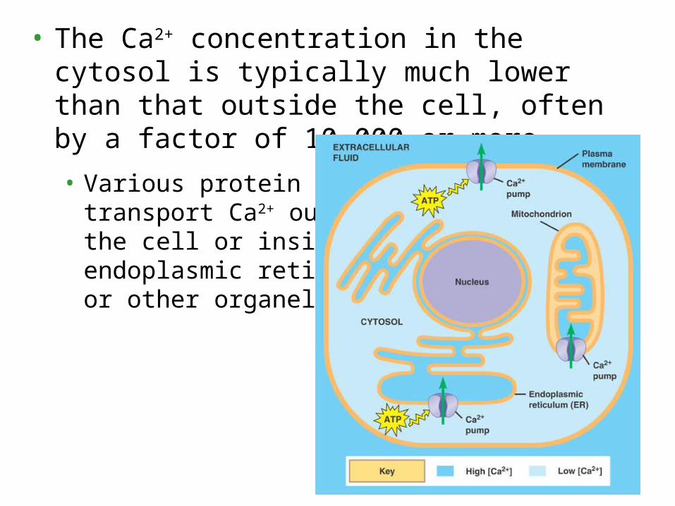
• The Ca2+ concentration in the cytosol is typically much lower than that outside the cell, often by a factor of 10,000 or more.
• Various protein pumps transport Ca2+ outside the cell or inside the endoplasmic reticulum or other organelles.
• Because cytosolic Ca2+ is so low, small changes in the absolute numbers of ions causes a relatively large percentage change in Ca2+ concentration.
• Signal-transduction pathways trigger the release of Ca2+ from the cell’s ER.
• Some pathways leading to release from the ER involve still other second messengers, diacylglycerol (DAG) and inositol trisphosphate (IP3).
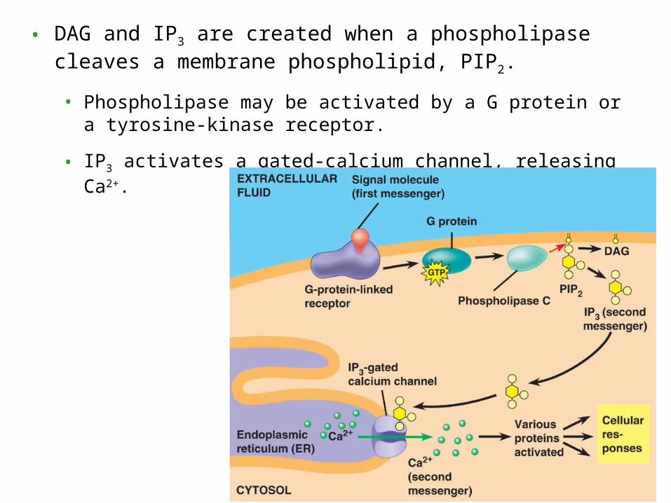
• DAG and IP3 are created when a phospholipase cleaves a membrane phospholipid, PIP2.
• Phospholipase may be activated by a G protein or a tyrosine-kinase receptor.
• IP3 activates a gated-calcium channel, releasing Ca2+.
• Ultimately, a signal-transduction pathway leads to the regulation of one or more cellular activities.
• This may be a change in an ion channel or a change in cell metabolism.
• For example, epinephrine helps regulate cellular energy metabolism by activating enzymes that catalyze the breakdown of glycogen.
In response to a signal, a cell may regulate activities in the cytoplasm or transcription in the nucleus
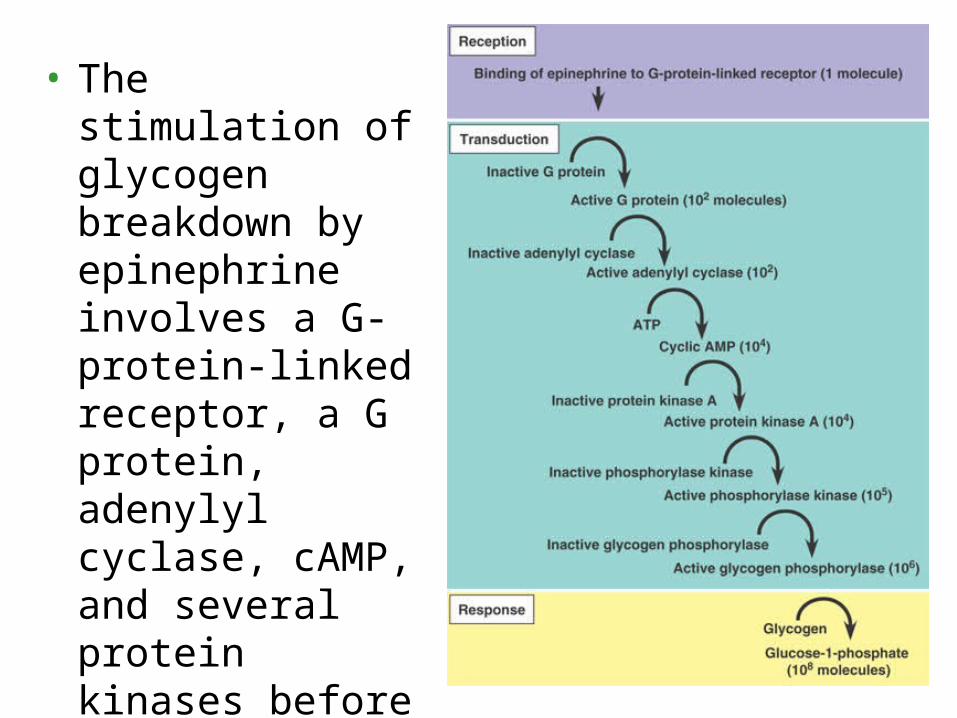
• The stimulation of glycogen breakdown by epinephrine involves a G-protein-linked receptor, a G protein, adenylyl cyclase, cAMP, and several protein kinases before glycogen phosphorylase is activated.
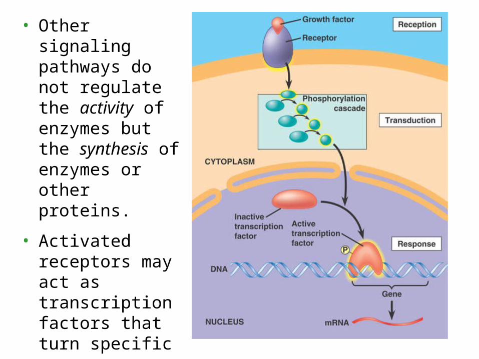
• Other signaling pathways do not regulate the activity of enzymes but the synthesis of enzymes or other proteins.
• Activated receptors may act as transcription factors that turn specific genes on or off in the nucleus.
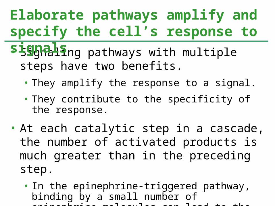
• Signaling pathways with multiple steps have two benefits.• They amplify the response to a signal.
• They contribute to the specificity of the response.
• At each catalytic step in a cascade, the number of activated products is much greater than in the preceding step.• In the epinephrine-triggered pathway, binding by a small number of epinephrine molecules can lead to the release of hundreds of millions of glucose molecules.
Elaborate pathways amplify and specify the cell’s response to signals
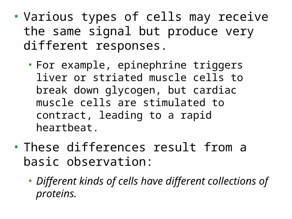
• Various types of cells may receive the same signal but produce very different responses.
• For example, epinephrine triggers liver or striated muscle cells to break down glycogen, but cardiac muscle cells are stimulated to contract, leading to a rapid heartbeat.
• These differences result from a basic observation:
• Different kinds of cells have different collections of proteins.
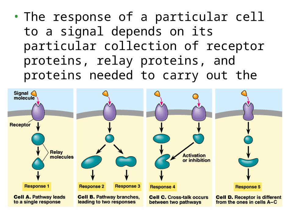
• The response of a particular cell to a signal depends on its particular collection of receptor proteins, relay proteins, and proteins needed to carry out the response.
• Two cells that respond differently to the same signal differ in one or more of the proteins that handle and respond to the signal.
• A single signal may follow a single pathway in one cell but trigger a branched pathway in another.
• Two pathways may converge to modulate a single response.
• Branching of pathways and interactions between pathways are important for regulating and coordinating a cell’s response to incoming information.
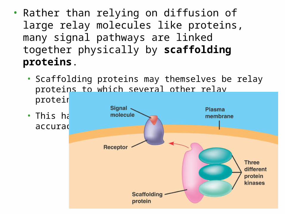
• Rather than relying on diffusion of large relay molecules like proteins, many signal pathways are linked together physically by scaffolding proteins.
• Scaffolding proteins may themselves be relay proteins to which several other relay proteins attach.
• This hardwiring enhances the speed and accuracy of signal transfer.


![Distinct signals and immune cells drive liver pathology ...€¦ · Research Article Distinct signals and immune cells drive liver pathology and glomerulonephritis in ABIN1[D485N]](https://static.documents.pub/doc/80x56/5f0ff74f7e708231d446c5df/distinct-signals-and-immune-cells-drive-liver-pathology-research-article-distinct.jpg)


