Page 1
Viruses 2013, 5, 374-405; doi:10.3390/v5010374
virusesISSN 1999-4915
www.mdpi.com/journal/viruses
Review
Cellular Aspects of Prion Replication In Vitro
Andrea Grassmann 1,†
, Hanna Wolf 1,†
, Julia Hofmann 1, James Graham
1 and Ina Vorberg
1,2,*
1 German Center for Neurodegenerative Diseases (DZNE e.V.), Ludwig-Erhard-Allee 2,
53175 Bonn, Germany; E-Mails: [email protected] (A.G.); [email protected] (H.W.);
[email protected] (J.H.); [email protected] (J.G.) 2
Rheinische Friedrich-Wilhelms-Universität Bonn, Bonn, Germany
† These authors contributed equally to this work.
* Author to whom correspondence should be addressed; E-Mail: [email protected] ;
Tel.: +49-228-43302-560; Fax.: +49-228-43302-689.
Received: 12 December 2012; in revised form: 7 January 2013 / Accepted: 16 January 2013 /
Published: 22 January 2013
Abstract: Prion diseases or transmissible spongiform encephalopathies (TSEs) are fatal
neurodegenerative disorders in mammals that are caused by unconventional agents
predominantly composed of aggregated misfolded prion protein (PrP).
Prions self-propagate by recruitment of host-encoded PrP into highly ordered
-sheet rich aggregates. Prion strains differ in their clinical, pathological and biochemical
characteristics and are likely to be the consequence of distinct abnormal prion protein
conformers that stably replicate their alternate states in the host cell. Understanding prion
cell biology is fundamental for identifying potential drug targets for disease intervention.
The development of permissive cell culture models has greatly enhanced our knowledge on
entry, propagation and dissemination of TSE agents. However, despite extensive research,
the precise mechanism of prion infection and potential strain effects remain enigmatic.
This review summarizes our current knowledge of the cell biology and propagation of
prions derived from cell culture experiments. We discuss recent findings on the trafficking
of cellular and pathologic PrP, the potential sites of abnormal prion protein synthesis and
potential co-factors involved in prion entry and propagation.
Keywords: prion; prion strains; transmissible spongiform encephalopathies;
glycosaminoglycans; LRP1; RPSA
OPEN ACCESS
Page 2
Viruses 2013, 5
375
1. Introduction
Prion diseases or transmissible spongiform encephalopathies (TSEs) are neurodegenerative
disorders that affect many mammalian species. TSEs include Creutzfeldt-Jakob disease, fatal familial
insomnia and Gerstmann-Sträussler-Scheinker syndrome in humans, scrapie in sheep and goats,
chronic wasting disease in deer and elk and bovine spongiform encephalopathy. In humans, prion
diseases can be sporadic, infectious or of genetic origin. Natural genetic prion diseases in animals have
not been reported until now. In animals, infection occurs mainly through the intestinal tract due to
ingestion of prions present in the food or the natural environment [1]. During TSE disease, an
abnormally folded conformer (PrPSc
) of the cellular prion protein (PrPC) accumulates in the central
nervous and lymphoreticular system of the infected host. According to the prion hypothesis, PrPSc
constitutes the major, if not only, component of the proteinaceous infectious particles [2,3]. The
conversion of the host-encoded PrPC to PrP
Sc is a post-translational process that involves a
conformational change from a predominantly -helical structure to a protein fold increased in ß-sheet.
PrPSc
is likely generated by a seeded polymerization reaction in which it serves as a template that binds
to normal PrPC and catalyzes its conformational conversion to an abnormal, aggregated isoform. PrP
aggregates consist of fibrils with a cross-ß-structure that is characteristic of amyloid. As the amyloid
fibril elongates and matures, it acquires an increase in conformational stability that is resistant to
denaturation by heating, detergents and proteases. Amyloid fibrils are associated with many other
neurodegenerative protein misfolding disorders, notably Alzheimer’s and Parkinson’s disease [4].
However, prion diseases are unconventional protein misfolding disorders because they constitute
infectious diseases that are often naturally transmitted within species and sometimes even across
species barriers.
The first prion disease studied was scrapie of sheep and goats. Seminal work on scrapie by Pattison
and Millson in 1961 laid the foundations for the hypothesis that prions exist as different strains [5].
At least 20 different prion strains have been isolated from scrapie that can be propagated in the same
inbred mouse line. Prion strains are distinguished by several semi-quantitative factors including
incubation time before disease onset, lesion profiles in the brain and the areas of deposition of
aggregated PrP [6,7]. PrPSc
molecules associated with prion strains differ in their biochemical and
biophysical properties. For example, PrPSc
molecules exhibit strain-specific glycosylation profiles, and
differ in their resistance to proteases as well as in their binding to conformation-specific
antibodies [8,9]. This led to the proposal that prion strains are enciphered by the specific fold of PrPSc
[3,10]. According to this theory, strain-specific PrPSc
conformations would be adopted and amplified
by the binding and subsequent conversion of PrPC, thereby preserving the strain-specific information
enciphered by the respective quaternary structures of PrPSc
.
2. The Cellular Prion Protein PrPC: Structure, Biosynthesis and Intracellular Trafficking
In 1985 researchers identified the Prnp gene encoding the prion protein [11,12] on chromosome
20 in humans and chromosome 2 in mice [13,14]. The Prnp gene is evolutionary highly conserved,
exhibiting a sequence homology of approximately 80% from amphibia to mammals [15–17]. The Prnp
gene contains two to three exons depending on the species, with the last exon encoding the open
Page 3
Viruses 2013, 5
376
reading frame [12]. Cellular prion protein is constitutively expressed in many tissues, including the
central and peripheral nervous system as well as the immune, lymphoreticular and intestinal
system [18]. A particularly high expression is found in neurons localized both at pre- and post-synaptic
sites [19] and in glial cells [20].
PrPC is synthesized on the rough endoplasmic reticulum (ER) and transits through the Golgi
apparatus to the cell surface (Figure 1A). Within the ER and Golgi, PrPC becomes glycosylated at two
asparagine residues [21]. Further post-translational modifications include the formation of a disulfide
bond between two cysteine residues (amino acid residues 179 and 214 in human PrP) [22] and the
attachment of a glycosyl-phosphatidyl-inositol (GPI) moiety at the carboxy-terminus of the protein
[23]. At the plasma membrane, PrPC is incorporated into lipid rafts and caveolae (raft structures with
caveolin-1), which are regions of the membrane enriched in cholesterol and sphingolipids [24,25].
Targeting to these lipid rafts is mediated by the amino-terminus of PrPC [26,27]. An early association
of PrPC with lipid rafts during its biosynthesis appears to be necessary for its correct folding [28].
Although PrPC is normally translocated to the plasma membrane, high concentrations have been
detected within multivesicular bodies [29]. Once on the plasma membrane, PrPC can undergo
proteolytic processing by metalloproteases, resulting in a membrane-attached carboxyterminal (C1)
and an extracellularly released amino-terminal fragment [30–33]. In addition, it has been observed that
a small percentage of full-length PrPC molecules is secreted, either in a soluble form [34,35] or in
association with exosomes [36,37]. Within the cell there is a minor sub-population of PrPC molecules
present in the cytosol [38]. Interestingly, using an inducible cell line, PrP23-230 was found in the
nucleus of these cells and in association with chromatin [39]. The physiological relevance of such
intranuclear localization so far is unclear.
Extensive research into the biological function of PrPC has resulted in a plethora of different
possible functions. So far, these include involvement in signaling cascades, neuronal survival,
apoptosis, oxidative stress, cell adhesion, differentiation, immunomodulation and more recently,
microRNA metabolism [40,41]. PrPC
has a high affinity for metals such as copper, zinc and manganese
through binding at its amino-terminus. Binding to PrPC mediates neuronal uptake of these metal ions
potentially through interaction with other receptors [42,43]. PrPC has also been proposed to act as a
cell surface scaffold protein that interacts with different partners. These mediate the activation of a
range of diverse signaling pathways that modulate neuritogenesis and synapse formation [40].
Interactions of PrPC
with the neural cell adhesion molecule NCAM or with the laminin receptor
precursor LRP/LR have been reported to elicit specific signaling cascades in neurons [44–46].
In non-neuronal cells, PrPC also plays an important role during embryogenesis or during stem-cell
proliferation and differentiation [47,48]. Interestingly, PrP has also been shown to bind both RNA
and DNA in vitro [49–52]. Evidence for a physiological role of these nucleic acid-protein
associations [41] is accumulating but needs further clarification.
Page 4
Viruses 2013, 5
377
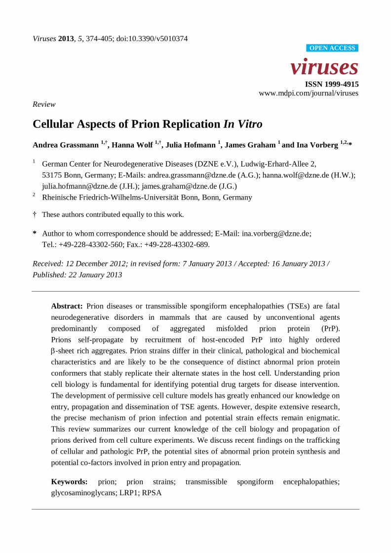
Figure 1. Localization of PrPC and PrP
Sc in L929 fibroblast cells. (A) Indirect
immunofluorescence (IF) staining of cellular PrP (green) in uninfected L929 cells. PrPC
predominantly resides at the cell surface with some intracellular localization. (B) Detection
of PrPSc
in L929 cells persistently infected with prion strain 22L by IF. In contrast to PrPC,
PrPSc
(green) primarily localizes intracellularly and partially co-localizes with the
lysosomal marker Lamp-1 (red). (A,B) Nuclei were counterstained with Hoechst (blue).
Scale bar: 5 µm.
PrPC is rapidly and constitutively endocytosed from the plasma membrane [53,54]. External stimuli
such as the binding of copper or stress-inducible protein 1 (STI1) to PrPC can stimulate its
internalization [55]. Endocytosis occurs via a dynamin-dependent pathway. PrPC transits through Rab5
positive early endosomes (EEs) before it is degraded via the endosomal/lysosomal pathway [54,56–
59]. Alternatively, endocytosed PrPC can transfer rapidly and directly to recycling endosomes (RE) and
back to the cell surface [54,56,60,61]. It has been proposed that the
dynamin-dependent endocytosis of PrPC is a GPI-anchor independent event mediated by the
interaction of other proteins with specific domains within PrPC [56].
Both, clathrin-dependent and -independent pathways have been described for PrPC
internalization [54,59,62–64] (Figure 2). Although PrPC may be endocytosed through rafts in some
cells [62,64,65], most studies demonstrate that PrPC translocates out of rafts prior to its internalization
via clathrin-coated pits in permanent cell cultures and primary neurons [54,60,66–69].
An amino-terminal, positively charged domain of PrPC is important for its endocytosis by
clathrin-coated vesicles [54,66]. PrPC has been detected in clathrin-coated vesicles using electron
microscopy [54,60,70]. Still, PrPC internalization in mature primary hippocampal neurons appears to
depend on rafts and cholesterol [71]. In agreement with this, Sarnataro et al. showed that lipid rafts and
clathrin-coated vesicles can co-operate in the internalization of PrPC [72]. The conflicting results
obtained in different cell culture models argue that the internalization of PrPC is a complex event that
PrPCA Bnot permeabilized
permeabilized
PrPSc Lamp-1
merge inset
Page 5
Viruses 2013, 5
378
may involve different receptors and co-receptors and more than one endocytic route depending on the
cell type or stimulus.
Figure 2. Cell biology of PrP in scrapie-infected cells. PrPC is synthesized in the
endoplasmic reticulum (ER) and passes through the secretory pathway to the cell surface,
where it resides in lipid rafts. In many cells, PrPC leaves lipid rafts prior to being
internalized by clathrin-dependent endocytosis (I). Clathrin-independent
raft/caveolae-dependent internalization (II) of PrPC has also been proposed for some cells.
PrPC can be degraded by lysosomes or rapidly recycled back to the cell surface by
recycling endosomes (RE). In cultured scrapie-infected cells the conversion of PrPC to
PrPSc
is believed to take place on the cell surface and/or in vesicles along the
endolysosomal pathway. After conversion PrPSc
can accumulate at the cell surface or in
intracellular vesicles (e.g. lysosomes).
3. Cell Surface Receptors for PrPC
Clathrin-coated vesicles mediate internalization of transmembrane proteins by interaction with
accessory proteins [73]. Since PrPC lacks a transmembrane domain capable of interacting with adaptor
proteins, co-internalization of PrPC with other proteins has been suggested. Several potential receptors
for co-internalization have been proposed, including the laminin receptor precursor LRP/LR, the
low-density lipoprotein receptor-related protein 1 (LRP1) and glycosaminoglycans (GAGs).
Comparative studies on the involvement of these receptors for PrPC endocytosis are lacking, so it is
still unclear which role these receptors play in certain cell types. Of note, also other so far unidentified
receptors might be involved.
The membrane-associated form of the ribosomal protein SA (RPSA), termed laminin receptor
precursor LRP/LR, has been characterized as a potential binding partner of PrP [74–77]. RPSA is a
multifunctional protein present on the cell surface or associated with cytosolic ribosomes. The 67 kDa
PrPC
PrPSc
Raft
Caveolin
Clathrin
Dynamin
EE: Early endosome
RE: Recycling endosome
LE: Late endosome
LYS: Lysosome
ER: Endoplasmic reticulum
EE
Nucleus
LELYS
RE
ER
Golgi
(I)(II)
Page 6
Viruses 2013, 5
379
membrane-bound form LRP/LR is a high affinity receptor for laminin derived from a 37 kDa
polypeptide (37LRP) precursor by homo- or heterodimerization through fatty acid acylation. LRP/LR
is expressed in a variety of tissues and cells, including neurons [78], and also binds growth factors,
toxins and pathogens. LRP/LR was first identified in a yeast two-hybrid system using a HeLa cDNA
library as prey and PrP as bait [76]. Interaction of PrP23-231 and LRP/LR, both ectopically expressed,
was confirmed in insect cells and N2a cells. Recombinant human PrP also bound to N2a and BHK
cells expressing LRP/LR [74]. Direct binding of recombinant PrP to LRP/LR is mediated through a
region in PrP encompassing amino-acid residues 144-179 in human PrP [77]. Recombinant PrP also
indirectly associates with LRP/LR on the surface of CHO cells through the interaction of both
molecules with the GAG heparan sulfate proteoglycan (HSPG). Although an association of PrP and
LRP/LR was confirmed in an interactome analysis of tagged-PrP expressed in neuroblastoma cells, co-
internalization of LRP/LR and endogenously expressed GPI-anchored PrPC has not been formally
demonstrated [79].
Another putative PrPC co-receptor, LRP1, belongs to the low-density lipoprotein (LDL) receptor
family and is abundantly expressed in neurons and hepatocytes [80]. LRP1 acts as a scavenger receptor
with two clusters of complement-type repeats with high affinity for at least 24 different ligands [80].
Ligands either directly adhere to LRP1 or initially bind to HSPG before being scavenged by LRP1
for endocytosis. Thus, HSPGs serve as a primary docking site for ligands acting as a ligand reservoir
and thereby regulating LRP1 activity. Recognition sites for cytosolic adaptor proteins in the
cytoplasmic tail of the light chain regulate receptor-mediated endocytosis by clathrin-coated pits
(CCPs) [80]. LRP1 transiently associates with rafts before it undergoes rapid endocytosis by CCPs.
Partial lipid raft localization has been demonstrated in some, but not all cell lines tested [81].
LDL receptor family members also physically and functionally interact with other cell surface
proteins, including GPI-anchored proteins and thereby modulate their activity [80]. Bridging of LRP1
to other cell surface receptors by ligands or cytosolic adaptor proteins has been shown to promote their
co-internalization [81]. PrPC and LRP1 have been shown to co-localize on the cell surface of SH-
SY5Y cells [82]. In neurons, association of PrPC with LRP1 in the secretory pathway assists in
trafficking of PrPC to the cell membrane [83]. Both, knock-down of LRP1 or the use of LRP
antagonists, inhibit endocytosis of PrPC [82,83]. A stretch of basic amino acid residues in the amino-
terminus of PrPC mediates binding to LRP1. HSPGs have been shown to be required for LRP1-protein
complex internalization in some cases [84].
The metabolism and trafficking of PrPC is influenced by the interaction with cell surface bound
GAGs. These are long unbranched polysaccharides, composed of repeating disaccharide units that are
highly sulfated. GAGs are either secreted or linked to core proteins to make an entity known as a
proteoglycan. Proteoglycans are abundantly expressed on cell surfaces and differ by their
polysaccharide backbone and the degree of sulfation. Heparin is a GAG mainly produced and secreted
by mast cells that is structurally closely related to heparan sulfate (HS). The most common
disaccharide unit within HS consists of glucuronic acid and N-acetylglucosamine. HS side chains are
covalently linked to either transmembrane syndecans or GPI-anchored glypicans. The basic amino acid
motif KKRPKP present in the amino-terminus of PrP (residues 23-28) and two additional regions
(residues 53-93 and 110-120) are important for the association of PrPC with HS [85–87].
Experimentally, brain-derived PrPC as well as recombinant PrP have been shown to bind to heparin or
Page 7
Viruses 2013, 5
380
another polyanion, Congo red, in vitro [88,89]. Recombinant PrP has also been shown to bind to
HSPG on the plasma membrane of CHO cells [90].
Whether HSPGs are important for the cell surface localization of PrPC remains controversial.
In N2a cells, degradation of HS by heparinases neither affected the cell surface expression of PrPC nor
influenced its raft association [91]. GAG mimetics could potentially modulate cell surface expression
of PrPC by competing for the binding site of endogenously expressed HSPGs, as exogenously added
soluble GAGs have been previously shown to influence cellular PrPC levels. Early studies
demonstrated that treatment of N2a cells with HS increased total cellular levels of PrPC [88].
While pentosan polysulfate (PPS), a GAG analogue, had no apparent effect on PrPC cell surface
expression in N2a cells at concentrations of 0.1 g/mL [89], a concentration of 100 g/mL drastically
reduced PrPC cell membrane localization [90]. Likewise, other polyanionic compounds such as Congo
red and dextran sulfate 500 kDa (DS500) at concentrations of 10 g/mL reduced the amount of cell
surface PrPC in N2a cells [90]. PPS treatment did not affect biosynthesis or trafficking through the
secretory pathway but instead enhanced the endocytosis rate of PrPC, resulting in a redistribution of a
proportion of PrPC into late endosomal vesicles. The amino-terminus, comprising residues 25-91, was
shown to be important for this. In contrast, GAG analogue suramin was shown to impair PrPC folding
in the secretory pathway, resulting in the re-routing of PrPC to acidic compartments [92].
4. Cellular Models for Studying PrPSc
Formation
Cell culture models replicating prion infectivity were already established in 1970 [93], even before
PrPSc
was identified as a surrogate marker and potential TSE agent. PrPSc
formation was first detected
in the murine neuroblastoma cell line N2a when exposed to mouse-adapted scrapie [94,95].
Subsequent infection experiments demonstrated susceptibility of N2a cells to several different mouse-
adapted scrapie strains [96]. Since then, several cell lines of neuronal and non-neuronal origin have
been identified to be susceptible to a stable infection with prions (Table 1). Once prions have
successfully infected a cell line, they can replicate persistently over multiple cell passages, with very
few exceptions [97], without any overt cytopathic effect. Cell lines that have been successfully
infected include microglial cells as well as epithelial cells, fibroblasts and myoblasts, which have all
been demonstrated to persistently replicate an array of prion strains in vitro [98–108]. Curiously, a
rabbit kidney epithelial cell line genetically engineered to express PrPC of different species was shown
to be susceptible to a variety of prion strains isolated from different sources [99–105,109]. Several
primary cell culture models for prion replication have been reported, some of which show cytopathic
effects upon infection [110–115].
Despite recent success with prion cell culture models (Table 1), prion infection of cells in vitro has
been notoriously difficult and often unsuccessful. Most cell lines expressing PrPC are resistant to prion
infection, and for many prion strains, suitable cell culture models have not been
established [95,116–118]. Importantly, ex vivo models for the propagation of prion strains of human
origin have only been reported once [119]. Infections with human strains were more successful when
prions had been previously adapted to mice [99,120]. Whilst expression of PrPC is necessary for prion
infection in vitro [121,122], the expression level of PrPC does not generally appear to influence
susceptibility [107,118,123]. Importantly, infection rates and prion titers in cell culture are usually low
Page 8
Viruses 2013, 5
381
and subsequent cloning of infected cells or pre-selection of clones is a necessary process to increase
the percentage of infected cells within a cell population [95,118,124–127]. Remarkably, persistent
prion infection is often lost over continuous passage. Changes in growth medium composition and
culture conditions can account for prion loss in cell culture [128,129]. Additionally, genetic
heterogeneity and chromosomal instability have been proposed to affect susceptibility of cell
populations over time [118,130].
Cell lines that are susceptible to some prion strains demonstrate a remarkable resistance to other
strains [107,118,125]. The mouse derived fibroblast cell line NIH/3T3 for example has been shown to
be susceptible only to mouse-adapted scrapie strain 22L, whilst the murine fibroblast cell line L929 is
capable of replicating the strains 22L, RML and ME7 [107,125]. The reason for the differences in
susceptibility to prion infection is unclear but points to substantial differences in the cell biology of
prion strain replication. So far, susceptibility of a cell line to any given prion strain can only be
determined empirically.
A major restriction in the analysis and understanding of prion cell biology is the specific detection
of the disease-associated isoform PrPSc
over the host-encoded isoform. There is a shortage of
antibodies that are suitable for the convincing and specific detection of PrPSc
by western blot or
immunofluorescence. Therefore, it is extremely difficult to investigate the uptake of PrPSc
, the
subcellular distribution and location of de novo synthesis. Presently, the protocols for the specific
detection of the misfolded isoform take advantage of the unique biochemical features of PrPSc
and
include treatments with denaturants to enhance immunoreactivity [131]. Moreover, newly generated
PrPSc
cannot be discriminated from the inoculated PrPSc
used, unless either the substrate PrPC or
template PrPSc
are tagged by antibody-specific epitopes or fluorescent labels. In most studies done so
far, cells overexpressed tagged PrPC [132–134]. Thus, either the presence of the tag or overexpression
of PrPC could influence the conversion process. Amino-acid residue substitutions in PrP often create
complications such as a transmission barrier. Tagging of PrPC at the amino-terminus with GFP has
been shown to compromise prion infection and PrPSc
formation in vivo and in vitro [135].
Alternatively, fluorescent labeling of purified prion preparations has been successfully used to study
prion uptake and intraneuronal transport in vitro. However, the uptake characteristics of labeled fibrils
show striking differences compared to those of untagged PrPSc
from crude brain homogenate
preparations [133,136]. More recently, 3F4-tagged PrPSc
derived from transgenic mice that were
infected with prions proved effective in studying prion uptake [133]. Of note, changes in the PrP
amino acid substitutions could affect prion strain characteristics and might thus not be suited to study
the cell biology of different prion strains.
Previously, prion cell culture systems relied on the detection of PrPSc
as a marker for infection and
prion titers were determined by inoculation of cell lysates into panels of mice [107]. A major
breakthrough came in determining the titers of standard prion strains with the development of the
“Standard Scrapie Cell Assay” (SSCA) [118]. The SSCA incorporates a highly susceptible N2a
subclone that is inoculated with serial dilutions of the prion strain RML as a standard. These infected
N2a cells are propagated in a microtiter format until de novo formed PrPSc
accumulates to detectable
levels. After three cell passages, defined numbers of cells are filtered onto nitrocellulose membranes
and PrPSc
positive cells are detected by immunoblot using an ELISPOT reader. The SSCA can also be
used as an endpoint assay (SCEPA) to quantify prion titers of individual samples by comparison with
Page 9
Viruses 2013, 5
382
the standard titration curve [137]. The SSCA was subsequently adapted to a panel of cell lines
exhibiting selective susceptibility to different strains [125,138].
Table 1. Cell culture models susceptible to transmissible spongiform encephalopathy
(TSE) agents.
Cell
designation Tissue of origin or cell type
Species
of origin Prion strain References
1. Neuronal or brain-derived cells
N2a neuroblastoma cell line* mouse
Chandler,RML, 139A,
22L, C506, Fukuoka-1,
FU CJD
[95,96,127,
139–144]
GT1 hypothalamic cell line mouse
Chandler,RML, 139A,
22L, kCJD, FU CJD ,
M1000
[96,97,99,120,
139,145]
SN56 cholinergic septal cell line mouse Chandler, ME7, 22L [146]
HpL3-4
hippocampal PrP-deficient
cell line,
upon ectopic expression of
moPrP*
mouse 22L [121,147]
CF10
brain derived PrP-deficient
cell line,
upon ectopic expression of moPrP
mouse 22L [122]
SMB prion-infected brain cell mouse Chandler, 139A, 22F,
79A [93,148,149]
CAD catecholaminergic cell line mouse RML, 22L, 22F, 79A,
139A, ME7 [125,150–152]
MG20 microglial cell line
overexpressing PrPC
tg20
mouse
Chandler, ME7, Obihiro,
mouse-adapted BSE [98]
PC12 pheochromocytoma cell line rat 139A, ME7 [153–155]
HaB brain-derived cell line hamster Sc237 [131]
SH-SY5Y neuroblastoma cell line human sCJD brain material [119]
MDB primary brain cells,
SV40 transformed mule deer CWD [129]
2. Primary neuronal or brain-derived cells
CGN cerebellar granule neurons
overexpressing ovine PrPC
tgov
mouse mo 127S [111]
CAS cerebellar astrocytes
overexpressing ovine PrPC
tgov
mouse mo 127S [111]
NSC neural stem cells mouse 22L, RML [112,113,115]
Page 10
Viruses 2013, 5
383
Table 1. Cont.
* some cells overexpress moPrPC-A or 3F4 antibody-epitope tagged moPrP
C
5. PrPSc
Uptake During the Infection Process
The prion infection process in vitro can be divided into four main steps: 1) Attachment of PrPSc
to
the cell; 2) uptake; 3) initiation of PrPSc
formation and establishment of productive infection; and 4)
persistent propagation. Most of the steps have been studied separately. The use of different prion
preparations, strains, and cell lines has complicated direct comparison of results. Consequently, the
following paragraphs can only give an overview of the possible infection processes.
Most cell lines in vitro are capable of taking up PrPSc
(Figure 3). Uptake of prion strains was
reported to be neither cell type nor strain dependent [133]. However, even within a cell population
exposed to scrapie brain homogenate, uptake is evident only in a subset of cells [133]. The observed
differences in the speed of internalization are at least in part due to variations in the PrPSc
sample
preparation [133,158]. Detergent extraction of PrPSc
prior to fluorescence labeling resulted in a slow
uptake over a number of days [136]. However, PrPSc
from crude brain homogenate preparations was
taken up rapidly within minutes to hours post prion exposure [132,133,159–164]. Several studies have
demonstrated that PrPSc
is readily taken up by cells known to be resistant to prion
Cell
designation Tissue of origin or cell type
Species
of origin Prion strain References
3. Non-neuronal cells
C2C12 skeletal myoblast cell line mouse 22L [108]
L fibroblasts fibroblast cell line mouse ME7, Chandler [106]
L929 fibroblast cell line mouse 22L, RML, ME7 [107]
NIH/3T3 fibroblast cell line mouse 22L [107]
MSC-80 Schwann cell line mouse Chandler [156]
MovS Schwann cell-like
from dorsal root ganglia
tgov
mouse
PG127, SSBP/1, scrapie
field isolates [104,157]
moRK13 epithelial cell line
expressing mouse PrPC
rabbit
Fukuoka-1, 22L,
Chandler, M1000, mo
sCJD
[99–101,120]
voRK13 epithelial cell line
expressing vole PrPC
rabbit vo BSE [100]
ovRK13/
RoV9
epithelial cell line
expressing ovine PrPC rabbit
PG127, LA404, SSBP/1,
scrapie field isolates [102–104]
elkRK13 epithelial cell line
expressing elk PrPC
rabbit CWD [105,109]
4. Primary non-neuronal cells
BM-derived
MSC
bone marrow derived
mesenchymal stem cell mouse Fukuoka-1 [110]
BM-derived
MSC-like
bone marrow derived
mesenchymal stem cell like mouse Fukuoka-1 [114]
Page 11
Viruses 2013, 5
384
infection [159,160,165,166], arguing that potential receptors and uptake mechanisms for PrPSc
are also
present in non-permissive cells.
As physical interaction between PrPC and PrP
Sc is required for the conversion of cellular prion
protein to its pathological isoform, PrPC might also serve as a receptor for PrP
Sc uptake. Interestingly,
overexpression of PrPC did not affect initial binding of PrP
Sc to CHO cells [159]. It was later shown
that cells devoid of PrPC also take up PrP
Sc, demonstrating that PrP
C is not generally required for PrP
Sc
uptake (Figure 3) [133,136,159,162]. But how does PrPSc
bind to the cell and how does it enter?
Three putative cell surface receptors have been characterized that could be involved in PrPSc
uptake.
LRP/LR has been found expressed in human small intestinal mucosa [167], suggesting that it could
mediate the initial PrPSc
uptake in the gut when the animal is first exposed to prions by food
contaminants. Importantly, PrPSc
uptake in human intestinal enterocytes in culture depended on both
prion preparations and strains [158]. Uptake of PrPSc
present in brain homogenate from mice infected
with bovine spongiform encephalopathy was reduced upon preincubation of cells with anti-LRP/LR
antibodies, suggesting that LRP/LR is involved in this process. Likewise, uptake of proteinase K
treated mouse-adapted scrapie prions into non-permissive BHK cells was dependent on the LRP/LR
receptor and HS [166]. Of note, establishment of prion infection in these systems has not been shown.
Jen and colleagues recently demonstrated that a specific inhibitor of LRP1 receptors and
siRNA-mediated knock-down both drastically impaired binding and uptake of both recombinant PrP
fibrils and PrPSc
in wildtype and PrP knock-out neurons [164]. Interestingly, addition of PrPSc
to the
cells slowed down endocytosis of endogenous PrPC, suggesting that PrP
Sc and PrP
C were competing
for the same binding site on LRP1. Further studies demonstrated that the binding of PrPSc
to LRP1 was
mediated by cluster 4 of LRP1 that is also implicated in endocytosis of PrPC [82,83,164].
Figure 3. Non-neuronal cells and PrP-deficient cells take up PrPSc
. Brain homogenate from
mice infected with the 22L prion strain is taken up by L929 fibroblast cells (left panel) and
PrP-deficient HpL3-4 cells (right panel). Cells were incubated with infected brain
homogenate for 18 hours prior to fixation, permeabilization, guanidine hydrochloride
treatment and immunofluorescence staining. Cells incubated with uninfected brain
homogenate (MOCK ctrl) served as control for specific detection of PrPSc
. PrPSc
uptake is
observed in both fibroblast cells and PrP-deficient cells. Monoclonal anti-PrP antibody:
4H11. Nuclei were counterstained with Hoechst (blue). Scale bar: 5 µm.
PrP0/0 cell line HpL3-4
PrPSc MOCK ctrlPrPSc
L929 cells
MOCK ctrl
Page 12
Viruses 2013, 5
385
Proteoglycans could represent the third class of PrPSc
receptors necessary for binding and early
uptake of exogenous PrPSc
. PrPSc
binds to the HS analog heparin and disulfonated Congo red
in vitro [159,168]. HS serves as a binding partner for PrPSc
in vivo, as proteinase K digested PrPSc
(hamster scrapie Sc237) poorly bound to mutant CHO cells lacking HS or GAGs [159]. Addition of
heparin, a natural HS analog, competitively inhibited binding of PrPSc
to N2a and wildtype CHO
cells [159]. GAG mimetics have also been shown to inhibit uptake of PrPSc
in cell culture.
Incubation of non-permissive CHO cells with heparan mimetic HM2602 drastically impaired uptake of
hamster prion rods (strain Sc237) [165]. Likewise, DS500 and HM2602 impaired entry of hamster
prion rods in N2a cells [160]. Of note, concentrations sufficient to inhibit PrPSc
accumulation in RML
infected N2a cells were inefficient in inhibiting PrPSc
uptake [160]. A candidate proteoglycan for PrPSc
binding and uptake is glypican-1 [169–171].
Although several putative receptors for PrPSc
endocytosis have been identified, the exact
mechanism of uptake has not been elucidated. Besides the classical endocytosis pathways of
clathrin-mediated endocytosis or raft-mediated endocytosis, PrPSc
could also be taken up by
macropinocytosis. Macropinocytosis is a relatively non-selective process that delivers its cargo to late
endosomal and lysosomal compartments. Studies on the uptake of fluorescently labeled detergent
extracted, proteinase K treated PrPSc
(Chandler scrapie strain) by SN56 cells revealed no
co-localization with raft marker choleratoxin [136]. Instead, extensive co-localization was observed
with fluorescent dextran, a marker for internalization by macropinocytosis. Addition of amiloride, an
inhibitor of macropinocytosis, to Rov cells (RK13 cells expressing ovine PrP) did not inhibit uptake of
exogenous PrPSc
, arguing that this internalization process is not involved in PrPSc
uptake at least in
these cells [162]. However, productive infection with RML prions was prevented in N2a cells upon
addition of macropinocytosis inhibitor EIPA for 48 hours during the infection process [172]. Whether
this treatment influenced external PrPSc
uptake or impaired de novo PrPSc
production has not been
shown. Future studies will need to clarify the role of macropinocytosis for PrPSc
uptake and
establishment of persistent infections.
In summary, the mechanism of PrPSc
internalization is not fully understood. PrPSc
uptake might not
be restricted to one pathway but could occur through multiple pathways [173] and host factors are
likely to influence the outcome of the infection process [133,174,175]. One important question that
needs to be addressed further is if the proposed uptake pathways also lead to a productive prion
infection. So far, it cannot be excluded that productive infection requires a distinct internalization route
and alternative uptake mechanisms might prevent chronic infection. Furthermore, it is unclear if
different prion strains utilize the same entry pathways for establishing chronic infections.
6. Early Steps of Prion Infection
The aforementioned studies so far demonstrated that PrPSc
can be taken up by a vast majority of
cells in vitro, independent of PrPC expression and receptors such as LRP/LR, LPR1 and proteoglycans
might contribute to PrPSc
internalization. But where exactly is PrPSc
formed, and is the uptake of PrPSc
necessary for a productive prion infection? Recent progress in studying the earliest events of prion
infection has been made by expressing tagged PrPC [132,134]. According to these studies, de novo
PrPSc
formation is a fast process, initiated within minutes [134] to hours post-exposure [132].
Page 13
Viruses 2013, 5
386
Remarkably, initial PrPSc
formation was independent of the scrapie strain and was even apparent in
cells that do not become persistently infected or with strains previously not shown to propagate in cell
culture. However, PrPSc
formation was often transient and did not result in a productive
infection [132]. These data demonstrate that (1) non-permissive cells can transiently produce PrPSc
,
(2) the establishment of a prion infection is initiated after the first round of PrPSc
formation
and (3) restricted susceptibility to certain strains is controlled by processes that take place after the
initial PrPSc
formation. Studies using myc-tagged PrPC expressed in N2a cells demonstrated that PrP
Sc
was formed on the plasma membrane within 2 minutes post prion exposure and was then rapidly
trafficked to the perinuclear region [134]. Lipid rafts appeared to be important for PrPSc
formation, as
treatment with the cholesterol sequestering drug filipin, abolished this process [134]. Neither de novo
PrPSc
formation nor PrPSc
accumulation in perinuclear compartments was abolished by inhibitors of
dynamin-dependent endocytosis, CCPs or macropinocytosis. Thus, these endocytic pathways are either
not involved in de novo formation and trafficking of PrPSc
or multiple pathways can be utilized for
PrPSc
uptake [134]. The involvement of the LRP1 receptor for the establishment of a productive
infection is unclear. Knock-down of LRP1 in sensory neurons during the acute infection step appeared
to decrease uptake of PrPSc
but had no influence on overall PrPSc
levels four weeks post infection, a
time point at which PrPSc
replication is usually not observed in untreated sensory neurons [164].
Further experiments will be necessary to prove if the LRP1 receptor is also contributing to the
establishment of a productive prion infection.
7. PrPSc
Formation in Persistently Infected Cells
The cellular compartments involved in PrPSc
formation and accumulation are still ill-defined. In cell
culture, PrPSc
accumulation has been reported mainly on the cell surface and within endocytic
compartments [176–178], but also within vesicles of the secretory pathway [179–181], and even in the
nucleus [182]. PrPSc
formation is a post-translational event that requires physical interaction between
PrPSc
and PrPC (Figure 2). Although both PrP
C and PrP
Sc are present on the plasma membrane of
infected N2a cells [180,183], PrPSc
localizes primarily intracellularly, with only minor amounts on the
cell surface (Figure 1B) [131]. Still, transport of PrPC to the plasma membrane is required for
conversion into the abnormal isoform [92,134,177,178,181,184]. Removal of PrPC from the plasma
membrane by phospholipase C diminishes PrPSc
accumulation in N2a cells [140,177,184]. Likewise,
impaired transport of PrPC to the cell surface by suramin cures chronically infected N2a cells and
prevents PrPSc
formation [92]. Lipid rafts appear to play an important role in the formation of
PrPSc
[58]. Detergent-resistant microdomains isolated from persistently infected N2a cells contain both
PrPC and PrP
Sc [25,179,181,184]. Inhibition of cellular cholesterol synthesis drastically impairs raft
formation and also influences cellular PrPSc
levels [24,179]. Filipin extraction of membrane cholesterol
also affects cellular PrPSc
levels in persistently infected N2a cells [65]. Mutant PrPC with a
transmembrane anchor that redistributes into non-raft regions is not converted to its abnormal isoform,
suggesting that raft association is required for conversion. Of note, changing the PrP amino acid
sequence by addition of a transmembrane anchor to PrP could also impair the conversion process
per se, and the convertibility of such PrP molecule has not been formally proven in vitro [24].
Page 14
Viruses 2013, 5
387
Cells expressing PrPC lacking the GPI moiety do not support sustained prion infection in vitro, arguing
that the anchor is necessary for efficient PrPSc
formation in cell culture [122].
The role of the secretory pathway for PrPSc
formation is unclear. Early studies reported that PrPSc
co-localized with Golgi markers [131]. It has been speculated that either PrPC or PrP
Sc are directly
translocated from the cell membrane to the ER by a Rab6 controlled retrograde pathway [185].
Interestingly, PrP mutants that are retained in the ER or Golgi apparatus can drastically interfere with
PrPSc
accumulation in RML infected N2a cells, suggesting that the mutants competitively inhibited
binding or conversion of wildtype PrPC in these compartments [186]. Alternatively, minute amounts of
PrPC trafficked correctly through the secretory pathway to the cell surface are capable of dominant
negative interference with the conversion of PrPC.
An important role in the conversion of PrPC to PrP
Sc in persistently infected cells is assigned to the
endocytic pathway [176–178]. In primary hippocampal neurons, PrPSc
was found at the cell surface
and in early as well as recycling endosomes [61]. The early recycling compartment was suggested to
be the primary location of prion conversion [187]. Recently, Zurzolo and co-workers studied the
intracellular localization of PrPSc
in three cell lines persistently infected with different prion strains and
detected more than 25% of the protein co-localized with a marker for the early recycling
compartment [187]. Others found that in chronically infected cell lines N2a and GT1, the majority of
PrPSc
accumulates intracellularly mainly localized within late endosomes and lysosomal
compartments [131,177,181,184,188,189]. In endosomal or lysosomal compartments, PrPSc
undergoes
an initial proteolytic cleavage, leading to PrPSc
lacking its amino-terminus [178,181,189,190].
Importantly, inhibition of amino-terminal trimming does not inhibit PrPSc
accumulation, arguing that
this step is not essential for PrPSc
biogenesis [176,178]. In conclusion, while it is unclear if PrPSc
replication mechanisms are the same for different strains and in different cell types, most studies argue
that PrPSc
formation in persistently infected cells takes place either on the cell surface or along
the endocytic pathway, with the majority of PrPSc
eventually accumulating in the lysosomal
compartment (Figure 2B).
8. GAGs As Co-Factors for PrPSc
Formation
The interaction of PrPC and PrP
Sc with receptors for binding and uptake is closely linked to the
conversion process. GAGs are not only involved in the binding and uptake of PrP, but also play an
important role for PrPSc
formation or stabilization. In vivo HS is a prominent component of cerebral
prion amyloid plaques and diffuse PrPSc
deposits [191]. Treatment of uninfected cells with lyases that
cleave GAG chains from endogenous proteoglycans prevents prion infection, arguing that GAGs are
essential for initiation of a productive prion infection [160]. However, GAGs also play an essential role
in PrPSc
accumulation in cells chronically infected with prions. Enzymatic digestion of cellular HS, but
not cellular chondroitin or dermatan sulfate, reduced PrPSc
levels in N2a cells chronically infected with
RML prions, suggesting that HS is a major co-factor necessary during PrPSc
biogenesis [91]. In line
with this, sodium chlorate and xyloside EDX, inhibitors for sulfation and proteoglycan glycosylation,
drastically reduced PrPSc
levels in N2a cells chronically infected with RML [88,91]. Most exogenously
added sulfated glycans interfere with PrPSc
accumulation in a variety of persistently infected cell
culture models, likely by binding to PrPSc
or PrPC and by competing for the interaction with
Page 15
Viruses 2013, 5
388
endogenous sulfated glycans required for PrPSc
formation and/or stabilization [88,89,192]. The degree
of sulfation, but also other properties such as the glycan backbone, positioning of sulfates, non-sulfate
substituents and glycan chain size are important for the anti-PrPSc
activity of GAG analogs [89,165].
Disulfonated Congo red and sulfated glycans such as low molecular weight heparin, dextran sulfate,
suramin and PPS all reduced PrPSc
accumulation in N2a cells persistently infected with RML or
Chandler [88,89,92,192]. Less sulfated HS, high molecular weight heparin, or other GAGs such as
dermatan sulfate, chondroitin sulfate and hyaluronic acid exerted no anti-PrPSc
activity [88]. HS side
chains on glypican-1 are likely important for facilitating PrP conversion, as siRNA knock-down of
glypican-1 significantly reduces total PrPSc
levels in N2a cells [170]. In conclusion, in vivo and in vitro
data argue that endogenous GAGs stimulate prion conversion, potentially by providing a scaffold for
PrPC/PrP
Sc clustering and interaction [193–195]. Exogenous GAGs competitively inhibit the
interaction of PrPSc
and PrPC with endogenous GAGs and thereby interfere with the
conversion process.
9. Cell-To-Cell Transmission of Prions
Under the right culturing conditions, prion-infected cells retain stable PrPSc
levels over multiple cell
divisions. PrPSc
accumulation in dividing cells is strongly influenced by the rate of PrPSc
synthesis,
degradation and cell division [196]. In persistently infected cells, prion infectivity is primarily
transmitted from mother to daughter cells [196]. Interestingly, an increase of infected cells during cell
propagation was observed in some [118,197] but not all cell cultures [107], arguing that at least in
some cultures, prions spread to neighboring cells. Two major routes have been described for
intercellular spread of prions in vitro. Several studies have reported release of PrPSc
and/or infectivity
into the cell culture medium (Table 2). Prions have also been found to be associated with exosomes
released from infected cells [36,37,99,198]. In NIH/3T3 cells, retroviral co-infection enhanced the
release of PrPSc
and prion infectivity into the cell culture supernatant. Prion proteins were released in
association with exosomes and viral particles, suggesting that retroviral co-infection could contribute
to prion spreading [198]. Kanu and colleagues showed that in SMB cells infected with Chandler
scrapie, cell-to-cell infection was dependent on close proximity or direct cell contact between donor
and recipient cell [149]. Culturing infected and uninfected cell populations separated by transwells
abolished infection of target cells. Likewise, conditioned medium was ineffective at transmitting
prions to recipient cells. For some cell lines, secretion of infectivity has been reported, but prions were
preferentially transmitted to nearby cells, suggesting that direct cell proximity promoted efficient
infection [197]. The fact that living cells were far more effective in transmitting infectivity than dead
cells argues that cell biological processes are involved in prion transmission. The exact mechanism of
direct cell-to-cell spread in SMB, Mov and Rov cells needs to be determined, but recent studies argue
that cytoplasmic bridges, so called tunneling nanotubes (TNTs), are involved in this process in CAD
cells persistently infected with 139A prions [199]. TNTs are actin and/or microtubule containing
cytoplasmic bridges that allow intercellular communication. These sometimes contradictory results
might be explained by the use of different cell types and prion strains. Indeed, the intercellular
transmission efficiency can differ significantly in different cell lines bearing comparable titers of the
Page 16
Viruses 2013, 5
389
same prion strain, arguing that the ability to propagate and to disseminate prions are distinct
phenomena [197].
Table 2. Routes of prion dissemination in cell culture.
Prion-infected donor cell
line
Prion
strain
Intercellular prion
spreading PrP
Sc secreted References
N2a 22L Yes, via conditioned
medium
Yes, associated with
exosomes [36]
N2a RML No or inefficient Not determined [97,196]
SMB Chandler Yes, via direct cell
contact Not determined [149]
HpL3-4* 22L Yes, via conditioned
medium Not determined [121]
NIH/3T3 22L Yes, via conditioned
medium
Yes, associated with
exosomes [198]
CAD 139A Yes, via TNTs Not determined [200]
GT1 RML Yes, via conditioned
medium Not determined [97]
GT1 FU CJD Yes, via conditioned
medium Not determined [201]
GT1 M1000 Yes Yes, associated with
exosomes [99]
ovRK13/ RoV9 PG127 Yes (inefficiently) Yes, associated with
exosomes [37,197]
moRK13 M1000 Yes Yes, associated with
exosomes [99]
Mov PG127 Yes, via close proximity
of cells
Yes, associated with
exosomes
[37,111,
197]
SN56 Chandler Yes, via conditioned
medium Yes [202]
* cells ectopically express 3F4 antibody-epitope tagged moPrPC
10. Other Protein Aggregates Can Spread and Propagate in Cell Culture
Over the last few years an increasing number of studies have shown that non-prion protein
aggregates associated with other neurodegenerative diseases can spread from cell to cell in a prion-like
manner [203]. The most studied amyloid proteins are Aβ and tau in Alzheimer`s disease (AD),
α-synuclein in Parkinson`s disease (PD), superoxide dismutase 1 (SOD1) in amyotrophic lateral
sclerosis (ALS), and polyglutamine-rich huntingtin fragments in Huntington`s disease (HD).
These proteins differ from PrP in their amino acid sequences, functions and cellular locations, but all
share the cross β-sheet conformation in their aggregated states. Although not infectious from a
classical point of view, protein aggregates accumulating during those diseases have been shown to
spread in tissues in vivo [204–207] and infect neighboring cells in vitro [208–211]. Spreading of
protein misfolding along interconnected brain regions argues for direct cell contact as a potential route
of transmission [212]. Co-cultures of donor and recipient cell lines demonstrated that tau, α-synuclein
Page 17
Viruses 2013, 5
390
and SOD1 could be transmitted via conditioned medium, sometimes in association with
exosomes [208–210,213,214]. A prerequisite of aggregate spreading is the presence of multiple seeds
that can be transmitted in the infection process. The high spreading efficiency of prions compared to
other amyloidogenic protein aggregates might, at least in part, be due to a more efficient aggregate
fragmentation process that produces new seeds [203]. Using a model system of mammalian cells
expressing the yeast prion protein Sup35 we have recently shown that the cytosol of mammalian cells
provides an environment for efficient aggregate replication (Figure 4) [215]. The efficiency at which
aggregate seeds are formed might differ depending on the protein aggregate, as Sup35 and SOD1
aggregates could be stably propagated over serial passages, while polyQ aggregates were diluted out
over time [211,215,216].
Figure 4. Propagation of cytosolic prions derived from the S. cerevisiae Sup35 prion
domain NM. N2a cells ectopically express the HA-tagged prion domain NM of Sup35,
which is the most well characterized yeast prion. The left image shows aggregated NM-HA
(green) after induction with recombinant NM fibrils, the right image shows the soluble
NM-HA (green). NM was stained with anti-HA antibody. F-Actin was stained with
fluorescently conjugated phalloidin (red). Nuclei were stained with Hoechst (blue).
Scale bar: 5 µm.
11. Concluding Remarks
Almost 25 years after the discovery of prion susceptible neuroblastoma cells, persistently infected
N2a cells still constitute the prototype cell-culture system for studying prions. Consequently, prion cell
biology has been mostly studied in permanent cell lines chronically infected with prion strains RML,
Chandler or 22L. Still, it is unclear if the identified pathways and co-factors are required for all prion
strains, or if different prion strains utilize different subcellular compartments for efficient propagation.
Clearly, pharmacological studies revealed significant differences in the anti-prion efficacy of
compounds tested against various prion strains in permanent cells and primary neurons [111,217].
Whilst some of the anti-prion effects might be directly attributed to their special binding properties to
PrPC or PrP
Sc [193], some might exert their effect more indirectly by affecting cellular metabolism.
Thus, thorough investigations with different strains propagated in the same cell line are necessary to
determine if prion strains utilize the same cellular pathways and co-factors for initial infection and
sustained propagation.
Page 18
Viruses 2013, 5
391
Acknowledgments
This work was funded by the DFG grant VO1277/1-3. We thank Hans Fried and Ireen König for
sharing expertise in microscopy and Donato DiMonte and Sybille Krauss for discussions and careful
review of this manuscript.
Conflict of Interest
The authors declare no conflict of interest.
References
1. Beekes, M.; McBride, P.A. The spread of prions through the body in naturally acquired
transmissible spongiform encephalopathies. FEBS J. 2007, 274, 588–605.
2. Bolton, D.C.; McKinley, M.P.; Prusiner, S.B. Identification of a protein that purifies with the
scrapie prion. Science 1982, 218, 1309–1311.
3. Prusiner, S.B. Novel proteinaceous infectious particles cause scrapie. Science 1982, 216,
136–144.
4. Chiti, F.; Dobson, C.M. Protein misfolding, functional amyloid, and human disease. Annu Rev.
Biochem. 2006, 75, 333–366.
5. Pattison, I.H.; Millson, G.C. Scrapie produced experimentally in goats with special reference to
the clinical syndrome. J. Comp. Pathol. 1961, 71, 101–109.
6. Bessen, R.A.; Marsh, R.F. Biochemical and physical properties of the prion protein from two
strains of the transmissible mink encephalopathy agent. J. Virol. 1992, 66, 2096–2101.
7. Safar, J.; Wille, H.; Itri, V.; Groth, D.; Serban, H.; Torchia, M.; Cohen, F.E.; Prusiner, S.B.
Eight prion strains have prp(sc) molecules with different conformations. Nat. Med. 1998, 4,
1157–1165.
8. Asante, E.A.; Linehan, J.M.; Desbruslais, M.; Joiner, S.; Gowland, I.; Wood, A.L.; Welch, J.;
Hill, A.F.; Lloyd, S.E.; Wadsworth, J.D.; et al. Bse prions propagate as either variant cjd-like
or sporadic cjd-like prion strains in transgenic mice expressing human prion protein. EMBO J.
2002, 21, 6358–6366.
9. Cali, I.; Castellani, R.; Alshekhlee, A.; Cohen, Y.; Blevins, J.; Yuan, J.; Langeveld, J.P.;
Parchi, P.; Safar, J.G.; Zou, W.Q.; et al. Co-existence of scrapie prion protein types 1 and 2 in
sporadic creutzfeldt-jakob disease: Its effect on the phenotype and prion-type characteristics.
Brain 2009, 132, 2643–2658.
10. Weissmann, C. Birth of a prion: Spontaneous generation revisited. Cell. 2005, 122, 165–168.
11. Chesebro, B.; Race, R.; Wehrly, K.; Nishio, J.; Bloom, M.; Lechner, D.; Bergstrom, S.;
Robbins, K.; Mayer, L.; Keith, J.M.; et al. Identification of scrapie prion protein-specific mrna
in scrapie-infected and uninfected brain. Nature 1985, 315, 331–333.
12. Basler, K.; Oesch, B.; Scott, M.; Westaway, D.; Walchli, M.; Groth, D.F.; McKinley, M.P.;
Prusiner, S.B.; Weissmann, C. Scrapie and cellular prp isoforms are encoded by the same
chromosomal gene. Cell. 1986, 46, 417–428.
Page 19
Viruses 2013, 5
392
13. Robakis, N.K.; Sawh, P.R.; Wolfe, G.C.; Rubenstein, R.; Carp, R.I.; Innis, M.A. Isolation of a
cdna clone encoding the leader peptide of prion protein and expression of the homologous gene
in various tissues. Proc. Natl. Acad. Sci. USA 1986, 83, 6377–6381.
14. Sparkes, R.S.; Simon, M.; Cohn, V.H.; Fournier, R.E.; Lem, J.; Klisak, I.; Heinzmann, C.;
Blatt, C.; Lucero, M.; Mohandas, T.; et al. Assignment of the human and mouse prion protein
genes to homologous chromosomes. Proc. Natl. Acad. Sci. USA 1986, 83, 7358–7362.
15. Oesch, B.; Westaway, D.; Prusiner, S.B. Prion protein genes: Evolutionary and functional
aspects. Curr. Top. Microbiol. Immunol. 1991, 172, 109–124.
16. Schatzl, H.M.; Da Costa, M.; Taylor, L.; Cohen, F.E.; Prusiner, S.B. Prion protein gene
variation among primates. J. Mol. Biol 1995, 245, 362–374.
17. Wopfner, F.; Weidenhofer, G.; Schneider, R.; von Brunn, A.; Gilch, S.; Schwarz, T.F.;
Werner, T.; Schatzl, H.M. Analysis of 27 mammalian and 9 avian prps reveals high
conservation of flexible regions of the prion protein. J. Mol. Biol. 1999, 289, 1163–1178.
18. Linden, R.; Martins, V.R.; Prado, M.A.; Cammarota, M.; Izquierdo, I.; Brentani, R.R.
Physiology of the prion protein. Physiol. Rev. 2008, 88, 673–728.
19. Brown, D.R.; Schmidt, B.; Kretzschmar, H.A. Role of microglia and host prion protein in
neurotoxicity of a prion protein fragment. Nature 1996, 380, 345–347.
20. Moser, M.; Colello, R.J.; Pott, U.; Oesch, B. Developmental expression of the prion protein
gene in glial cells. Neuron 1995, 14, 509–517.
21. Haraguchi, T.; Fisher, S.; Olofsson, S.; Endo, T.; Groth, D.; Tarentino, A.; Borchelt, D.R.;
Teplow, D.; Hood, L.; Burlingame, A.; et al. Asparagine-linked glycosylation of the scrapie
and cellular prion proteins. Arch. Biochem. Biophys. 1989, 274, 1–13.
22. Turk, E.; Teplow, D.B.; Hood, L.E.; Prusiner, S.B. Purification and properties of the cellular
and scrapie hamster prion proteins. Eur. J. Biochem. 1988, 176, 21–30.
23. Stahl, N.; Baldwin, M.A.; Hecker, R.; Pan, K.M.; Burlingame, A.L.; Prusiner, S.B.
Glycosylinositol phospholipid anchors of the scrapie and cellular prion proteins contain sialic
acid. Biochemistry 1992, 31, 5043–5053.
24. Taraboulos, A.; Scott, M.; Semenov, A.; Avrahami, D.; Laszlo, L.; Prusiner, S.B.
Cholesterol depletion and modification of cooh-terminal targeting sequence of the prion protein
inhibit formation of the scrapie isoform. J. Cell. Biol 1995, 129, 121–132.
25. Vey, M.; Pilkuhn, S.; Wille, H.; Nixon, R.; DeArmond, S.J.; Smart, E.J.; Anderson, R.G.;
Taraboulos, A.; Prusiner, S.B. Subcellular colocalization of the cellular and scrapie prion
proteins in caveolae-like membranous domains. Proc. Natl. Acad. Sci. USA 1996, 93,
14945–14949.
26. Baron, G.S.; Wehrly, K.; Dorward, D.W.; Chesebro, B.; Caughey, B. Conversion of raft
associated prion protein to the protease-resistant state requires insertion of prp-res (prp(sc)) into
contiguous membranes. EMBO J. 2002, 21, 1031–1040.
27. Walmsley, A.R.; Watt, N.T.; Taylor, D.R.; Perera, W.S.; Hooper, N.M. Alpha-cleavage of the
prion protein occurs in a late compartment of the secretory pathway and is independent of lipid
rafts. Mol. Cell. Neurosci. 2009, 40, 242–248.
Page 20
Viruses 2013, 5
393
28. Sarnataro, D.; Campana, V.; Paladino, S.; Stornaiuolo, M.; Nitsch, L.; Zurzolo, C.
Prp(c) association with lipid rafts in the early secretory pathway stabilizes its cellular
conformation. Mol. Biol. Cell. 2004, 15, 4031–4042.
29. Mironov, A., Jr.; Latawiec, D.; Wille, H.; Bouzamondo-Bernstein, E.; Legname, G.;
Williamson, R.A.; Burton, D.; DeArmond, S.J.; Prusiner, S.B.; Peters, P.J. Cytosolic prion
protein in neurons. J. Neurosci. 2003, 23, 7183–7193.
30. Vincent, B.; Paitel, E.; Frobert, Y.; Lehmann, S.; Grassi, J.; Checler, F. Phorbol ester-regulated
cleavage of normal prion protein in hek293 human cells and murine neurons. J. Biol. Chem.
2000, 275, 35612–35616.
31. Vincent, B.; Paitel, E.; Saftig, P.; Frobert, Y.; Hartmann, D.; De Strooper, B.; Grassi, J.;
Lopez-Perez, E.; Checler, F. The disintegrins adam10 and tace contribute to the constitutive
and phorbol ester-regulated normal cleavage of the cellular prion protein. J. Biol. Chem. 2001,
276, 37743–37746.
32. Parkin, E.T.; Watt, N.T.; Turner, A.J.; Hooper, N.M. Dual mechanisms for shedding of the
cellular prion protein. J. Biol. Chem. 2004, 279, 11170–11178.
33. Alfa Cisse, M.; Sunyach, C.; Slack, B.E.; Fisher, A.; Vincent, B.; Checler, F. M1 and m3
muscarinic receptors control physiological processing of cellular prion by modulating adam17
phosphorylation and activity. J. Neurosci. 2007, 27, 4083–4092.
34. Borchelt, D.R.; Rogers, M.; Stahl, N.; Telling, G.; Prusiner, S.B. Release of the cellular prion
protein from cultured cells after loss of its glycoinositol phospholipid anchor. Glycobiology
1993, 3, 319–329.
35. Starke, R.; Harrison, P.; Drummond, O.; Macgregor, I.; Mackie, I.; Machin, S. The majority of
cellular prion protein released from endothelial cells is soluble. Transfusion 2003, 43, 677–678;
author reply 678.
36. Alais, S.; Simoes, S.; Baas, D.; Lehmann, S.; Raposo, G.; Darlix, J.L.; Leblanc, P.
Mouse neuroblastoma cells release prion infectivity associated with exosomal vesicles.
Biol. Cell 2008, 100, 603–615.
37. Fevrier, B.; Vilette, D.; Archer, F.; Loew, D.; Faigle, W.; Vidal, M.; Laude, H.; Raposo, G.
Cells release prions in association with exosomes. Proc. Natl. Acad. Sci. USA 2004, 101,
9683–9688.
38. Rane, N.S.; Yonkovich, J.L.; Hegde, R.S. Protection from cytosolic prion protein toxicity by
modulation of protein translocation. EMBO J. 2004, 23, 4550–4559.
39. Crozet, C.; Vezilier, J.; Delfieu, V.; Nishimura, T.; Onodera, T.; Casanova, D.; Lehmann, S.;
Beranger, F. The truncated 23–230 form of the prion protein localizes to the nuclei of inducible
cell lines independently of its nuclear localization signals and is not cytotoxic. Mol. Cell.
Neurosci. 2006, 32, 315–323.
40. Linden, R.; Cordeiro, Y.; Lima, L.M. Allosteric function and dysfunction of the prion protein.
Cell. Mol. Life Sci. 2012, 69, 1105–1124.
41. Gibbings, D.; Leblanc, P.; Jay, F.; Pontier, D.; Michel, F.; Schwab, Y.; Alais, S.; Lagrange, T.;
Voinnet, O. Human prion protein binds argonaute and promotes accumulation of microrna
effector complexes. Nat. Struct. Mol. Biol. 2012, 19, 517–524, S511.
Page 21
Viruses 2013, 5
394
42. Cheng, F.; Lindqvist, J.; Haigh, C.L.; Brown, D.R.; Mani, K. Copper-dependent
co-internalization of the prion protein and glypican-1. J. Neurochem. 2006, 98, 1445–1457.
43. Watt, N.T.; Taylor, D.R.; Kerrigan, T.L.; Griffiths, H.H.; Rushworth, J.V.; Whitehouse, I.J.;
Hooper, N.M. Prion protein facilitates uptake of zinc into neuronal cells. Nat. Commun. 2012,
3, 1134.
44. Graner, E.; Mercadante, A.F.; Zanata, S.M.; Forlenza, O.V.; Cabral, A.L.; Veiga, S.S.;
Juliano, M.A.; Roesler, R.; Walz, R.; Minetti, A.; et al. Cellular prion protein binds laminin
and mediates neuritogenesis. Brain Res. Mol. 2000, 76, 85–92.
45. Zanata, S.M.; Lopes, M.H.; Mercadante, A.F.; Hajj, G.N.; Chiarini, L.B.; Nomizo, R.;
Freitas, A.R.; Cabral, A.L.; Lee, K.S.; Juliano, M.A.; et al. Stress-inducible protein 1 is a cell
surface ligand for cellular prion that triggers neuroprotection. EMBO J. 2002, 21, 3307–3316.
46. Santuccione, A.; Sytnyk, V.; Leshchyns'ka, I.; Schachner, M. Prion protein recruits its neuronal
receptor ncam to lipid rafts to activate p59fyn and to enhance neurite outgrowth. J. Cell. Biol.
2005, 169, 341–354.
47. Zhang, C.C.; Steele, A.D.; Lindquist, S.; Lodish, H.F. Prion protein is expressed on long-term
repopulating hematopoietic stem cells and is important for their self-renewal. Proc. Natl. Acad.
Sci. USA 2006, 103, 2184–2189.
48. Steele, A.D.; Emsley, J.G.; Ozdinler, P.H.; Lindquist, S.; Macklis, J.D. Prion protein (prpc)
positively regulates neural precursor proliferation during developmental and adult mammalian
neurogenesis. Proc. Natl. Acad. Sci. USA 2006, 103, 3416–3421.
49. Gabus, C.; Auxilien, S.; Pechoux, C.; Dormont, D.; Swietnicki, W.; Morillas, M.;
Surewicz, W.; Nandi, P.; Darlix, J.L. The prion protein has DNA strand transfer properties
similar to retroviral nucleocapsid protein. J. Mol. Biol. 2001, 307, 1011–1021.
50. Gabus, C.; Derrington, E.; Leblanc, P.; Chnaiderman, J.; Dormont, D.; Swietnicki, W.;
Morillas, M.; Surewicz, W.K.; Marc, D.; Nandi, P.; et al. The prion protein has rna binding and
chaperoning properties characteristic of nucleocapsid protein ncp7 of hiv-1. J. Biol. Chem.
2001, 276, 19301–19309.
51. Cordeiro, Y.; Machado, F.; Juliano, L.; Juliano, M.A.; Brentani, R.R.; Foguel, D.; Silva, J.L.
DNA converts cellular prion protein into the beta-sheet conformation and inhibits prion peptide
aggregation. J. Biol. Chem. 2001, 276, 49400–49409.
52. Lima, L.M.; Cordeiro, Y.; Tinoco, L.W.; Marques, A.F.; Oliveira, C.L.; Sampath, S.;
Kodali, R.; Choi, G.; Foguel, D.; Torriani, I.; et al. Structural insights into the interaction
between prion protein and nucleic acid. Biochemistry 2006, 45, 9180–9187.
53. Morris, R.J.; Parkyn, C.J.; Jen, A. Traffic of prion protein between different compartments on
the neuronal surface, and the propagation of prion disease. FEBS Lett. 2006, 580, 5565–5571.
54. Sunyach, C.; Jen, A.; Deng, J.; Fitzgerald, K.T.; Frobert, Y.; Grassi, J.; McCaffrey, M.W.;
Morris, R. The mechanism of internalization of glycosylphosphatidylinositol-anchored prion
protein. EMBO J. 2003, 22, 3591–3601.
55. Caetano, F.A.; Lopes, M.H.; Hajj, G.N.; Machado, C.F.; Pinto Arantes, C.; Magalhaes, A.C.;
Vieira Mde, P.; Americo, T.A.; Massensini, A.R.; Priola, S.A.; et al. Endocytosis of prion
protein is required for erk1/2 signaling induced by stress-inducible protein 1. J. Neurosci 2008,
28, 6691–6702.
Page 22
Viruses 2013, 5
395
56. Magalhaes, A.C.; Silva, J.A.; Lee, K.S.; Martins, V.R.; Prado, V.F.; Ferguson, S.S.;
Gomez, M.V.; Brentani, R.R.; Prado, M.A. Endocytic intermediates involved with the
intracellular trafficking of a fluorescent cellular prion protein. J. Biol. Chem. 2002, 277,
33311–33318.
57. Prado, M.A.; Alves-Silva, J.; Magalhaes, A.C.; Prado, V.F.; Linden, R.; Martins, V.R.;
Brentani, R.R. Prpc on the road: Trafficking of the cellular prion protein. J. Neurochem. 2004,
88, 769–781.
58. Campana, V.; Sarnataro, D.; Zurzolo, C. The highways and byways of prion protein trafficking.
Trends Cell. Biol. 2005, 15, 102–111.
59. Stuermer, C.A.; Langhorst, M.F.; Wiechers, M.F.; Legler, D.F.; Von Hanwehr, S.H.;
Guse, A.H.; Plattner, H. Prpc capping in t cells promotes its association with the lipid raft
proteins reggie-1 and reggie-2 and leads to signal transduction. FASEB J. 2004, 18, 1731–1733.
60. Shyng, S.L.; Huber, M.T.; Harris, D.A. A prion protein cycles between the cell surface and an
endocytic compartment in cultured neuroblastoma cells. J. Biol. Chem. 1993, 268,
15922–15928.
61. Godsave, S.F.; Wille, H.; Kujala, P.; Latawiec, D.; DeArmond, S.J.; Serban, A.; Prusiner, S.B.;
Peters, P.J. Cryo-immunogold electron microscopy for prions: Toward identification of a
conversion site. J. Neurosci. 2008, 28, 12489–12499.
62. Peters, P.J.; Mironov, A., Jr.; Peretz, D.; van Donselaar, E.; Leclerc, E.; Erpel, S.;
DeArmond, S.J.; Burton, D.R.; Williamson, R.A.; Vey, M.; et al. Trafficking of prion proteins
through a caveolae-mediated endosomal pathway. J. Cell. Biol. 2003, 162, 703–717.
63. Baumann, M.H.; Kallijarvi, J.; Lankinen, H.; Soto, C.; Haltia, M. Apolipoprotein e includes a
binding site which is recognized by several amyloidogenic polypeptides. Biochem. J. 2000,
349, 77–84.
64. Kang, Y.S.; Zhao, X.; Lovaas, J.; Eisenberg, E.; Greene, L.E. Clathrin-independent
internalization of normal cellular prion protein in neuroblastoma cells is associated with the
arf6 pathway. J. Cell. Sci. 2009, 122, 4062–4069.
65. Marella, M.; Lehmann, S.; Grassi, J.; Chabry, J. Filipin prevents pathological prion protein
accumulation by reducing endocytosis and inducing cellular prp release. J. Biol. Chem. 2002,
277, 25457–25464.
66. Taylor, D.R.; Watt, N.T.; Perera, W.S.; Hooper, N.M. Assigning functions to distinct regions
of the n-terminus of the prion protein that are involved in its copper-stimulated,
clathrin-dependent endocytosis. J. Cell Sci. 2005, 118, 5141–5153.
67. Watt, N.T.; Hooper, N.M. Reactive oxygen species (ros)-mediated beta-cleavage of the prion
protein in the mechanism of the cellular response to oxidative stress. Biochem. Soc. Trans.
2005, 33, 1123–1125.
68. Martins, V.R.; Graner, E.; Garcia-Abreu, J.; de Souza, S.J.; Mercadante, A.F.; Veiga, S.S.;
Zanata, S.M.; Neto, V.M.; Brentani, R.R. Complementary hydropathy identifies a cellular prion
protein receptor. Nat. Med. 1997, 3, 1376–1382.
69. Pauly, P.C.; Harris, D.A. Copper stimulates endocytosis of the prion protein. J. Biol. Chem.
1998, 273, 33107–33110.
Page 23
Viruses 2013, 5
396
70. Madore, N.; Smith, K.L.; Graham, C.H.; Jen, A.; Brady, K.; Hall, S.; Morris, R. Functionally
different gpi proteins are organized in different domains on the neuronal surface. EMBO J.
1999, 18, 6917–6926.
71. Galvan, C.; Camoletto, P.G.; Dotti, C.G.; Aguzzi, A.; Ledesma, M.D. Proper axonal
distribution of prp(c) depends on cholesterol-sphingomyelin-enriched membrane domains and
is developmentally regulated in hippocampal neurons. Mol. Cell. Neurosci. 2005, 30, 304–315.
72. Sarnataro, D.; Caputo, A.; Casanova, P.; Puri, C.; Paladino, S.; Tivodar, S.S.; Campana, V.;
Tacchetti, C.; Zurzolo, C. Lipid rafts and clathrin cooperate in the internalization of prp in
epithelial frt cells. PLoS One 2009, 4, e5829.
73. Kirchhausen, T. Clathrin. Annu Rev. Biochem. 2000, 69, 699–727.
74. Gauczynski, S.; Peyrin, J.M.; Haik, S.; Leucht, C.; Hundt, C.; Rieger, R.; Krasemann, S.;
Deslys, J.P.; Dormont, D.; Lasmezas, C.I.; et al. The 37-kda/67-kda laminin receptor acts as the
cell-surface receptor for the cellular prion protein. EMBO J. 2001, 20, 5863–5875.
75. Gauczynski, S.; Hundt, C.; Leucht, C.; Weiss, S. Interaction of prion proteins with cell surface
receptors, molecular chaperones, and other molecules. Adv. Protein. Chem. 2001, 57, 229–272.
76. Rieger, R.; Edenhofer, F.; Lasmezas, C.I.; Weiss, S. The human 37-kda laminin receptor
precursor interacts with the prion protein in eukaryotic cells. Nat. Med. 1997, 3, 1383–1388.
77. Hundt, C.; Peyrin, J.M.; Haik, S.; Gauczynski, S.; Leucht, C.; Rieger, R.; Riley, M.L.;
Deslys, J.P.; Dormont, D.; Lasmezas, C.I.; et al. Identification of interaction domains of the
prion protein with its 37-kda/67-kda laminin receptor. EMBO J. 2001, 20, 5876–5886.
78. Douville, P.J.; Harvey, W.J.; Carbonetto, S. Isolation and partial characterization of high
affinity laminin receptors in neural cells. J. Biol. Chem. 1988, 263, 14964–14969.
79. Watts, J.C.; Huo, H.; Bai, Y.; Ehsani, S.; Jeon, A.H.; Shi, T.; Daude, N.; Lau, A.; Young, R.;
Xu, L.; et al. Interactome analyses identify ties of prp and its mammalian paralogs to
oligomannosidic n-glycans and endoplasmic reticulum-derived chaperones. PLoS Pathog.
2009, 5, e1000608.
80. Nykjaer, A.; Willnow, T.E. The low-density lipoprotein receptor gene family: A cellular swiss
army knife? Trends Cell. Biol. 2002, 12, 273–280.
81. Wu, L.; Gonias, S.L. The low-density lipoprotein receptor-related protein-1 associates
transiently with lipid rafts. J. Cell. Biochem. 2005, 96, 1021–1033.
82. Taylor, D.R.; Hooper, N.M. The low-density lipoprotein receptor-related protein 1 (lrp1)
mediates the endocytosis of the cellular prion protein. Biochem. J. 2007, 402, 17–23.
83. Parkyn, C.J.; Vermeulen, E.G.; Mootoosamy, R.C.; Sunyach, C.; Jacobsen, C.; Oxvig, C.;
Moestrup, S.; Liu, Q.; Bu, G.; Jen, A.; et al. Lrp1 controls biosynthetic and endocytic
trafficking of neuronal prion protein. J. Cell. Sci. 2008, 121, 773–783.
84. Wang, S.; Herndon, M.E.; Ranganathan, S.; Godyna, S.; Lawler, J.; Argraves, W.S.; Liau, G.
Internalization but not binding of thrombospondin-1 to low density lipoprotein receptor-related
protein-1 requires heparan sulfate proteoglycans. J. Cell. Biochem. 2004, 91, 766–776.
85. Yin, S.; Yu, S.; Li, C.; Wong, P.; Chang, B.; Xiao, F.; Kang, S.C.; Yan, H.; Xiao, G.; Grassi, J.;
et al. Prion proteins with insertion mutations have altered n-terminal conformation and
increased ligand binding activity and are more susceptible to oxidative attack. J. Biol. Chem.
2006, 281, 10698–10705.
Page 24
Viruses 2013, 5
397
86. Pan, T.; Wong, B.S.; Liu, T.; Li, R.; Petersen, R.B.; Sy, M.S. Cell-surface prion protein
interacts with glycosaminoglycans. Biochem. J. 2002, 368, 81–90.
87. Warner, R.G.; Hundt, C.; Weiss, S.; Turnbull, J.E. Identification of the heparan sulfate binding
sites in the cellular prion protein. J. Biol. Chem. 2002, 277, 18421–18430.
88. Gabizon, R.; Meiner, Z.; Halimi, M.; Ben-Sasson, S.A. Heparin-like molecules bind
differentially to prion-proteins and change their intracellular metabolic fate. J. Cell. Physiol.
1993, 157, 319–325.
89. Caughey, B.; Raymond, G.J. Sulfated polyanion inhibition of scrapie-associated prp
accumulation in cultured cells. J. Virol. 1993, 67, 643–650.
90. Shyng, S.L.; Lehmann, S.; Moulder, K.L.; Harris, D.A. Sulfated glycans stimulate endocytosis
of the cellular isoform of the prion protein, prpc, in cultured cells. J. Biol. Chem. 1995, 270,
30221–30229.
91. Ben-Zaken, O.; Tzaban, S.; Tal, Y.; Horonchik, L.; Esko, J.D.; Vlodavsky, I.; Taraboulos, A.
Cellular heparan sulfate participates in the metabolism of prions. J. Biol. Chem. 2003, 278,
40041–40049.
92. Gilch, S.; Winklhofer, K.F.; Groschup, M.H.; Nunziante, M.; Lucassen, R.; Spielhaupter, C.;
Muranyi, W.; Riesner, D.; Tatzelt, J.; Schatzl, H.M. Intracellular re-routing of prion protein
prevents propagation of prp(sc) and delays onset of prion disease. EMBO J. 2001, 20,
3957–3966.
93. Clarke, M.C.; Haig, D.A. Multiplication of scrapie agent in cell culture. Res. Vet. Sci 1970, 11,
500–501.
94. Caughey, B.; Race, R.E.; Ernst, D.; Buchmeier, M.J.; Chesebro, B. Prion protein biosynthesis
in scrapie-infected and uninfected neuroblastoma cells. J. Virol. 1989, 63, 175–181.
95. Butler, D.A.; Scott, M.R.; Bockman, J.M.; Borchelt, D.R.; Taraboulos, A.; Hsiao, K.K.;
Kingsbury, D.T.; Prusiner, S.B. Scrapie-infected murine neuroblastoma cells produce
protease-resistant prion proteins. J. Virol 1988, 62, 1558–1564.
96. Nishida, N.; Harris, D.A.; Vilette, D.; Laude, H.; Frobert, Y.; Grassi, J.; Casanova, D.;
Milhavet, O.; Lehmann, S. Successful transmission of three mouse-adapted scrapie strains to
murine neuroblastoma cell lines overexpressing wild-type mouse prion protein. J. Virol. 2000,
74, 320–325.
97. Schatzl, H.M.; Laszlo, L.; Holtzman, D.M.; Tatzelt, J.; DeArmond, S.J.; Weiner, R.I.;
Mobley, W.C.; Prusiner, S.B. A hypothalamic neuronal cell line persistently infected with
scrapie prions exhibits apoptosis. J. Virol. 1997, 71, 8821–8831.
98. Iwamaru, Y.; Takenouchi, T.; Ogihara, K.; Hoshino, M.; Takata, M.; Imamura, M.;
Tagawa, Y.; Hayashi-Kato, H.; Ushiki-Kaku, Y.; Shimizu, Y.; et al. Microglial cell line
established from prion protein-overexpressing mice is susceptible to various murine prion
strains. J. Virol. 2007, 81, 1524–1527.
99. Vella, L.J.; Sharples, R.A.; Lawson, V.A.; Masters, C.L.; Cappai, R.; Hill, A.F. Packaging of
prions into exosomes is associated with a novel pathway of prp processing. J. Pathol. 2007,
211, 582–590.
Page 25
Viruses 2013, 5
398
100. Courageot, M.P.; Daude, N.; Nonno, R.; Paquet, S.; Di Bari, M.A.; Le Dur, A.; Chapuis, J.;
Hill, A.F.; Agrimi, U.; Laude, H.; et al. A cell line infectible by prion strains from different
species. J. Gen. Virol. 2008, 89, 341–347.
101. Lawson, V.A.; Vella, L.J.; Stewart, J.D.; Sharples, R.A.; Klemm, H.; Machalek, D.M.;
Masters, C.L.; Cappai, R.; Collins, S.J.; Hill, A.F. Mouse-adapted sporadic human creutzfeldt-
jakob disease prions propagate in cell culture. Int. J. Biochem. Cell. Biol. 2008, 40, 2793–2801.
102. Vilette, D.; Andreoletti, O.; Archer, F.; Madelaine, M.F.; Vilotte, J.L.; Lehmann, S.; Laude, H.
Ex vivo propagation of infectious sheep scrapie agent in heterologous epithelial cells expressing
ovine prion protein. Proc. Natl. Acad. Sci. USA 2001, 98, 4055–4059.
103. Sabuncu, E.; Petit, S.; Le Dur, A.; Lan Lai, T.; Vilotte, J.L.; Laude, H.; Vilette, D.
Prp polymorphisms tightly control sheep prion replication in cultured cells. J. Virol. 2003, 77,
2696–2700.
104. Neale, M.H.; Mountjoy, S.J.; Edwards, J.C.; Vilette, D.; Laude, H.; Windl, O.; Saunders, G.C.
Infection of cell lines with experimental and natural ovine scrapie agents. J. Virol. 2010, 84,
2444–2452.
105. Bian, J.; Napier, D.; Khaychuck, V.; Angers, R.; Graham, C.; Telling, G. Cell-based
quantification of chronic wasting disease prions. J. Virol. 2010, 84, 8322–8326.
106. Clarke, M.C.; Millson, G.C. Infection of a cell line of mouse l fibroblasts with scrapie agent.
Nature 1976, 261, 144–145.
107. Vorberg, I.; Raines, A.; Story, B.; Priola, S.A. Susceptibility of common fibroblast cell lines to
transmissible spongiform encephalopathy agents. J. Infect. Dis. 2004, 189, 431–439.
108. Dlakic, W.M.; Grigg, E.; Bessen, R.A. Prion infection of muscle cells in vitro. J. Virol. 2007,
81, 4615–4624.
109. Kim, H.J.; Tark, D.S.; Lee, Y.H.; Kim, M.J.; Lee, W.Y.; Cho, I.S.; Sohn, H.J.; Yokoyama, T.
Establishment of a cell line persistently infected with chronic wasting disease prions. J. Vet.
Med. Sci. 2012, 74, 1377–1380.
110. Akimov, S.; Vasilyeva, I.; Yakovleva, O.; McKenzie, C.; Cervenakova, L. Murine bone
marrow stromal cell culture with features of mesenchymal stem cells susceptible to
mouse-adapted human tse agent, fukuoka-1. Folia. Neuropathol. 2009, 47, 205–214.
111. Cronier, S.; Laude, H.; Peyrin, J.M. Prions can infect primary cultured neurons and astrocytes
and promote neuronal cell death. Proc. Natl. Acad. Sci. USA 2004, 101, 12271–12276.
112. Giri, R.K.; Young, R.; Pitstick, R.; DeArmond, S.J.; Prusiner, S.B.; Carlson, G.A.
Prion infection of mouse neurospheres. Proc. Natl. Acad. Sci. USA 2006, 103, 3875–3880.
113. Milhavet, O.; Casanova, D.; Chevallier, N.; McKay, R.D.; Lehmann, S. Neural stem cell model
for prion propagation. Stem. Cell. 2006, 24, 2284–2291.
114. Cervenakova, L.; Akimov, S.; Vasilyeva, I.; Yakovleva, O.; McKenzie, C.; Cervenak, J.;
Piccardo, P.; Asher, D.M. Fukuoka-1 strain of transmissible spongiform encephalopathy agent
infects murine bone marrow-derived cells with features of mesenchymal stem cells.
Transfusion 2011, 51, 1755–1768.
115. Herva, M.E.; Relano-Gines, A.; Villa, A.; Torres, J.M. Prion infection of differentiated
neurospheres. J. Neurosci. Meth. 2010, 188, 270–275.
Page 26
Viruses 2013, 5
399
116. Polymenidou, M.; Trusheim, H.; Stallmach, L.; Moos, R.; Julius, C.; Miele, G.;
Lenz-Bauer, C.; Aguzzi, A. Canine mdck cell lines are refractory to infection with human and
mouse prions. Vaccine 2008, 26, 2601–2614.
117. Gibson, P.E.; Bell, T.M.; Field, E.J. Failure of the scrapie agent to replicate in l5178y mouse
leukaemic cells. Res. Vet. Sci. 1972, 13, 95–96.
118. Klohn, P.C.; Stoltze, L.; Flechsig, E.; Enari, M.; Weissmann, C. A quantitative, highly
sensitive cell-based infectivity assay for mouse scrapie prions. Proc. Natl. Acad. Sci. USA
2003, 100, 11666–11671.
119. Ladogana, A.; Liu, Q.; Xi, Y.G.; Pocchiari, M. Proteinase-resistant protein in human
neuroblastoma cells infected with brain material from creutzfeldt-jakob patient. Lancet 1995,
345, 594–595.
120. Lewis, V.; Hill, A.F.; Haigh, C.L.; Klug, G.M.; Masters, C.L.; Lawson, V.A.; Collins, S.J.
Increased proportions of c1 truncated prion protein protect against cellular m1000 prion
infection. J. Neuropathol. Exp. Neurol. 2009, 68, 1125–1135.
121. Maas, E.; Geissen, M.; Groschup, M.H.; Rost, R.; Onodera, T.; Schatzl, H.; Vorberg, I.M.
Scrapie infection of prion protein-deficient cell line upon ectopic expression of mutant prion
proteins. J. Biol. Chem. 2007, 282, 18702–18710.
122. McNally, K.L.; Ward, A.E.; Priola, S.A. Cells expressing anchorless prion protein are resistant
to scrapie infection. J. Virol. 2009, 83, 4469–4475.
123. Bosque, P.J.; Ryou, C.; Telling, G.; Peretz, D.; Legname, G.; DeArmond, S.J.; Prusiner, S.B.
Prions in skeletal muscle. Proc. Natl. Acad. Sci. USA 2002, 99, 3812–3817.
124. Bosque, P.J.; Prusiner, S.B. Cultured cell sublines highly susceptible to prion infection.
J. Virol. 2000, 74, 4377–4386.
125. Mahal, S.P.; Baker, C.A.; Demczyk, C.A.; Smith, E.W.; Julius, C.; Weissmann, C. Prion strain
discrimination in cell culture: The cell panel assay. Proc. Natl. Acad. Sci. USA 2007, 104,
20908–20913.
126. Bach, C.; Gilch, S.; Rost, R.; Greenwood, A.D.; Horsch, M.; Hajj, G.N.; Brodesser, S.;
Facius, A.; Schadler, S.; Sandhoff, K.; et al. Prion-induced activation of cholesterogenic gene
expression by srebp2 in neuronal cells. J. Biol. Chem. 2009, 284, 31260–31269.
127. Race, R.E.; Fadness, L.H.; Chesebro, B. Characterization of scrapie infection in mouse
neuroblastoma cells. J. Gen. Virol 1987, 68, 1391–1399.
128. Kocisko, D.A.; Caughey, B. Searching for anti-prion compounds: Cell-based high-throughput
in vitro assays and animal testing strategies. Methods Enzymol. 2006, 412, 223–234.
129. Raymond, G.J.; Olsen, E.A.; Lee, K.S.; Raymond, L.D.; Bryant, P.K., 3rd; Baron, G.S.;
Caughey, W.S.; Kocisko, D.A.; McHolland, L.E.; Favara, C.; et al. Inhibition of
protease-resistant prion protein formation in a transformed deer cell line infected with chronic
wasting disease. J. Virol. 2006, 80, 596–604.
130. Chasseigneaux, S.; Pastore, M.; Britton-Davidian, J.; Manie, E.; Stern, M.H.; Callebert, J.;
Catalan, J.; Casanova, D.; Belondrade, M.; Provansal, M.; et al. Genetic heterogeneity versus
molecular analysis of prion susceptibility in neuroblasma n2a sublines. Arch. Virol. 2008, 153,
1693–1702.
Page 27
Viruses 2013, 5
400
131. Taraboulos, A.; Serban, D.; Prusiner, S.B. Scrapie prion proteins accumulate in the cytoplasm
of persistently infected cultured cells. J. Cell. Biol. 1990, 110, 2117–2132.
132. Vorberg, I.; Raines, A.; Priola, S.A. Acute formation of protease-resistant prion protein does
not always lead to persistent scrapie infection in vitro. J. Biol. Chem. 2004, 279, 29218–29225.
133. Greil, C.S.; Vorberg, I.M.; Ward, A.E.; Meade-White, K.D.; Harris, D.A.; Priola, S.A.
Acute cellular uptake of abnormal prion protein is cell type and scrapie-strain independent.
Virology 2008, 379, 284–293.
134. Goold, R.; Rabbanian, S.; Sutton, L.; Andre, R.; Arora, P.; Moonga, J.; Clarke, A.R.;
Schiavo, G.; Jat, P.; Collinge, J.; et al. Rapid cell-surface prion protein conversion revealed
using a novel cell system. Nat. Commun. 2011, 2, 281.
135. Bian, J.; Nazor, K.E.; Angers, R.; Jernigan, M.; Seward, T.; Centers, A.; Green, M.;
Telling, G.C. Gfp-tagged prp supports compromised prion replication in transgenic mice.
Biochem. Biophys. Res. Comm. 2006, 340, 894–900.
136. Magalhaes, A.C.; Baron, G.S.; Lee, K.S.; Steele-Mortimer, O.; Dorward, D.; Prado, M.A.;
Caughey, B. Uptake and neuritic transport of scrapie prion protein coincident with infection of
neuronal cells. J. Neurosci. 2005, 25, 5207–5216.
137. Mahal, S.P.; Demczyk, C.A.; Smith, E.W., Jr.; Klohn, P.C.; Weissmann, C. Assaying prions in
cell culture: The standard scrapie cell assay (ssca) and the scrapie cell assay in end point format
(scepa). Methods Mol. Biol. 2008, 459, 49–68.
138. Arellano-Anaya, Z.E.; Savistchenko, J.; Mathey, J.; Huor, A.; Lacroux, C.; Andreoletti, O.;
Vilette, D. A simple, versatile and sensitive cell-based assay for prions from various species.
PLoS One 2011, 6, e20563.
139. Arjona, A.; Simarro, L.; Islinger, F.; Nishida, N.; Manuelidis, L. Two creutzfeldt-jakob disease
agents reproduce prion protein-independent identities in cell cultures. Proc. Natl. Acad. Sci.
USA 2004, 101, 8768–8773.
140. Enari, M.; Flechsig, E.; Weissmann, C. Scrapie prion protein accumulation by scrapie-infected
neuroblastoma cells abrogated by exposure to a prion protein antibody. Proc. Natl. Acad. Sci.
USA 2001, 98, 9295–9299.
141. Markovits, P.; Dautheville, C.; Dormont, D.; Dianoux, L.; Latarjet, R. In vitro propagation of
the scrapie agent. I. Transformation of mouse glia and neuroblastoma cells after infection with
the mouse-adapted scrapie strain c-506. Acta. Neuropathol. 1983, 60, 75–80.
142. Ostlund, P.; Lindegren, H.; Pettersson, C.; Bedecs, K. Up-regulation of functionally impaired
insulin-like growth factor-1 receptor in scrapie-infected neuroblastoma cells. J. Biol. Chem.
2001, 276, 36110–36115.
143. Race, R. The scrapie agent in vitro. Curr Top. Microbiol. Immunol. 1991, 172, 181–193.
144. Scott, M.R.; Kohler, R.; Foster, D.; Prusiner, S.B. Chimeric prion protein expression in
cultured cells and transgenic mice. Protein. Sci. 1992, 1, 986–997.
145. Miyazawa, K.; Emmerling, K.; Manuelidis, L. High cjd infectivity remains after prion protein
is destroyed. J. Cell. Biochem. 2011, 112, 3630–3637.
146. Baron, T.G.; Biacabe, A.G.; Bencsik, A.; Langeveld, J.P. Transmission of new bovine prion to
mice. Emerg. Infect. Dis. 2006, 12, 1125–1128.
Page 28
Viruses 2013, 5
401
147. Nunziante, M.; Ackermann, K.; Dietrich, K.; Wolf, H.; Gadtke, L.; Gilch, S.; Vorberg, I.;
Groschup, M.; Schatzl, H.M. Proteasomal dysfunction and endoplasmic reticulum stress
enhance trafficking of prion protein aggregates through the secretory pathway and increase
accumulation of pathologic prion protein. J. Biol. Chem. 2011, 286, 33942–33953.
148. Birkett, C.R.; Hennion, R.M.; Bembridge, D.A.; Clarke, M.C.; Chree, A.; Bruce, M.E.;
Bostock, C.J. Scrapie strains maintain biological phenotypes on propagation in a cell line in
culture. EMBO J. 2001, 20, 3351–3358.
149. Kanu, N.; Imokawa, Y.; Drechsel, D.N.; Williamson, R.A.; Birkett, C.R.; Bostock, C.J.;
Brockes, J.P. Transfer of scrapie prion infectivity by cell contact in culture. Curr. Biol. 2002,
12, 523–530.
150. Dron, M.; Dandoy-Dron, F.; Farooq Salamat, M.K.; Laude, H. Proteasome inhibitors promote
the sequestration of prpsc into aggresomes within the cytosol of prion-infected cad neuronal
cells. J. Gen. Virol. 2009, 90, 2050–2060.
151. Browning, S.; Baker, C.A.; Smith, E.; Mahal, S.P.; Herva, M.E.; Demczyk, C.A.; Li, J.;
Weissmann, C. Abrogation of complex glycosylation by swainsonine results in strain- and cell-
specific inhibition of prion replication. J. Biol. Chem. 2011, 286, 40962–40973.
152. Julius, C.; Hutter, G.; Wagner, U.; Seeger, H.; Kana, V.; Kranich, J.; Klohn, P.C.;
Weissmann, C.; Miele, G.; Aguzzi, A. Transcriptional stability of cultured cells upon prion
infection. J. Mol. Biol. 2008, 375, 1222–1233.
153. Rubenstein, R.; Deng, H.; Race, R.E.; Ju, W.; Scalici, C.L.; Papini, M.C.; Kascsak, R.J.;
Carp, R.I. Demonstration of scrapie strain diversity in infected pc12 cells. J. Gen. Virol 1992,
73, 3027–3031.
154. Rubenstein, R.; Carp, R.I.; Callahan, S.M. In vitro replication of scrapie agent in a neuronal
model: Infection of pc12 cells. J. Gen. Virol. 1984, 65, 2191–2198.
155. Rubenstein, R.; Deng, H.; Scalici, C.L.; Papini, M.C. Alterations in neurotransmitter-related
enzyme activity in scrapie-infected pc12 cells. J. Gen. Virol. 1991, 72, 1279–1285.
156. Follet, J.; Lemaire-Vieille, C.; Blanquet-Grossard, F.; Podevin-Dimster, V.; Lehmann, S.;
Chauvin, J.P.; Decavel, J.P.; Varea, R.; Grassi, J.; Fontes, M.; et al. Prp expression and
replication by schwann cells: Implications in prion spreading. J. Virol. 2002, 76, 2434–2439.
157. Archer, F.; Bachelin, C.; Andreoletti, O.; Besnard, N.; Perrot, G.; Langevin, C.; Le Dur, A.;
Vilette, D.; Baron-Van Evercooren, A.; Vilotte, J.L.; et al. Cultured peripheral neuroglial cells
are highly permissive to sheep prion infection. J. Virol. 2004, 78, 482–490.
158. Morel, E.; Andrieu, T.; Casagrande, F.; Gauczynski, S.; Weiss, S.; Grassi, J.; Rousset, M.;
Dormont, D.; Chambaz, J. Bovine prion is endocytosed by human enterocytes via the 37
kda/67 kda laminin receptor. Am. J. Pathol. 2005, 167, 1033–1042.
159. Hijazi, N.; Kariv-Inbal, Z.; Gasset, M.; Gabizon, R. Prpsc incorporation to cells requires
endogenous glycosaminoglycan expression. J. Biol. Chem. 2005, 280, 17057–17061.
160. Horonchik, L.; Tzaban, S.; Ben-Zaken, O.; Yedidia, Y.; Rouvinski, A.; Papy-Garcia, D.;
Barritault, D.; Vlodavsky, I.; Taraboulos, A. Heparan sulfate is a cellular receptor for purified
infectious prions. J. Biol. Chem. 2005, 280, 17062–17067.
161. Mohan, J.; Hopkins, J.; Mabbott, N.A. Skin-derived dendritic cells acquire and degrade the
scrapie agent following in vitro exposure. Immunology 2005, 116, 122–133.
Page 29
Viruses 2013, 5
402
162. Paquet, S.; Daude, N.; Courageot, M.P.; Chapuis, J.; Laude, H.; Vilette, D. Prpc does not
mediate internalization of prpsc but is required at an early stage for de novo prion infection of
rov cells. J. Virol. 2007, 81, 10786–10791.
163. Langevin, C.; Gousset, K.; Costanzo, M.; Richard-Le Goff, O.; Zurzolo, C. Characterization of
the role of dendritic cells in prion transfer to primary neurons. Biochem. J. 2010, 431, 189–198.
164. Jen, A.; Parkyn, C.J.; Mootoosamy, R.C.; Ford, M.J.; Warley, A.; Liu, Q.; Bu, G.;
Baskakov, I.V.; Moestrup, S.; McGuinness, L.; et al. Neuronal low-density lipoprotein
receptor-related protein 1 binds and endocytoses prion fibrils via receptor cluster 4. J. Cell. Sci.
2010, 123, 246–255.
165. Schonberger, O.; Horonchik, L.; Gabizon, R.; Papy-Garcia, D.; Barritault, D.; Taraboulos, A.
Novel heparan mimetics potently inhibit the scrapie prion protein and its endocytosis.
Biochem. Biophys. Res. Commun. 2003, 312, 473–479.
166. Gauczynski, S.; Nikles, D.; El-Gogo, S.; Papy-Garcia, D.; Rey, C.; Alban, S.; Barritault, D.;
Lasmezas, C.I.; Weiss, S. The 37-kda/67-kda laminin receptor acts as a receptor for infectious
prions and is inhibited by polysulfated glycanes. J. Infect. Dis. 2006, 194, 702–709.
167. Shmakov, A.N.; Bode, J.; Kilshaw, P.J.; Ghosh, S. Diverse patterns of expression of the 67-kd
laminin receptor in human small intestinal mucosa: Potential binding sites for prion proteins?
J. Pathol. 2000, 191, 318–322.
168. Prusiner, S.B.; McKinley, M.P.; Bowman, K.A.; Bolton, D.C.; Bendheim, P.E.; Groth, D.F.;
Glenner, G.G. Scrapie prions aggregate to form amyloid-like birefringent rods. Cell 1983, 35,
349–358.
169. Lofgren, K.; Wahlstrom, A.; Lundberg, P.; Langel, U.; Graslund, A.; Bedecs, K.
Antiprion properties of prion protein-derived cell-penetrating peptides. FASEB J. 2008, 22,
2177–2184.
170. Taylor, D.R.; Whitehouse, I.J.; Hooper, N.M. Glypican-1 mediates both prion protein lipid raft
association and disease isoform formation. PLoS Pathog. 2009, 5, e1000666.
171. Hooper, N.M. Glypican-1 facilitates prion conversion in lipid rafts. J. Neurochem. 2011, 116,
721–725.
172. Wadia, J.S.; Schaller, M.; Williamson, R.A.; Dowdy, S.F. Pathologic prion protein infects cells
by lipid-raft dependent macropinocytosis. PLoS One 2008, 3, e3314.
173. Taylor, D.R.; Hooper, N.M. The prion protein and lipid rafts. Mol. Membr. Biol. 2006, 23,
89–99.
174. Deleault, N.R.; Harris, B.T.; Rees, J.R.; Supattapone, S. Formation of native prions from
minimal components in vitro. Proc. Natl. Acad. Sci. USA 2007, 104, 9741–9746.
175. Abid, K.; Morales, R.; Soto, C. Cellular factors implicated in prion replication. FEBS Lett.
2010, 584, 2409–2414.
176. Caughey, B.; Raymond, G.J.; Ernst, D.; Race, R.E. N-terminal truncation of the
scrapie-associated form of prp by lysosomal protease(s): Implications regarding the site of
conversion of prp to the protease-resistant state. J. Virol. 1991, 65, 6597–6603.
177. Borchelt, D.R.; Taraboulos, A.; Prusiner, S.B. Evidence for synthesis of scrapie prion proteins
in the endocytic pathway. J. Biol. Chem. 1992, 267, 16188–16199.
Page 30
Viruses 2013, 5
403
178. Taraboulos, A.; Raeber, A.J.; Borchelt, D.R.; Serban, D.; Prusiner, S.B. Synthesis and
trafficking of prion proteins in cultured cells. Mol. Biol. Cell. 1992, 3, 851–863.
179. Naslavsky, N.; Stein, R.; Yanai, A.; Friedlander, G.; Taraboulos, A. Characterization of
detergent-insoluble complexes containing the cellular prion protein and its scrapie isoform.
J. Biol. Chem. 1997, 272, 6324–6331.
180. Stahl, N.; Baldwin, M.A.; Burlingame, A.L.; Prusiner, S.B. Identification of glycoinositol
phospholipid linked and truncated forms of the scrapie prion protein. Biochemistry 1990, 29,
8879–8884.
181. Veith, N.M.; Plattner, H.; Stuermer, C.A.; Schulz-Schaeffer, W.J.; Burkle, A.
Immunolocalisation of prpsc in scrapie-infected n2a mouse neuroblastoma cells by light and
electron microscopy. Eur. J. Cell Biol. 2009, 88, 45–63.
182. Mange, A.; Crozet, C.; Lehmann, S.; Beranger, F. Scrapie-like prion protein is translocated to
the nuclei of infected cells independently of proteasome inhibition and interacts with
chromatin. J. Cell Sci. 2004, 117, 2411–2416.
183. Lehmann, S.; Harris, D.A. Mutant and infectious prion proteins display common biochemical
properties in cultured cells. J. Biol. Chem. 1996, 271, 1633–1637.
184. Caughey, B.; Raymond, G.J. The scrapie-associated form of prp is made from a cell surface
precursor that is both protease- and phospholipase-sensitive. J. Biol. Chem. 1991, 266,
18217–18223.
185. Beranger, F.; Mange, A.; Goud, B.; Lehmann, S. Stimulation of prp(c) retrograde transport
toward the endoplasmic reticulum increases accumulation of prp(sc) in prion-infected cells.
J. Biol. Chem. 2002, 277, 38972–38977.
186. Vorberg, I.; Chan, K.; Priola, S.A. Deletion of beta-strand and alpha-helix secondary structure
in normal prion protein inhibits formation of its protease-resistant isoform. J. Virol. 2001, 75,
10024–10032.
187. Marijanovic, Z.; Caputo, A.; Campana, V.; Zurzolo, C. Identification of an intracellular site of
prion conversion. PLoS Pathog. 2009, 5, e1000426.
188. McKinley, M.P.; Taraboulos, A.; Kenaga, L.; Serban, D.; Stieber, A.; DeArmond, S.J.;
Prusiner, S.B.; Gonatas, N. Ultrastructural localization of scrapie prion proteins in cytoplasmic
vesicles of infected cultured cells. Lab. Invest. 1991, 65, 622–630.
189. Pimpinelli, F.; Lehmann, S.; Maridonneau-Parini, I. The scrapie prion protein is present in
flotillin-1-positive vesicles in central- but not peripheral-derived neuronal cell lines.
Eur. J. Neurosci. 2005, 21, 2063–2072.
190. McKinley, M.P.; Meyer, R.K.; Kenaga, L.; Rahbar, F.; Cotter, R.; Serban, A.; Prusiner, S.B.
Scrapie prion rod formation in vitro requires both detergent extraction and limited proteolysis.
J. Virol. 1991, 65, 1340–1351.
191. Snow, A.D.; Kisilevsky, R.; Willmer, J.; Prusiner, S.B.; DeArmond, S.J. Sulfated
glycosaminoglycans in amyloid plaques of prion diseases. Acta. Neuropathol. 1989, 77,
337–342.
192. Adjou, K.T.; Simoneau, S.; Sales, N.; Lamoury, F.; Dormont, D.; Papy-Garcia, D.;
Barritault, D.; Deslys, J.P.; Lasmezas, C.I. A novel generation of heparan sulfate mimetics for
the treatment of prion diseases. J. Gen. Virol. 2003, 84, 2595–2603.
Page 31
Viruses 2013, 5
404
193. Wong, C.; Xiong, L.W.; Horiuchi, M.; Raymond, L.; Wehrly, K.; Chesebro, B.; Caughey, B.
Sulfated glycans and elevated temperature stimulate prp(sc)-dependent cell-free formation of
protease-resistant prion protein. EMBO J. 2001, 20, 377–386.
194. Murayama, Y.; Yoshioka, M.; Masujin, K.; Okada, H.; Iwamaru, Y.; Imamura, M.;
Matsuura, Y.; Fukuda, S.; Onoe, S.; Yokoyama, T.; et al. Sulfated dextrans enhance in vitro
amplification of bovine spongiform encephalopathy prp(sc) and enable ultrasensitive detection
of bovine prp(sc). PLoS One 2010, 5.
195. Yokoyama, T.; Takeuchi, A.; Yamamoto, M.; Kitamoto, T.; Ironside, J.W.; Morita, M.
Heparin enhances the cell-protein misfolding cyclic amplification efficiency of variant
creutzfeldt-jakob disease. Neurosci. Lett. 2011, 498, 119–123.
196. Ghaemmaghami, S.; Phuan, P.W.; Perkins, B.; Ullman, J.; May, B.C.; Cohen, F.E.;
Prusiner, S.B. Cell division modulates prion accumulation in cultured cells. Proc. Natl. Acad.
Sci. USA 2007, 104, 17971–17976.
197. Paquet, S.; Langevin, C.; Chapuis, J.; Jackson, G.S.; Laude, H.; Vilette, D.
Efficient dissemination of prions through preferential transmission to nearby cells. J. Gen.
Virol. 2007, 88, 706–713.
198. Leblanc, P.; Alais, S.; Porto-Carreiro, I.; Lehmann, S.; Grassi, J.; Raposo, G.; Darlix, J.L.
Retrovirus infection strongly enhances scrapie infectivity release in cell culture. EMBO J.
2006, 25, 2674–2685.
199. Gousset, K.; Zurzolo, C. Tunnelling nanotubes: A highway for prion spreading? Prion 2009, 3,
94–98.
200. Gousset, K.; Schiff, E.; Langevin, C.; Marijanovic, Z.; Caputo, A.; Browman, D.T.;
Chenouard, N.; de Chaumont, F.; Martino, A.; Enninga, J.; et al. Prions hijack tunnelling
nanotubes for intercellular spread. Nat. Cell. Biol. 2009, 11, 328–336.
201. Nishida, N.; Katamine, S.; Manuelidis, L. Reciprocal interference between specific cjd and
scrapie agents in neural cell cultures. Science 2005, 310, 493–496.
202. Baron, G.S.; Magalhaes, A.C.; Prado, M.A.; Caughey, B. Mouse-adapted scrapie infection of
sn56 cells: Greater efficiency with microsome-associated versus purified prp-res. J. Virol.
2006, 80, 2106–2117.
203. Krammer, C.; Schatzl, H.M.; Vorberg, I. Prion-like propagation of cytosolic protein
aggregates: Insights from cell culture models. Prion 2009, 3, 206–212.
204. Luk, K.C.; Kehm, V.M.; Zhang, B.; O'Brien, P.; Trojanowski, J.Q.; Lee, V.M. Intracerebral
inoculation of pathological alpha-synuclein initiates a rapidly progressive neurodegenerative
alpha-synucleinopathy in mice. J. Exp. Med. 2012, 209, 975–986.
205. Meyer-Luehmann, M.; Coomaraswamy, J.; Bolmont, T.; Kaeser, S.; Schaefer, C.; Kilger, E.;
Neuenschwander, A.; Abramowski, D.; Frey, P.; Jaton, A.L.; et al. Exogenous induction of
cerebral beta-amyloidogenesis is governed by agent and host. Science 2006, 313, 1781–1784.
206. Desplats, P.; Lee, H.J.; Bae, E.J.; Patrick, C.; Rockenstein, E.; Crews, L.; Spencer, B.;
Masliah, E.; Lee, S.J. Inclusion formation and neuronal cell death through neuron-to-neuron
transmission of alpha-synuclein. Proc. Natl. Acad. Sci. USA 2009, 106, 13010–13015.
Page 32
Viruses 2013, 5
405
207. Clavaguera, F.; Bolmont, T.; Crowther, R.A.; Abramowski, D.; Frank, S.; Probst, A.; Fraser,
G.; Stalder, A.K.; Beibel, M.; Staufenbiel, M.; et al. Transmission and spreading of tauopathy
in transgenic mouse brain. Nat. Cell. Biol 2009, 11, 909–913.
208. Munch, C.; O'Brien, J.; Bertolotti, A. Prion-like propagation of mutant superoxide dismutase-1
misfolding in neuronal cells. Proc. Natl. Acad. Sci. USA 2011, 108, 3548–3553.
209. Kfoury, N.; Holmes, B.B.; Jiang, H.; Holtzman, D.M.; Diamond, M.I. Trans-cellular
propagation of tau aggregation by fibrillar species. J. Biol. Chem. 2012, 287, 19440–19451.
210. Hansen, C.; Angot, E.; Bergstrom, A.L.; Steiner, J.A.; Pieri, L.; Paul, G.; Outeiro, T.F.;
Melki, R.; Kallunki, P.; Fog, K.; et al. Alpha-synuclein propagates from mouse brain to grafted
dopaminergic neurons and seeds aggregation in cultured human cells. J. Clin. Invest. 2011,
121, 715–725.
211. Ren, P.H.; Lauckner, J.E.; Kachirskaia, I.; Heuser, J.E.; Melki, R.; Kopito, R.R.
Cytoplasmic penetration and persistent infection of mammalian cells by polyglutamine
aggregates. Nat. Cell. Biol. 2009, 11, 219–225.
212. Braak, H.; Del Tredici, K.; Rub, U.; de Vos, R.A.; Jansen Steur, E.N.; Braak, E. Staging of
brain pathology related to sporadic parkinson's disease. Neurobiol. Aging 2003, 24, 197–211.
213. Lee, H.J.; Patel, S.; Lee, S.J. Intravesicular localization and exocytosis of alpha-synuclein and
its aggregates. J. Neurosci. 2005, 25, 6016–6024.
214. Luk, K.C.; Kehm, V.; Carroll, J.; Zhang, B.; O'Brien, P.; Trojanowski, J.Q.; Lee, V.M.
Pathological alpha-synuclein transmission initiates parkinson-like neurodegeneration in
nontransgenic mice. Science 2012, 338, 949–953.
215. Krammer, C.; Kryndushkin, D.; Suhre, M.H.; Kremmer, E.; Hofmann, A.; Pfeifer, A.;
Scheibel, T.; Wickner, R.B.; Schatzl, H.M.; Vorberg, I. The yeast sup35nm domain propagates
as a prion in mammalian cells. Proc. Natl. Acad. Sci. USA 2009, 106, 462–467.
216. Munch, C.; Bertolotti, A. Self-propagation and transmission of misfolded mutant sod1: Prion or
prion-like phenomenon? Cell. Cycle. 2011, 10, 1711.
217. Kocisko, D.A.; Engel, A.L.; Harbuck, K.; Arnold, K.M.; Olsen, E.A.; Raymond, L.D.;
Vilette, D.; Caughey, B. Comparison of protease-resistant prion protein inhibitors in cell
cultures infected with two strains of mouse and sheep scrapie. Neurosci. Lett. 2005, 388,
106–111.
© 2013 by the authors; licensee MDPI, Basel, Switzerland. This article is an open access article
distributed under the terms and conditions of the Creative Commons Attribution license
(http://creativecommons.org/licenses/by/3.0/).