Cell Host & Microbe Article Cellular Inhibitor of Apoptosis Protein cIAP2 Protects against Pulmonary Tissue Necrosis during Influenza Virus Infection to Promote Host Survival Ian Gae ¨ l Rodrigue-Gervais, 1,6 Katherine Labbe ´, 2,6 Maryse Dagenais, 3 Jeremy Dupaul-Chicoine, 3 Claudia Champagne, 1 Alexandre Morizot, 1 Alexander Skeldon, 3 Erik L. Brincks, 5 Silvia M. Vidal, 4 Thomas S. Griffith, 5 and Maya Saleh 1,2,3, * 1 Department of Medicine 2 Department of Microbiology and Immunology 3 Department of Biochemistry 4 Department of Human Genetics McGill University, Montre ´ al, QC H3A 1B1, Canada 5 Department of Urology, Center for Immunology, University of Minnesota, Minneapolis, MN 55455, USA 6 These authors contributed equally to this work *Correspondence: [email protected]http://dx.doi.org/10.1016/j.chom.2013.12.003 SUMMARY Cellular inhibitors of apoptosis proteins (cIAPs) are essential regulators of cell death and immunity. The corresponding contributions of IAPs to infectious disease outcomes are relatively unexplored. We find that mice deficient in cIAP2 exhibit increased susceptibility and mortality to influenza A virus infec- tion. The lethality was not due to impaired antiviral immune functions, but rather because of death- receptor-induced programmed necrosis of airway epithelial cells that led to severe bronchiole epithelial degeneration, despite control of viral replication. Pharmacological inhibition of RIPK1 or genetic dele- tion of Ripk3, both kinases involved in programmed necrosis, rescued cIAP2-deficient mice from influ- enza-induced lethality. Genetic deletion of the death receptor agonists Fas ligand or TRAIL from the hematopoietic compartment also reversed the susceptibility of cIAP2-deficient mice. Thus, cIAP2- dependent antagonism of RIPK3-mediated pro- grammed necrosis critically protects the host from influenza infection through maintenance of pulmo- nary tissue homeostasis rather than through path- ogen control by the immune system. INTRODUCTION Infection with influenza A virus causes an acute, highly conta- gious respiratory illness that triggers cell death of infected cells and airway epithelial destruction. Cell death is an integral part of the immune response to influenza (Yatim and Albert, 2011). For example, apoptosis is thought to clear infected cells to limit viral propagation and to control runaway immunopathological responses. However, inappropriate cell death can disrupt the architecture and integrity of the bronchoalveolar network, leading to barrier disruption and compromised arterial blood oxygenation following lethal influenza infection in the mouse model (Sanders et al., 2013). In humans, both necrosis and apoptosis of distal pulmonary epithelia were described as a classical feature of H5N1 or H1N1 influenza-induced acute res- piratory distress syndrome (Korteweg and Gu, 2008; Mauad et al., 2010). Viruses can trigger not only apoptosis but also an alternative form of caspase-independent cell death called programmed necrosis (or necroptosis). Necroptosis, which depends on the kinase activity of the receptor-interacting proteins (RIPK)1 and RIPK3 (He et al., 2009), is emerging as a backup host defense mechanism that restricts virus persistence in instances when apoptosis is disabled by the virus, as exempli- fied by vaccinia virus or the murine cytomegalovirus (Cho et al., 2009; Upton et al., 2010, 2012). Whether necroptosis plays a similar role in host resistance to RNA viruses is not clear (Berger and Danthi, 2013). While cell death is effective in curbing viral spread by removing intracellular replication niches, it places a cost on the host by exacerbating tissue injury following infection and, as such, must be tightly controlled. Apoptosis and necroptosis are complementary cell death pathways controlled by common signaling adaptors, kinases, caspases, and ubiquitin ligases. Of these, the cellular inhibitors of apoptosis proteins (cIAPs), through their E3 ubiquitin ligase activity, are critical upstream regulators of both forms of cell death (Vanlangenakker et al., 2011b). However, the exact con- tribution of individual IAP in antagonizing cell death in vivo has been difficult to ascertain because of their apparent functional redundancy in mammals. Indeed, Birc2(cIAP1) /, Birc3(cIAP2) /and Birc4(XIAP) /mice are all viable and display limited cell-death-related phenotypes in opposition to Birc2 /Birc4 /or Birc2 /Birc3 /mice that die by embryonic day E10.5 (Moulin et al., 2012). This lethality was delayed to some extent by codeletion of Ripk1 or Ripk3, highlighting that cIAPs function in vivo as arbiters of cell fate through repression of RIP kinase activity. Mechanistically, cIAP1 and cIAP2 mediate the ubiquitination of RIPK1, which prevents the formation of a large 2 MDa RIPK1-dependent platform, named the ‘‘ripopto- some’’ (also referred to as necrosome) (Feoktistova et al., 2011; Tenev et al., 2011). In response to different stressors such as viral infections or engagement of death receptors Cell Host & Microbe 15, 23–35, January 15, 2014 ª2014 Elsevier Inc. 23
Transcript
Cell Host & Microbe
Article
Cellular Inhibitor of Apoptosis Protein cIAP2Protects against Pulmonary Tissue Necrosis duringInfluenza Virus Infection to Promote Host SurvivalIan Gael Rodrigue-Gervais,1,6 Katherine Labbe,2,6 Maryse Dagenais,3 Jeremy Dupaul-Chicoine,3 Claudia Champagne,1
Alexandre Morizot,1 Alexander Skeldon,3 Erik L. Brincks,5 Silvia M. Vidal,4 Thomas S. Griffith,5 and Maya Saleh1,2,3,*1Department of Medicine2Department of Microbiology and Immunology3Department of Biochemistry4Department of Human Genetics
McGill University, Montreal, QC H3A 1B1, Canada5Department of Urology, Center for Immunology, University of Minnesota, Minneapolis, MN 55455, USA6These authors contributed equally to this work
Cellular inhibitors of apoptosis proteins (cIAPs) areessential regulators of cell death and immunity. Thecorresponding contributions of IAPs to infectiousdisease outcomes are relatively unexplored. Wefind that mice deficient in cIAP2 exhibit increasedsusceptibility and mortality to influenza A virus infec-tion. The lethality was not due to impaired antiviralimmune functions, but rather because of death-receptor-induced programmed necrosis of airwayepithelial cells that led to severe bronchiole epithelialdegeneration, despite control of viral replication.Pharmacological inhibition of RIPK1 or genetic dele-tion of Ripk3, both kinases involved in programmednecrosis, rescued cIAP2-deficient mice from influ-enza-induced lethality. Genetic deletion of thedeath receptor agonists Fas ligand or TRAIL fromthe hematopoietic compartment also reversed thesusceptibility of cIAP2-deficient mice. Thus, cIAP2-dependent antagonism of RIPK3-mediated pro-grammed necrosis critically protects the host frominfluenza infection through maintenance of pulmo-nary tissue homeostasis rather than through path-ogen control by the immune system.
INTRODUCTION
Infection with influenza A virus causes an acute, highly conta-
gious respiratory illness that triggers cell death of infected cells
and airway epithelial destruction. Cell death is an integral part
of the immune response to influenza (Yatim and Albert, 2011).
For example, apoptosis is thought to clear infected cells to limit
viral propagation and to control runaway immunopathological
responses. However, inappropriate cell death can disrupt the
architecture and integrity of the bronchoalveolar network,
leading to barrier disruption and compromised arterial blood
Cell H
oxygenation following lethal influenza infection in the mouse
model (Sanders et al., 2013). In humans, both necrosis and
apoptosis of distal pulmonary epithelia were described as a
classical feature of H5N1 or H1N1 influenza-induced acute res-
piratory distress syndrome (Korteweg and Gu, 2008; Mauad
et al., 2010). Viruses can trigger not only apoptosis but also an
alternative form of caspase-independent cell death called
programmed necrosis (or necroptosis). Necroptosis, which
depends on the kinase activity of the receptor-interacting
proteins (RIPK)1 and RIPK3 (He et al., 2009), is emerging as a
backup host defense mechanism that restricts virus persistence
in instances when apoptosis is disabled by the virus, as exempli-
fied by vaccinia virus or the murine cytomegalovirus (Cho et al.,
2009; Upton et al., 2010, 2012). Whether necroptosis plays a
similar role in host resistance to RNA viruses is not clear (Berger
and Danthi, 2013). While cell death is effective in curbing viral
spread by removing intracellular replication niches, it places a
cost on the host by exacerbating tissue injury following infection
and, as such, must be tightly controlled.
Apoptosis and necroptosis are complementary cell death
pathways controlled by common signaling adaptors, kinases,
caspases, and ubiquitin ligases. Of these, the cellular inhibitors
of apoptosis proteins (cIAPs), through their E3 ubiquitin ligase
activity, are critical upstream regulators of both forms of cell
death (Vanlangenakker et al., 2011b). However, the exact con-
tribution of individual IAP in antagonizing cell death in vivo
has been difficult to ascertain because of their apparent
functional redundancy in mammals. Indeed, Birc2(cIAP1)�/�,Birc3(cIAP2)�/� and Birc4(XIAP)�/� mice are all viable and
display limited cell-death-related phenotypes in opposition to
Birc2�/�Birc4�/� orBirc2�/�Birc3�/�mice that die by embryonic
day E10.5 (Moulin et al., 2012). This lethality was delayed to
some extent by codeletion of Ripk1 or Ripk3, highlighting that
cIAPs function in vivo as arbiters of cell fate through repression
of RIP kinase activity. Mechanistically, cIAP1 and cIAP2 mediate
the ubiquitination of RIPK1, which prevents the formation of a
large �2 MDa RIPK1-dependent platform, named the ‘‘ripopto-
some’’ (also referred to as necrosome) (Feoktistova et al.,
2011; Tenev et al., 2011). In response to different stressors
such as viral infections or engagement of death receptors
ost & Microbe 15, 23–35, January 15, 2014 ª2014 Elsevier Inc. 23
and/or pulmonary tissue homeostasis to viral infections, wild-
type (WT) and Birc3�/� mice were infected intranasally with a
sublethal dose of H1N1 influenza A virus Puerto Rico/8/34 strain
(referred to as PR/8) and monitored for pathologic and virologic
parameters. After exposure to PR/8, over 75%ofWTmice recov-
ered by day 13 postinfection (p.i.) (Figure 1A), despite a loss of
25% of their original body weight (Figure 1B). In contrast,
Birc3�/� mice had increased mortality (Figure 1A; p % 0.0001)
and considerably greater weight loss (Figure 1B), despite both
genotypes not exhibiting differences in viral replication (Fig-
ure 1C) or viral titers in the lung (Figure 1D). To determine the
cellular compartment in which cIAP2 expression was required
for host protection, we generated bone marrow chimeras in
which only the hematopoietic (WT/Birc3�/�) or radio-resistantcompartment (Birc3�/�/WT) expressed cIAP2. The failure of
WT marrow to rescue Birc3�/� mice from PR/8-induced lethality
suggested that cIAP2 is primarily required in the radio-resistant
compartment (Figure 1E). Notwithstanding the ubiquitous role
of the cIAPs in immune signaling, the lethality of Birc3�/� mice
after PR/8 infection was not linked to appreciable defects in
inflammation or the innate and adaptive immune responses (Fig-
ure S1 available online; related to Figure 1). With the exception of
CXCL1, which was moderately higher in Birc3�/� mice (Fig-
ure S1A; p < 0.05), whole-lung levels of IL-1b, IL-6, CCL2, and
IFN-lwere similar between genotypes on all daysmeasured (Fig-
ure S1A). Secretion of these cytokines into the bronchoalveolar
lavage fluid (BALF) on day 3 p.i. was also not different between
genotypes (Figure S1B). Correspondingly, there was similar
recruitment of leukocytes into the lung on days 3 and 7 p.i. (Fig-
ure S1C) and the frequencies of macrophages, neutrophils, NK
cells, B cells, and T cells in the infected lung were not different
24 Cell Host & Microbe 15, 23–35, January 15, 2014 ª2014 Elsevier I
(Figure S1D). Secretion of IFN-b and CXCL10 (two antiviral de-
fense genes) by BMDCs from Birc3�/� mice was equivalent to
that of WT BMDCs when infected in vitro with Sendai virus or
transfected with 50 triphosphate dsRNA (Figures S1E and S1F),
indicating that there is no intrinsic defect in RIG-I signaling in
the absence of cIAP2. Indeed, secreted levels of IFN-a and
CXCL10 into the BALF of WT and Birc3�/� mice were similar
between genotypes (Figures 1F and 1G). Consistent with intact
antiviral immune responses capable of controlling PR/8 infection,
the total number of influenza DbNP366-specific CD8+ T cells pre-
sent in the BALF, lung parenchyma and spleen of Birc3�/� mice
was similar to that of WT mice (Figure S1G–S1J). Additionally,
influenza-specific CD8+ T cells from Birc3�/� mice were as
capable of producing IFN-g as those ofWTmice upon ex vivo re-
stimulationwith influenza peptides (Figure S1K). Finally, although
influenza-specific IgG antibody levels were reduced in the sera of
Birc3�/� mice on days 7 and 11 p.i. (Figure S1L), this decrease
did not appear to influence viral clearance in this genotype, as
the lung viral titers from the same animals were not increased
compared to WT mice (Figures 1D and 2E).
Impaired Airway Integrity Following Influenza Infectionin Birc3–/– MiceHistopathological examination of lung sections from naive mice
(Figure 2A), compared to those from infected WT mice on days
7 and 11 p.i. (Figure 2B), revealed peribronchiolar inflammation
and epithelial hyperplasia characteristic of ongoing tissue dam-
age and repair responses during PR/8 infection (Monticelli et al.,
2011). In contrast, lungs from infected Birc3�/� mice had large
regions of hemorrhage, as well as more pronounced bronchiolar
epithelial damage with extensive sloughing of the epithelial lining
(Figure 2B) and, in many cases, obstruction of the airway by
exudate and dead cells in the bronchiolar lumen. These histo-
pathological features were unlikely to be due to preexisting
differences in the airway epithelium of Birc3�/� mice, as their
bronchioles prior to infection had normal epithelial cell lining
similar to naive WT mice (Figure 2A). The observed changes to
the airway architecture of Birc3�/� mice following infection
were linked to impaired epithelial integrity by day 11 p.i.
compared to WT mice, as measured by increased translocation
into the BALF of intravenously injected BSA-fluorescein isothio-
cyanate (FITC) (Figure 2C). Probing for the epithelial cell marker
CD326 confirmed that a fraction of the cells shed into the bron-
chiolar lumen were of epithelial origin and that this phenomenon
was generally confined to Birc3�/� mice (Figure 2D). Moreover,
there was no significant difference in the pathogen burden, as
measured by immunofluorescence staining for PR/8 antigens
(Figure 2E), in the samemice that showed loss of epithelial integ-
rity (Figures 2B and 2C), reinforcing the idea that lethality and
tissue damage are independent of viral control. Collectively,
these observations indicate a role for cIAP2 in maintaining tissue
homeostasis in the lung following acute viral infection.
Caspase-Independent Cell Death of Birc3–/– AirwayEpithelial CellsBecause histological analysis of hematoxylin and eosin
(H&E)-stained lung sections taken from Birc3�/� mice revealed
signs of necrotizing bronchitis (Figure 2), we turned our attention
to the effect cIAP2 deletion has on cell death following PR/8
nc.
Figure 1. Nonhematopoietic Expression of cIAP2 Is Required for Survival after Pulmonary Challenge with Influenza A Virus
(A and B) (A) Survival and (B) body weight changes of PR/8-infected Birc3�/� (n = 20) and WT (n = 33) mice compared to mock-inoculated WT mice (n = 15)
(p < 0.0001; log rank). Data are pooled from two independent experiments; each line in (B) represents an individual mouse.
(C) Influenza M1 gene expression (mean ±SEM) by qPCR at 3 or 5 days p.i. Data are pooled from two independent experiments (n = 8–10 mice per time point).
(D) Viral titers were quantified on 3, 7, and 11 days p.i. by plaque assay (mean ±SD; n = 3–4 mice per genotype per time point).
(E) Survival rate of chimeric mice after infection with PR/8 virus. Log rank statistical significance is depicted in the table below the graph.
(F and G) BALF IFN-a (F) and CXCL10 (G) during infection. Data are pooled from two independent experiments (mean ±SEM; n = 8–10mice per genotype per time
point). See also Figure S1.
Cell Host & Microbe
cIAP2 Maintains Lung Homeostasis during Infection
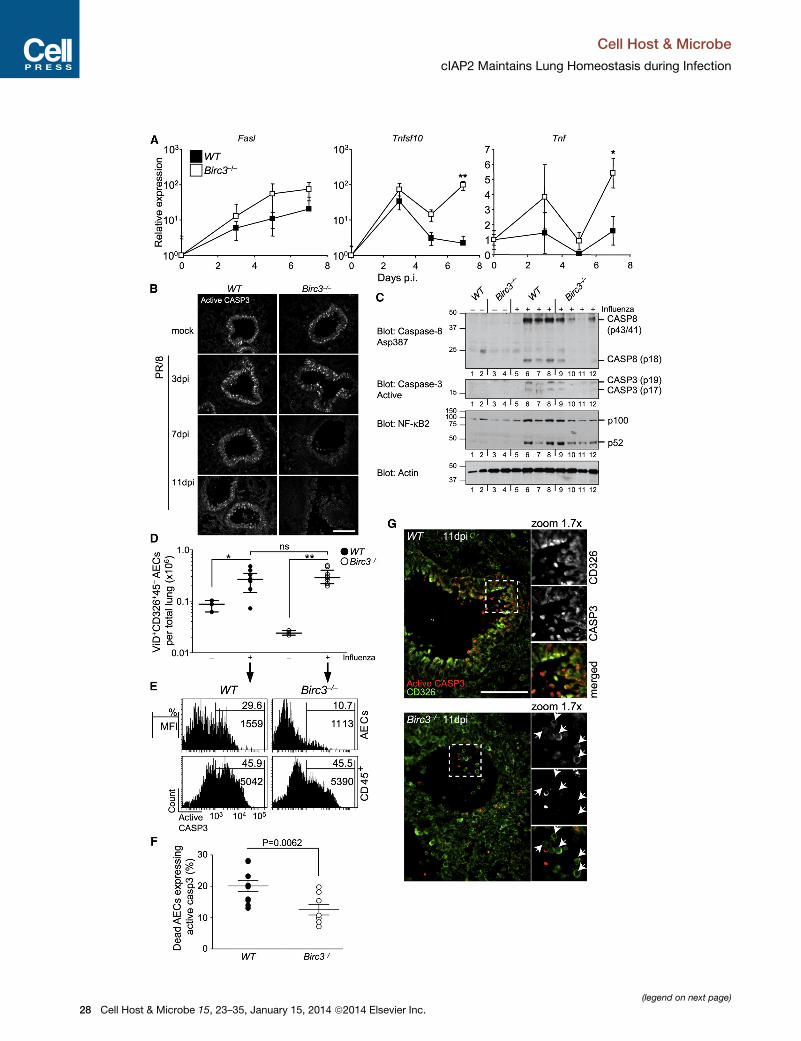
infection. Real-time qPCR analysis of death ligands revealed that
the expression of Fasl, Tnfsf10, and Tnf in the lung was induced
by infection in both Birc3�/� andWTmice but that Birc3�/� mice
exhibited higher levels of these transcripts than WT mice at later
time points (Figure 3A). However, when we examined apoptotic
caspase activation by immunofluorescence and immunoblot
analysis in lungs of infected Birc3�/� mice, we found reduced
levels of processed caspase-3 (Figures 3B and 3C) and cas-
pase-8 (Figure 3C) compared to WT controls. This difference
was observed despite the presence of inflammation, as
Cell H
measured by NF-kB2 activation (Figure 3C). Correspondingly,
the proportion of apoptotic pulmonary epithelial cells
was reduced in Birc3�/� mice compared to WTmice, as demon-
strated by fluorescence-activated cell sorting (FACS)
quantification of active caspase-3 in dying CD326+CD45�
CD31�Ter119� airway epithelial cells (AECs) isolated from colla-
genase-digested lungs of PR/8 infected mice at day 7 p.i. (Fig-
ures 3D–3F). Analysis of tissue sections by immunofluorescence
showed that CD326+ AECs in the bronchiole lumen of Birc3�/�
mice were negative for active caspase-3 (Figure 3G). No
ost & Microbe 15, 23–35, January 15, 2014 ª2014 Elsevier Inc. 25
Figure 2. Respiratory Tissue Damage Following Influenza Infection in Birc3–/– Mice
(A and B) H&E sections through themain bronchiole of the left lobe showing pulmonary hemorrhage (star) and epithelial shedding (black arrows) and degeneration
(black arrow head). Scale bar indicates 25 mm. Insets correspond to the boxed regions. Images shown are representative of four to five mice per genotype for
three independent experiments.
(C) BSA-FITC arbitrary fluorescent units in BALF (mean ±SD; n = 4–5 mice per genotype per time point). One-way ANOVA with Bonferroni’s post-test (*p < 0.05).
(D and E) Epithelial cell marker CD326 (D) or PR/8 virus antigens (E) immunofluorescence. White arrowheads point to epithelial cell shedding. Images shown are
representative of two to three mice from two independent experiments. Scale bar indicates 50 mm.
Cell Host & Microbe
cIAP2 Maintains Lung Homeostasis during Infection
differences in CD45+ immune cell (Figure 3E, bottom) or
CD31+CD45� lung endothelial cell (Figure S2A, related to Fig-
ure 3) apoptosis could be detected in infected Birc3�/� mice
compared with WT mice, suggesting that modulation of cell
death in the absence of cIAP2 is cell-type specific.
The NLRP3-caspase-1 inflammasome has been reported to
protect the host from influenza-induced acute injury to the respi-
ratory epithelium (Thomas et al., 2009). On the other hand,
hyperactivation of the inflammasome pathway through loss of
Ripk2 was recently linked to influenza-induced airway neutro-
philia, necrotic cell death, tissue damage, and lethality (Lupfer
et al., 2013). Because cIAP2 is required for efficient activation
of caspase-1 (Labbe et al., 2011) and RIPK2 ubiquitination (Ber-
trand et al., 2009), we next asked what role, if any, activation of
caspase-1 and its ensuing pyroptotic cell death program might
play in the lethality to influenza in Birc3�/� mice. At the sublethal
dose of PR/8 used in this study, Casp1�/�/Casp11�/� and
Nlrp3�/� mice proved as resistant to infection as WT mice (Fig-
ure S2B, related to Figure 3). In contrast, deficiency in either
the inflammasome adaptor ASC or overall IL-1 signaling
26 Cell Host & Microbe 15, 23–35, January 15, 2014 ª2014 Elsevier I
(Il1r1�/�) impaired protection to PR/8 infection (Figure S2B).
However, the frequency of necrotic AECs in both of these geno-
types was either equivalent to or lower than WT mice (Fig-
ure S2C), suggesting that the pathogenic mechanisms at play
in Asc�/� and Il1r1�/� mice are distinct from those observed in
Birc3�/� mice and are acting independently of the canonical
inflammasome pathway in this model. Moreover, contrary to
Birc3�/� mice, infection of Ripk2�/� mice resulted in slightly
higher BALF IL-1b and IL-6 levels (Figures S2D and S2E) and
with previous data (Lupfer et al., 2013). In addition, Ripk2�/�
mice exhibited normal AEC apoptosis (Figure S2G). These re-
sults argue that the Birc3�/� mice phenotype is independent of
a deregulation of the autophagy processes controlled by the
NOD2-RIPK2 axis that lead to aberrant caspase-1 activation
and lethality in that system. Altogether, these findings suggest
that cIAP2 likely confers protection to influenza independently
of its function in caspase-1 activation.
Caspase-11-induced pyroptotic cell death, while a defense
strategy against bacterial pathogens that engage type I
nc.
Cell Host & Microbe
cIAP2 Maintains Lung Homeostasis during Infection
interferons (Rathinam et al., 2012), can also contribute to lethal
inflammatory disease independently of caspase-1 (Broz et al.,
2012). Caspase-11 was induced by PR/8 infection in both WT
and Birc3�/� mice (Figure S2H). To assess to what extent
caspase-11 might influence PR/8-induced lethality, we ob-
tained, from two independent colonies, Birc3�/� mice that
were either carriers or not of a passenger intronic inactivating
deletion in the Casp11 gene (Kenneth et al., 2012) and inter-
crossed them to each other. The presence or absence of cas-
pase-11 did not alter the susceptibility of Birc3�/� mice, as the
mortality rates between Birc3�/�Casp11+/+, Birc3�/�Casp11+/�
and Birc3�/�Casp11�/� littermates were not different (Fig-
ure S2I). Additionally, caspase-11 deficiency did not render
mice more susceptible to intranasal infection with a sublethal
dose of a second influenza A strain (H3N2, Figure S2J), unlike
H3N2-infected Birc3�/� mice, which had reduced survival (Fig-
ure S2J) as well as increased AEC necrosis on day 7 p.i. (Figures
S2K and S2L). These results exclude a strain-inherent property
related to either increased infectivity and/or pathogenicity of
PR/8 in Birc3�/� mice and suggest that the activity of either
caspase-1 or caspase-11 is unlikely to account for the observed
lethality. Collectively, these findings demonstrate that a fraction
of the AEC cell death induced by influenza infection proceeds
independently of the activation of both apoptotic and inflamma-
tory caspases in Birc3�/� mice.
Necroptosis Inhibition inBirc3–/– Mice Improves SurvivalTo test whether necroptosis influenced the progression of influ-
enza-induced lethality, we used the chemical inhibitor Necrosta-
tin (Nec)-1 to block the kinase activity of RIPK1 in vivo, because
RIPK1-deficient mice exhibit early postnatal lethality (Kelliher
et al., 1998). Nec-1 prolonged the survival of Birc3�/� mice (Fig-
ure 4A; p = 0.0016) and diminished epithelial cell necrosis and
bronchial degeneration within the airways (Figure 4C), in marked
contrast with the tissue damage observed in vehicle-control-
treated mice (Figure 4B). Concordantly, genetic deletion of
Ripk3 protected Birc3�/� mice from PR/8-induced lethality to
levels similar to those of Ripk3�/� and WT mice (Figure 4D),
and it restored airway epithelial architecture without impacting
hemorrhage (Figure 4E). This rescue was dependent on Ripk3
deletion from the radio-resistant compartment of Birc3�/�
mice, since reconstitution of Birc3�/�/Ripk3�/� mice with
Ripk3-sufficient hematopoietic cells did not modify resistance
to PR/8 (Table 1). Viral clearance was not affected inmice lacking
Ripk3, as demonstrated by quantification of PR/8 titers in lung
tissues (Figures 4F and 4G). These experiments provided
genetic evidence that necroptosis is not required for effective
antiviral host defense against influenza but, rather, is a patholog-
ical feature that drives elevated morbidity and mortality in
infected Birc3�/� mice. To elucidate the potential mechanisms
by which cIAP2 deficiency skews the cell death programs in
AECs toward necroptosis following PR/8 infection, we immuno-
blotted lung homogenates for all known components that enable
RIPK3-dependent necrosis. The short isoform of FLIP (FLIPS)
inhibits apoptosis while actively promoting RIPK3-dependent
necrosis, whereas FLIP long (FLIPL) antagonizes both types of
cell death (Green et al., 2011). Because of higher FLIPS expres-
sion and a decreased FLIPL:FLIPS ratio in lungs of viral-infected
Birc3�/� mice (Figure 4H), we hypothesized that lack of cIAP2
Cell H
might promote the formation of the necrosome complex consist-
ing of RIPK1, RIPK3, and FADDduring influenza infection. To test
this, we immunoprecipitated endogenous FADD from lung ex-
tracts and examined the assembly of the necrosome. Although
RIPK3 was constitutively associated with FADD, as previously
reported (Cho et al., 2009), influenza infection led to additional
recruitment of RIPK3 to this FADD complex in Birc3�/� mice
compared to WT infected mice (Figure 4I, lanes 10–14 and
5–9; p = 0.0317), whereas virus-dependent interaction of
RIPK1 with FADD was remarkably similar between genotypes
(Figure 4I). In naivemice, a RIPK1-FADD complex spontaneously
assembled upon cIAP2 depletion (Figure 4I, lanes 3 and 4 and 1
and 2), as noted by others (Feoktistova et al., 2011; Tenev et al.,
2011). Our data suggest that cIAP2 deficiency favors the as-
sembly of the necrosome during infection, which translates to
a skewing of cell death toward necroptosis. Pharmacological
depletion of cIAPs in C57BL/6 by treatment with a SMAC
mimetic also resulted in reduced survival to PR/8 infection (Fig-
ures S3A and S3B, related to Figure 4) and a notable increase in
AEC necrosis on day 7 p.i. (Figures S3C and S3D). Altogether,
these data indicate that, following influenza virus infection,
cIAP2 expression in the lung determines, in a cell-specific
fashion, the sensitivity to necroptosis.
Role of Death Ligands in the Development ofNecroptosis-Induced Lethality in Birc3–/– MiceOne function of cIAPs is to act as negative regulators of caspase-
independent Fas-induced cell death (Geserick et al., 2009). We
therefore reasoned that cIAP2 deficiency might affect lung
homeostasis following infection by disturbing DR-mediated
signaling in AECs, rather than through direct virus-induced
sensing or cytopathic effects. Because of the observation that
influenza-induced acute lung injury stems from the absence of
cIAP2 expression in the nonhematopoietic compartment (Fig-
ure 1E and Table 1), and because abrogation of death ligand
expression by immune cells attenuates lung epithelial apoptosis
following influenza virus pneumonia (Herold et al., 2008), we
explored the role of DR signals through bone marrow transplan-
tation experiments. To this end, marrow from Faslgld, Tnfsf10�/�,Tnftm1Gkl, and Ltatm1Dch were transferred into irradiated Birc3�/�
mice to block specific DR signaling (Table 1). Micewith disrupted
Tnf (Figure 5A) or lymphotoxin-a (Figure 5B) signaling developed
influenza-induced lethality with identical kinetics and severity
compared to Birc3�/� mice transplanted with WT marrow
(WT/Birc3�/�), whereas genetic mutation of FasL (Figure 5C)
from the immune compartment had a stronger effect than TRAIL
deficiency (Figure 5D and Table 1) in protecting Birc3�/� mice
following PR/8 infection. To address whether direct infection of
AECs was required for necrosis induction in vivo, we analyzed
by FACS the proportion of uninfected AECs (measured as nega-
tive for PR/8 nucleoprotein; Figure 5E) that on day 7 p.i. stained
positive for viability dye uptake without caspase-3 activation
and/or phosphatidylserine translocation. Necrosis was induced
in both uninfected and infected AECs from PR/8-challenged
mice; however, the frequency of necrotic cells in Birc3�/� mice
exceeded that in WT mice (Figure 5F), regardless of whether
the cells were infected or not, revealing that skewing of AEC
cell death in the absence of cIAP2 is linked to airway inflamma-
tion rather than direct virus infection of AECs. These data
ost & Microbe 15, 23–35, January 15, 2014 ª2014 Elsevier Inc. 27
(legend on next page)
Cell Host & Microbe
cIAP2 Maintains Lung Homeostasis during Infection
28 Cell Host & Microbe 15, 23–35, January 15, 2014 ª2014 Elsevier Inc.
Cell Host & Microbe
cIAP2 Maintains Lung Homeostasis during Infection
suggest that FasL and (to a lesser extent) TRAIL signaling, as
opposed to direct viral cytopathic effects, are major determi-
nants of mortality triggered by cIAP2 deficiency during influenza
infection.
DISCUSSION
A cell’s decision to self-destruct in response to a virus is irrevers-
ible, and, therefore, in order to be an effective antiviral defense
strategy, it must be tightly controlled to maintain tissue homeo-
stasis during infectious disease. Similar to cell-fate decisions
regulated by XIAP in the liver (Jost et al., 2009), this work iden-
tifies a biological role for cIAP2 as a gatekeeper of pathological
DR-induced activation of cell death during respiratory infection.
We show that cIAP2 regulates death signaling following influenza
infection at the level of the assembly of the necrosome and the
stoichiometry of FLIP isoforms in the lung. RIPK3-dependent
programmed necrosis is the favored cell death mechanism
when the cellular apoptosis machinery is inhibited by FLIPS
(Feoktistova et al., 2011). Other IAPs appear to play nonredun-
dant roles in the infected lung as they were unable to compen-
sate for cIAP2-deficiency, which increased susceptibility to
DR-induced necroptosis independently of pathogen burden or
inflammatory response.
In line with previous in vitro work (Feoktistova et al., 2011;
Tenev et al., 2011), spontaneous assembly of the RIPK1-FADD
ripoptosome under steady-state conditions is insufficient for
cell death induction in vivo. Conceivably, its formation in the
absence of cIAP2 in the lung only preconditions AECs to become
more sensitive to death ligands, with the death-inducing sensi-
tivity of this complex once formed being modulated by the
expression levels of FLIP isoforms, as reported by others (Feok-
tistova et al., 2011; Oberst et al., 2011). Whereas necroptosis
acts as an antiviral mechanism against some DNA viruses (Cho
et al., 2009; Upton et al., 2010), it is dispensable for the control
of influenza replication, arguing for case-specific use of this
death modality during immune defenses to viruses. It is aston-
ishing that a minor switch in the decision of the type of death
by which AECs self-destruct can account for the extensive
epithelium loss that is characteristic ofBirc3�/� lungs. But a rela-
tively small increase of �10% in AEC death is sufficient to result
in considerable cell loss over time and leads to severe virus-
induced acute lung injury (Sanders et al., 2013). Necrotic loss
of alveolar type II epithelial cells, which are sensitive to FasL (Ha-
mann et al., 1998) and have an important role in the endogenous
repair capacity of the alveolar epithelium (Wang et al., 2010),
Figure 3. Caspase-Independent Cell Death of Birc3–/– AECs during Infl
(A) Tnfa, Tnfsf10, and Fasl expression in infected lungs (*p < 0.05, **p < 0.01; Stud
n = 8–10 mice per genotype per time point).
(B) Active caspase-3-stained lung sections from mock- or PR/8-infected mice (s
genotype for two independent experiments.
(C) Immunoblot analysis of lung tissue at 7 days p.i. (representative of two indep
(D and E) Representative FACS plots of day 7 airway epithelium dead cell number
Live/Dead stain (ViD) and (E) active caspase-3 staining; n = 8–10 mice total per g
(F) FACS quantification of caspase-dependent AEC cell death (mean ±SEM; Man
mice total for each genotype).
(G) CD326-positive cells in the bronchiole lumen of Birc3�/� mice are negative
representative of two to three mice per genotype for two independent experime
Cell H
could compromise repopulation of the injured epithelial lining
and accelerate AEC depletion. Our data also caution that deple-
tion of the cIAPs by synthetic IAP antagonists could adversely
affect individuals who develop respiratory viral infections. Taken
together, these observations favor a model in which cell death
initiation integrates the different stresses that converge on
lowering a cIAP expression threshold that otherwise effectively
(A) Survival rate of infected Birc3�/� mice treated with Nec-1 or vehicle intrape
experiments.
(B and C) H&E-stained lung cross-sections of Birc3�/� vehicle-treated (B) and N
within the bronchioles (arrow head: erosion). Insets correspond to the boxed re
mouse from two independent experiments with n = 3–4 mice per group.
(D) Survival rate of Birc3�/�, Birc3�/�Ripk3�/�, Ripk3�/� and WT mice to PR/8 in
(E) H&E-stained lung cross-sections on 11 days p.i. Data are representative of tw
scale.
(F) Viral loads by plaque assay (mean ±SD; n = 4 mice per genotype per time po
(G) Lung sections stained for PR/8 antigens (scale bar indicates 50 mm).
(H) Immunoblot analysis of necrosome component expression in lung tissue from
individual mouse. Densitometry analysis using ImageJ software (mean ±SEM; n =
infected mice per genotype; *p = 0.0315, Mann-Whitney test).
(I) Formation of the necrosome on 7 days p.i. Asterix denotes (IgGH) heavy chains.
FADD ratio is shown at the bottom. See also Figure S3.
Cell H
with the concept that insufficient host defense to infectious dis-
ease can be due to an impaired ability to tolerate tissue damage
(Medzhitov et al., 2012). In this context, we envisage that the
transcriptional induction of cIAP2 by inflammatory triggers
(Wang et al., 1998) may have evolved to ensure survival of critical
cell types in response to strong DR signals induced by inflamma-
tion. The fact that the lethality from viral-induced necrosis in the
context of cIAP2 deficiency is found for two different influenza
strains points to a more general mechanism that is not specific
to a single virus and, thus, is likely of broader relevance for our
understanding of the mechanisms that are engaged during
airway inflammation to maintain tissue homeostasis and host
fitness. Indeed, DR-mediated lung epithelial injury is not
restricted to influenza virus, as it also occurs during infection
with other respiratory RNA viruses (Bem et al., 2010). Therefore,
continued characterization of cytoprotective tolerance mecha-
nisms, like the one described here during influenza infection,
may provide novel entry points for therapeutics against acute
lung injury.
EXPERIMENTAL PROCEDURES
Reagents
ELISA kits for anti-CXCL10 (DY466) were from R&D and VeriKine anti-IFNa
(42120) were from PBL; anti-caspase-8 Asp387 (9429) and anti-cFLIP (3210)
from Cell Signaling Technology; anti-FADD (M-19) was from Santa Cruz;
anti-RIPK1 (610459) was from BD Transduction Laboratories; anti-RIPK3
(PSC-2283) was from Axxora; anti-active caspase-3 (AF835) was from R&D;
Influenza-Induced Lethality in Birc3–/– Mice
ritoneally daily starting on 3 days p.i. Data are pooled from two independent
ec-1-treated (C) mice on 7 days p.i. Black arrow indicates epithelial shedding
gions (enlarged by an 8.53 zoom). Each section corresponds to an individual
fection. Data are pooled from two independent experiments.
o independent experiments with n = 3–4 mice per group. All H&E are to 25 mm
int).
PR/8-infected mice (lysates used in immunoprecipitation in (I). Each lane is an
3 independent experiments with each two to three uninfected and four to five
Each lane is an individual mouse. Densitometry analysis of RIPK1 and RIPK3 to
ost & Microbe 15, 23–35, January 15, 2014 ª2014 Elsevier Inc. 31
Figure 5. FasL and TRAIL Are Required for Influenza-Induced
Lethality in Birc3–/– Mice
(A–D) Survival rate of chimeric mice after PR/8 infection. Data are pooled from
two independent experiments in (D).
(E) Representative FACS plots of infected (NP+) versus noninfected
(NP�) AECs.(F) FACS quantification of cell-death type (necrosis, open circles; apoptosis,
closed circles) after 7 days p.i. in infected (NP+) versus uninfected (NP�) AECsfrom the same mouse. Necrosis (open circles) is defined as annexin V�, activecaspase-3� and ViD+; apoptosis (closed circles) is defined as annexin V+,
active caspase-3+, and ViD+/�. Lines and bars represent the mean and SD
(n = 5–6 mice per genotype, *p < 0.05; **p < 0.001; Student’s two-tailed t test).
Cell Host & Microbe
cIAP2 Maintains Lung Homeostasis during Infection
32 Cell Host & Microbe 15, 23–35, January 15, 2014 ª2014 Elsevier I
anti-influenza A,B (M149) was from Takara Bio; anti-CD326 (G8.8), anti-active
caspase-3-PE (C92-605), and streptavidin APCCy7 were from BD Biosci-
ences; Alexa Fluor-594 or Alexa Fluor647 anti-rabbit or rat antibodies were
from Molecular Probes/Life Technologies; anti-CD31-APC (390), anti-CD45-
PECy7 (30-F11), anti-CD326-PerCP-eFluor710 (G8.8), and anti-Ter119 were
from eBioscience; anti-influenza A virus nucleoprotein-FITC (431) was from
Abcam; PR/8 was from Charles River; Nec-1 (N9037), BSA-FITC (A9771),
and protein G sepharose beads (P3296) were from Sigma-Aldrich; Smac-
mimetic LCL-161 (A-1147)was from Active Biochem; and SURFLO I.V.
20G31’ catheter (SR-0X2025CA) was from Terumo (Elkton).
Mice
All mice were maintained at McGill University and were previously described
(Conte et al., 2006; Glaccum et al., 1997; Kobayashi et al., 2002; Kuida
et al., 1995; Mariathasan et al., 2004, 2006; Newton et al., 2004).
Birc3�/�Ripk3�/� mice were generated through intercrossing. B6Smn.C3-