KChIPs and Kv4 � Subunits as Integral Components ofA-Type Potassium Channels in Mammalian Brain
Kenneth J. Rhodes,1 Karen I. Carroll,1 M. Amy Sung,1 Lisa C. Doliveira,1 Michael M. Monaghan,1 Sharon L. Burke,1
Brian W. Strassle,1 Lynn Buchwalder,2 Milena Menegola,4 Jie Cao,3 W. Frank An,3 and James S. Trimmer2,4
1Neuroscience, Wyeth Discovery Research, Princeton, New Jersey 08543, 2Department of Biochemistry and Cell Biology, State University of New York,Stony Brook, New York 11794, 3Millennium Pharmaceuticals, Cambridge, Massachusetts 02139, and 4Department of Pharmacology, School of Medicine,University of California, Davis, California 95616
Voltage-gated potassium (Kv) channels from the Kv4, or Shal-related, gene family underlie a major component of the A-type potassiumcurrent in mammalian central neurons. We recently identified a family of calcium-binding proteins, termed KChIPs (Kv channel inter-acting proteins), that bind to the cytoplasmic N termini of Kv4 family � subunits and modulate their surface density, inactivation kinetics,and rate of recovery from inactivation (An et al., 2000). Here, we used single and double-label immunohistochemistry, together withcircumscribed lesions and coimmunoprecipitation analyses, to examine the regional and subcellular distribution of KChIPs1– 4 and Kv4family � subunits in adult rat brain. Immunohistochemical staining using KChIP-specific monoclonal antibodies revealed that the KChIPpolypeptides are concentrated in neuronal somata and dendrites where their cellular and subcellular distribution overlaps, in an isoform-specific manner, with that of Kv4.2 and Kv4.3. For example, immunoreactivity for KChIP1 and Kv4.3 is concentrated in the somata anddendrites of hippocampal, striatal, and neocortical interneurons. Immunoreactivity for KChIP2, KChIP4, and Kv4.2 is concentrated inthe apical and basal dendrites of hippocampal and neocortical pyramidal cells. Double-label immunofluorescence labeling revealed thatthroughout the forebrain, KChIP2 and KChIP4 are frequently colocalized with Kv4.2, whereas in cortical, hippocampal, and striatalinterneurons, KChIP1 is frequently colocalized with Kv4.3. Coimmunoprecipitation analyses confirmed that all KChIPs coassociate withKv4 � subunits in brain membranes, indicating that KChIPs 1– 4 are integral components of native A-type Kv channel complexes and arelikely to play a major role as modulators of somatodendritic excitability.
IntroductionIn mammalian central neurons, certain rapidly inactivating, orA-type, voltage-gated potassium (Kv) currents are concentratedin somatodendritic membranes (Sheng et al., 1992; Bekkers,2000a,b; Korngreen and Sakmann, 2000) where they shapepostsynaptic responses to excitatory input (Johnston et al., 2000,2003; Storm, 2000), regulate amplitude and duration of back-propagating action potentials (Hoffman et al., 1997), and mod-ulate dendritic excitability in response to second messenger acti-vation (Nakamura et al., 1997; Hoffman and Johnston, 1998;Holmqvist et al., 2001) and long-term potentiation (Frick et al.,2004). Immunohistochemical analyses (Sheng et al., 1992;Maletic-Savatic et al., 1995; Tsaur et al., 1997; Varga et al., 2000;for review, see Trimmer and Rhodes, 2004), pharmacological
sensitivity (Wu and Barish, 1992; Holmqvist et al., 2001), andelectrophysiological characteristics (Serodio et al., 1994, 1996;Serodio and Rudy, 1998) suggest that a major component of thesomatodendritic A-type Kv current is formed by � subunits fromthe Shal or Kv4 family (Gutman et al., 2003). However, A-typecurrents from Kv4 � subunits expressed in heterologous cells andneurons differ. That coexpression of Kv4 � subunits with brainmRNA in heterologous cells gives rise to A-type currents with amore “native” phenotype (Rudy et al., 1988; Chabala et al., 1993;Serodio et al., 1994) prompted our search for accessory Kv4 chan-nel subunits.
We used the cytoplasmic N terminus of Kv4.3 as bait in a yeasttwo-hybrid screen of a rat midbrain cDNA library and identifiedtwo members of a calcium-binding protein family, KChIPs (Kvchannel interacting proteins) 1 and 2, that are highly expressed inthe CNS and interact with Kv4 but not other Kv � subunits (An etal., 2000). Coexpression of KChIP1, KChIP2, or a third familymember, KChIP3, dramatically increases density, slows inactiva-tion kinetics, and speeds the rate of recovery from inactivation ofKv4 channels expressed in heterologous cells (An et al., 2000;Bahring et al., 2001; Holmqvist et al., 2002; Shibata et al., 2003;Patel et al., 2004). KChIP4 can have distinct effects, depending ona splice variant-specific unique N terminus (Holmqvist et al.,2002; Shibata et al., 2003). Kv4 � subunits and KChIPs form
Received March 4, 2004; revised July 29, 2004; accepted July 29, 2004.This work was supported by Wyeth Research and by National Institutes of Health Grant NS42225 (J.S.T.). We
thank Dr. Pranab Chanda and Wade Edris for preparation and purification of KChIP fusion proteins, Dr. Gail Mandelfor the use of the confocal microscope and luminometer, and Drs. Mark Bowlby, John Moyer, James Barrett, PeterDiStefano, and Kevin Willis for support and encouragement during the course of this work.
Correspondence should be addressed to Dr. James S. Trimmer, Department of Pharmacology, 1311 Tupper Hall,School of Medicine, University of California, One Shields Avenue, Davis, CA 95616-8635. E-mail: [email protected].
K. J. Rhodes’s present address: Neurological Disorders, Johnson and Johnson Pharmaceutical Research and De-velopment, Raritan, NJ 08869.
The Journal of Neuroscience, September 8, 2004 • 24(36):7903–7915 • 7903
Kv44KChIP4 octomeric complexes (Kimet al., 2004a), with the KChIPs interactingwith the Kv4 N termini (Scannevin et al.,2004; Zhou et al., 2004) in a large cytoplas-mic structure in which the KChIPs appearto enwrap the “hanging gondola” formedby the Kv4 N termini (Kim et al., 2004b).
Here, we describe in detail the distribu-tion, colocalization, and coassociation ofKv4.2, Kv4.3, and KChIPs 1, 2, 3 (An et al.,2000), and 4 (Holmqvist et al., 2002) inadult rat brain. We generated and charac-terized monoclonal antibodies (mAbs) specific for each Kv4 orKChIP polypeptide and used these reagents for immunohisto-chemical and multiple-label immunofluorescence and coimmu-noprecipitation analyses to map the loci of KChIP/Kv4 interac-tion. We also used circumscribed ibotenic acid lesions within thehippocampal formation and immunohistochemistry (Mon-aghan et al., 2001) to confirm the somatodendritic localization ofKChIP and Kv4 immunoreactivity.
Materials and MethodsMaterials. Reagents were molecular biology grade from Sigma (St. Louis,MO) or Roche Diagnostics (Indianapolis, IN), except where noted oth-erwise. Alexa-488 and Alexa-594 fluorophore-conjugated isotype-specific antibodies were purchased from Molecular Probes (Eugene,OR). Biotin-conjugated anti-mouse isotype-specific antibodies werepurchased from Southern Biotechnology (Atlanta, GA). Precast, neutralpH (NuPAGE) gels were used for all SDS-PAGE experiments and werepurchased from Invitrogen (Carlsbad, CA).
Generation and characterization of KChIP and Kv4 antibodies. Mousemonoclonal and affinity-purified rabbit polyclonal antibodies were gen-erated and purified essentially as described previously (Trimmer, 1991;Rhodes et al., 1995, 1996; Bekele-Arcuri et al., 1996). Peptide or fusionprotein sequences used to generate antibodies against Kv4.2, Kv4.3,KChIP1, KChIP2, KChIP3, or KChIP4 are listed in Table 1. Rabbits wereimmunized with purified recombinant glutathione S-transferase (GST)fusion proteins or with synthetic peptides [Quality Controlled Biochemi-cals (Hopkinton, MA) or Research Genetics (Huntsville, AL)] conju-gated to keyhole limpet hemocyanin. Antibody titers were determined byELISA against the cognate synthetic peptide or fusion protein immuno-gen. In cases in which GST fusion proteins were used as the immunogen,titers against the GST fusion protein were compared with titers againstGST protein alone. Rabbit polyclonal antibodies were affinity purified onimmobilized peptide or fusion protein affinity columns using standardprocedures (Rhodes et al., 1995).
BALB/c mice used for the production of mAbs were obtained fromTaconic Farms (Germantown, NY). The myeloma cell line SP2/0 wasgenerously provided by Dr. I. S. Trowbridge (The Salk Institute, La Jolla,CA). BALB/c mice were first immunized with an intraperitoneal injec-tion containing 100 �g of Kv4 or KChIP immunogen resuspended in50% RIBI adjuvant system MPL�TDM emulsion R-700 (RIBI Immu-nochem, Hamilton, MT) in phosphate-buffered saline (PBS; 150 mM
NaCl and 10 mM sodium phosphate, pH 7.4). Mice were subsequentlyimmunized with 50 �g of immunogen on days 14 and 21. Sera werecollected on days 10, 17, and 21 and screened for immunoreactivityagainst the Kv4 or KChIP immunogen by ELISA using a goat anti-mouseIgG-specific secondary antibody. The mouse that displayed the moremature immune response received 50 �g of immunogen in PBS intrave-nously on days 28 –30 and was killed for splenectomy on day 31.
Hybridomas were produced by standard methodology (Trimmer etal., 1985; Bekele-Arcuri et al., 1996). In brief, spleen cells were dissociatedand fused with polyethylene glycol to SP2/0 mouse myeloma cells at aratio of 10:1 spleen cells to myeloma cells, and the resultant fusion mix-ture was plated into 10 96-well tissue culture plates. Hybridomas wereselected for growth in media containing hypoxanthine, aminopterin, andthymidine for 14 d; media containing hypoxanthine and thymidine were
used for the third week. Ten to 14 d after the fusion, tissue culture super-natants were screened by ELISA against COS-1 cells transiently trans-fected with cDNAs encoding the appropriate full-length Kv4 or KChIPpolypeptide. Bound mAb was detected by luminometry. In each case,large pools of positive clones were obtained for additional analysis (K57:48; K75:48; K55:96; K60:36; K66:60). Samples exhibiting positive reac-tions were expanded, and the resulting culture supernatants werescreened by immunofluorescence staining of COS-1 cells transientlytransfected with cDNAs encoding the appropriate full-length Kv4 orKChIP polypeptide. The same sample set was also screened for specificimmunoreactivity on immunoblots of rat brain membranes and for im-munoperoxidase staining of rat brain sections. Specificity for distinctKv4 or KChIP isoforms was verified by immunofluorescence stainingand immunoblot analyses of COS-1 cells transiently transfected withcDNAs encoding the entire set of full-length Kv4 or KChIP polypeptides,by competition experiments to show elimination of staining of brainsections by preincubation with the appropriate peptide/fusion proteinimmunogen, and by verification that cellular immunocytochemicalstaining patterns matched gene expression patterns obtained from in situhybridization analyses. Selected hybridomas were then subcloned by lim-iting dilution, reassayed, and grown in BALB/c mice for production ofascites fluid as described previously (Trimmer et al., 1985). Immuno-globulins were purified by ammonium sulfate precipitation, followed byDEAE chromatography, as described (Trimmer et al., 1985).
Immunoprecipitation. Immunoprecipitation reactions were per-formed at 4°C using detergent lysates of crude membranes (Trimmer,1991) isolated from freshly dissected adult rat brain. In brief, membranesderived from whole brain or individual brain regions as indicated (0.5 mgof membrane protein/tube) were solubilized in radioimmunoprecipita-tion assay (RIPA) buffer [1% IGEPAL CA-630, 0.5% deoxycholic acid,0.1% SDS, 0.15 M NaCl, and 50 mM Tris-HCl, pH 7.4, containing pro-tease inhibitor mixture (Roche Diagnostics)]. Affinity-purified rabbitpolyclonal or mouse mAbs specific for each antigen were added, and thevolume was adjusted with RIPA buffer to 0.1 ml per reaction tube. Sixtymicroliters of 50% slurry of protein A agarose (Pierce, Rockford, IL) wereadded to each tube, and the samples were incubated at 4°C overnight ona rocker table. After incubation, protein A agarose was centrifuged at14,000 � g for 20 sec, and the resulting pellets were washed by resuspen-sion and centrifugation four times with lysis buffer (1% Triton X-100,0.15 M NaCl, 1 mM EDTA, 10 mM sodium azide, and 10 mM Tris-HCl, pH8.0). The final pellets were resuspended in 80 �l of 2� reducing samplebuffer.
SDS-polyacrylamide gels and immunoblotting. Pellets from immuno-precipitation reactions were heated to 50°C for 10 min, vortexed, andcentrifuged to pellet the agarose. Thirty microliters of each sample weresize fractionated on 4 –12% gradient (for analysis of Kv4 � subunits) or10% (for analysis for KChIP proteins) precast NuPAGE gels. After elec-trophoretic transfer to nitrocellulose membrane, the resulting blots wereblocked in Tris-buffered saline (20 mM Tris, pH 7.6, and 0.137 M NaCl)containing 5% nonfat dried milk and 0.1% Tween 20. The blots wereincubated in purified antibody diluted in the blocking agent overnight at4°C, washed three times for 10 min in TBS–Tween, and then incubatedfor 1 hr at room temperature in a blocking agent containing HRP-conjugated secondary antibody (Jackson ImmunoResearch, West Grove,PA). After another three 10 min washes in TBS–Tween, the membraneswere incubated in substrate for enhanced chemiluminescence (ECL; Am-
Table 1. Sequences of peptide and fusion protein antigens used to generate Kv4- and KChIP-specific antibodies
Kv4.2-E 209 –225 CGSSPGHIKELPSGERY K57/27.1 Baldwin et al., 1991Kv4.3-C GST-415– 636 C-terminal tail K75/30.1 Tsaur et al., 1997KChIP1 GST-KChIP1 Full-length GST fusion K55/7.1 An et al., 2000Pan-KChIP GST-KChIP1 Full-length GST fusion K55/82.1* An et al., 2000KChIP2 GST-KChIP2 Full-length GST fusion K60/73.1 An et al., 2000KChIP3 GST-KChIP3 Full-length GST fusion K66/36.1 An et al., 2000KChIP4 GST-KChIP4 Full-length GST fusion 1G2 Holmqvist et al., 2002
C denotes cysteine residue added for conjugation to KLH or BSA. *This antibody recognizes all KChIP family members.
7904 • J. Neurosci., September 8, 2004 • 24(36):7903–7915 Rhodes et al. • KChIP and Kv4 Localization in Rat Brain
ersham, Arlington Heights, IL) for 1 min. The excess ECL substrate wasremoved and the blots exposed to Hyperfilm-ECL film (Amersham).
Experimental localization of channel subunits. All surgical procedureswere approved by the Wyeth Institutional Animal Care and Use Com-mittee and were in accordance with the NIH Guide for the Care and Use ofLaboratory Animals. Before surgery, animals were anesthetized deeplywith sodium pentobarbital (50 mg/kg, i.p.) and secured in a stereotaxiccarrier (David Kopf Instruments, Tajunga, CA). Ibotenic acid lesions ofhippocampal subfields were performed as described previously (Mon-aghan et al., 2001). In brief, ibotenic acid (0.1– 0.4 �l of a 10 �g/�lsolution in 0.1 M sodium phosphate buffer, pH 7.4) was injected directlyinto the target structure using a 2 �l Hamilton microsyringe mounted ina Kopf microsyringe microdrive. In some of these animals, injectionswere made at two or three depths, with each injection separated in thedorsoventral axis by 2.5 mm.
Immunohistochemistry. Animals that sustained surgery were killed 7 dpostoperatively. At the time of death, all animals were anesthetizeddeeply with sodium pentobarbital (60 mg/kg, i.p.) and then perfusedthrough the ascending aorta with 0.1 M NaPO4 buffer, pH 7.4, followedby fixative containing freshly depolymerized 4% paraformaldehyde in0.1 M NaPO4 buffer. The remaining procedures for light microscopicimmunohistochemistry were described in detail previously (Rhodes etal., 1995, 1996). All of the immunohistochemical and immunofluores-cence data presented here were collected on sections processed using thepurified mouse mAbs described in Results, with the exception of stainingfor Kv2.1, which used an affinity-purified rabbit polyclonal antibody(“KC”) described previously (Trimmer, 1991; Rhodes et al., 1995, 1996;
Monaghan et al., 2001). Briefly, 40-�m-thickhorizontal sections were incubated overnight at4°C in antibody vehicle containing purifiedmouse mAb. Detection of antibody–antigencomplexes was accomplished using the ABCElite peroxidase reaction kit (Vector Laborato-ries, Burlingame, CA) and visualized using anickel-enhanced diaminobenzidine procedure(Tago et al., 1986; Rhodes et al., 1995). Double-label immunofluorescence labeling was per-formed as described previously (Rhodes et al.,1997), except that isotype-specific anti-mouseantibodies conjugated to Alexa fluorophores(Molecular Probes) were used as the detectingantibodies.
Diaminobenzidine-stained and immunoflu-orescence sections were analyzed and imagedusing an Axiophot photomicroscope and con-focal microscope (Zeiss, Thornwood, NY)equipped with argon lasers. Black-and-white 35mm film negatives containing photomicro-graphs of stained sections were digitized using aNikon LS1000 35 mm film scanner. Thescanned and/or digital images were arrangedand labeled in Adobe Photoshop with only mi-nor adjustments of image brightness andcontrast.
ResultsCharacterization of anti-KChIP and antiKv4 mAbsThe KChIP polypeptides have variable Ntermini but share 70% identity through-out their C-terminal 185 amino acids (Anet al., 2000; Holmqvist et al., 2002). Ini-tially, we focused our efforts to generateKChIP-specific antibodies on the variableN termini. Although we were successful inobtaining high-titer rabbit polyclonal an-tibodies using this approach, whereasthese antibodies were specific for the targetprotein, these antibodies did not yield
crisp immunohistochemical staining of brain sections. In a sec-ond attempt to generate high-quality reagents for immunohisto-chemistry, we generated GST fusion proteins encoding each full-length KChIP polypeptide and used these as immunogens togenerate mouse mAbs. To identify anti-KChIP mAbs that reactedspecifically with the desired KChIP polypeptide and not otherantigens, hybridoma supernatants from the KChIP immuniza-tions were screened by ELISA versus the cognate immunogen, byimmunofluorescence staining versus a panel of transiently trans-fected COS cells expressing the entire set of full-length Kv4or KChIP polypeptides (data not shown), and subsequently byimmunoblotting versus cell lysates prepared from transientlytransfected COS cells (supplemental material, available at www.jneurosci.org). Using this strategy, from large pools of positivemAbs (see Materials and Methods for details) we identified andsubsequently purified mAbs that specifically recognized the cog-nate Kv4 or KChIP antigen and not other proteins.
A representative assay of specificity is shown in the supple-mental material (available at www.jneurosci.org). By immuno-blotting samples from transfected mammalian cells, each of theselected mAbs recognized a major band corresponding to theappropriate Kv4 or KChIP polypeptide and did not cross-reactwith other highly related family members. In the immunoblots
Figure 1. Immunohistochemical localization of Kv4 � subunits and KChIPs in the hippocampal formation. Photomicrographsof coronal sections taken to show the areal and laminar distribution of Kv4.2 ( A), Kv4.3 ( B), KChIP1 ( C), KChIP2 ( D), KChIP3 ( E),and KChIP4 ( F) immunoreactivity in the rat hippocampus are shown. Immunoreactivity for Kv4.2, KChIP2, KChIP3, or KChIP4 isconcentrated in dendritic fields, including the apical dendritic fields of dentate granule cells and the apical and basal dendrites ofCA and subicular pyramidal cells. In the dentate gyrus and CA3 subfields, immunoreactivity for Kv4.3 is concentrated in granuleand pyramidal cell dendrites. However, in these subfields, Kv4.3 immunoreactivity is also concentrated in the somata and den-drites of large multipolar interneurons. In all hippocampal subfields, immunoreactivity for KChIP1 is concentrated in the cellbodies and throughout the dendritic trees of large multipolar interneurons. DG, Dentate gyrus.
Rhodes et al. • KChIP and Kv4 Localization in Rat Brain J. Neurosci., September 8, 2004 • 24(36):7903–7915 • 7905
probed with antibodies against Kv4.2 orKv4.3, we always observed immunoreactiv-ity with high molecular weight bands. Thesebands represent dimers and larger aggregatesof Kv4.2 or Kv4.3 (supplemental material,available at www.jneurosci.org) (Sheng et al.,1993; An et al., 2000). We confirmed thatthese higher molecular weight bands are ag-gregated Kv4 � subunits by stripping andreprobing the immunoblots with anti-Kv4antibodies directed at epitopes elsewhere inthe Kv4 sequence. These disparate anti-Kv4 antibodies recognized these same highmolecular weight bands identified by theanti-Kv4 mAbs (data not shown), con-firming that the high molecular weight ag-gregates contain Kv4 � subunits. In theimmunoblots probed with anti-KChIP3mAbs, we always observed immunoreac-tivity with a lower molecular weight (�20kDa) band (supplemental material, avail-able at www.jneurosci.org). Presumably,this band represents a KChIP3 N-terminalcleavage product, as reported previously(Tekirian et al., 2001). This lower molecu-lar weight band is observed only in lysatesprepared from COS cells transfected withKChIP3, making it is highly unlikely thatthis second band indicates cross-reactivityof the anti-KChIP3 mAb with anotherprotein.
To further verify the specificity of theanti-KChIP antibody reagents used forimmunohistochemistry, competition ex-periments were performed in which eachanti-KChIP antibody was incubated witheither the cognate recombinant KChIPprotein antigen or with one of the otherrecombinant KChIP polypeptides. We observed that immuno-histochemical staining was completely inhibited by incubationwith the cognate KChIP antigen but not with recombinant pro-teins encoding other KChIP family members (data not shown).We also verified that cellular immunocytochemical staining pat-terns matched gene expression patterns obtained from in situhybridization analyses. It is also important to note that in ourscreen of anti-KChIP hybridomas we identified several hybrid-omas that secreted mAbs recognizing more than one, or even all,KChIP polypeptides (so-called “pan-KChIP” antibodies) (Table1) (An et al., 2000). Table 1 lists the mAbs that were used for theimmunohistochemical analyses described below.
ImmunohistochemistryBecause a comprehensive description of the patterns of KChIPand Kv4 � subunit immunoreactivity is well beyond the scope orintent of this report, we focused our analyses and descriptions onbrain regions and cell types in which somatodendritic A-currentshave been the most intensely studied: the hippocampal forma-tion, neocortex, and striatum. We reasoned that the wealth ofpublished electrophysiological data would allow us to draw rea-sonable inferences about the relationships between the Kv4 andKChIP staining patterns and the underlying currents in thesebrain regions.
As described in more detail below, immunoreactivity for Kv4
� subunits and KChIPs is concentrated primarily in the dendritesand somata of central neurons. The staining associated with la-beled dendrites tends to be quite uniform, with little evidence oflocal concentrations of immunoreactivity at synapses or on den-dritic spines. For comparison and contrast with the pattern ofKv4 and KChIP immunoreactivity, in most of the figures weshow immunohistochemical staining for Kv2.1, a delayed recti-fier Kv channel expressed in the somata and proximal dendritesof rat central neurons (Trimmer, 1991; Rhodes et al., 1995, 1996;Scannevin et al., 1996; Du et al., 1998).
The hippocampal formationThe staining pattern for Kv4.2, Kv4.3, and all four KChIPpolypeptides across hippocampal subfields and laminas is shownin Figure 1. Higher magnification micrographs of the stainingpatterns within individual hippocampal subfields are shown inFigures 3–5. Overall, there is a high density of immunoreactivityand a close correspondence in the staining patterns for Kv4.2,Kv4.3, and KChIPs 2, 3, and 4 in the dentate gyrus and CA sub-fields. There is also a close correspondence between the stainingpatterns for Kv4.3 and KChIP1, particularly in the dentate hilusand CA1 subfields, where immunoreactivity for these two Kvchannel subunits is concentrated in the somata and dendrites oflarge multipolar interneurons.
In the dentate gyrus, immunoreactivity for Kv4.2, Kv4.3, and
Figure 2. Distribution of Kv4 � subunits and KChIPs in the dentate gyrus. In the dentate gyrus (cellular architecture shown bythe Nissl stain in A), a high density of immunoreactivity for Kv4.2 ( B), Kv4.3 ( C), and KChIPs 2– 4 (E–G, respectively) is concen-trated throughout the molecular layer, where the staining appears to be concentrated in the dendrites of dentate granule cells.This extensive dendritic staining contrasts with the distribution of immunoreactivity for Kv2.1 ( H ), which is confined to the somaand proximal dendrites only. There is also a moderate to high density of immunoreactivity for Kv4.2, Kv4.3, and KChIPs 2– 4 in theinfragranular zone, where the staining is concentrated in the dendritic fields of hilar neurons. In the infragranular zone, immu-noreactivity for Kv4.3 and KChIP1 is concentrated in the somata and dendrites of large interneurons, presumably dentate mossycells, the dendrites of which extend across the entire width of the molecular layer. ig, Infragranular layer; gc, granule cell layer; ml,molecular layer.
7906 • J. Neurosci., September 8, 2004 • 24(36):7903–7915 Rhodes et al. • KChIP and Kv4 Localization in Rat Brain
KChIPs 2, 3, and 4 is concentrated throughout in the molecularlayer, where the staining appears to be associated with the den-drites of dentate granule cells (Fig. 2). Surprisingly, there is littleor no staining for these subunits in the perinuclear cytoplasm ofdentate granule cells despite the high levels of expression ofKv4.2, Kv4.3, KChIP2, KChIP3, and KChIP4 mRNA in dentategranule cell somata (Serodio and Rudy, 1998, their Fig. 3; M. M.Monaghan, B. W. Strassle, M. A. Sung, W. F. An, and K. J.Rhodes, unpublished observations). Across the molecular layerof the dentate gyrus, the density of Kv4 and KChIP immunore-activity is not uniform; there is a greater density of immunoreac-tivity in the outer two-thirds of the molecular layer comparedwith the inner third (Fig. 2 and supplemental material, availableat www.jneurosci.org). This staining pattern suggests that there isa greater density of Kv4-mediated A-type currents in distal den-drites of dentate granule cells compared with the more proximaldendrites. Immunoreactivity for KChIP1, together with Kv4.3, is
concentrated in the somata and dendritesof large multipolar interneurons locatedwithin the infragranular zone and granulecell layer of the dentate gyrus. The den-drites of these large interneurons are veryintensely labeled, and there is also a highdensity of immunoreactivity for Kv4.3 andKChIP1 in the perinuclear cytoplasm ofthese cells.
In the CA3 subfield, there is a moderateto high density of immunoreactivity forKv4.2, Kv4.3, KChIP2, and KChIP4 and alow density of immunoreactivity forKChIP3 (Fig. 3). The immunoreactivityfor these subunits is concentrated in thestratum oriens and stratum radiatum,where the staining appears to be associatedwith the apical and basal dendrites of CA3pyramidal cells. There is little or no stain-ing for these subunits within the stratumlucidum (mossy fiber zone) or, surpris-ingly, in the stratum moleculare. As in thedentate gyrus, there is very little, if any,Kv4 or KChIP immunoreactivity withinthe perinuclear cytoplasm of CA3 pyrami-dal cells. In CA3, immunoreactivity forKChIP1 and Kv4.3 is concentrated in thesomata and dendrites of large multipolarinterneurons. These interneurons havethick dendrites that are very intensely la-beled for Kv4.3 and KChIP1.
In the CA2 and CA1 subfields, immu-noreactivity for Kv4.2, KChIP2, KChIP3,and KChIP4 is concentrated in the stratumoriens and stratum radiatum and appearsto be associated with the apical and basaldendritic arbors of pyramidal cells (Fig. 4).As described for CA3, there is little stainingin the perinuclear cytoplasm of CA1 orCA2 pyramidal cells. Interestingly, as inCA3, at the junction of the stratum radia-tum and stratum moleculare, there is avery abrupt decrease in the intensity ofimmunoreactivity for Kv4.2, KChIP2,KChIP3, and KChIP4, suggesting thatthere is a far lower density of Kv4 channels,
and presumably A-type currents, in the far distal and apical den-dritic tufts of CA1 (and CA3) pyramidal cells compared the apicaland basal dendrites in the stratum radiatum and oriens or the finedendritic branches contained within the stratum radiatum. Asubtle gradient in Kv4.2 expression is also observed in the stratumradiatum of CA1 (Fig. 2 and supplemental material, available atwww.jneurosci.org).
In stark contrast to CA3, in the CA1 subfield, immunoreac-tivity for Kv4.3 is concentrated exclusively in large multipolarinterneurons; there is little, if any, detectable Kv4.3 staining in theapical or basal dendrites of CA1 pyramidal cells. The cellularpatterns of staining for Kv4.2 and Kv4.3 protein in the CA sub-fields match well to that predicted from previous in situ hybrid-ization studies (Serodio and Rudy, 1998). This dramatic differ-ence in the distribution of Kv4.3 protein between CA3 and CA1suggests that there is a shift in the � subunit composition of Kv4channels in CA3 pyramidal cell dendrites versus those in CA1,
Figure 3. Distribution of Kv4 and KChIP immunoreactivity in the CA3 subfield. In the CA3 subfield (cellular architecture shownby the Nissl stain in A), immunoreactivity for Kv4.2 ( B), Kv4.3 ( C), KChIP2 ( E), and KChIP4 ( G) is concentrated in the apical andbasal dendrites and fine dendritic branches of pyramidal cells. There is little, if any, observable immunoreactivity for thesesubunits in the somata of CA3 pyramidal cells. Although there is also immunoreactivity for KChIP3 ( F) in CA3 pyramidal celldendrites, the density of KChIP3 immunoreactivity is far lower than KChIPs 2 or 4. Interestingly, there is a greater density of Kv4and KChIP immunoreactivity in the proximal two-thirds of the dendritic fields of CA3 pyramidal cells (stratum radiatum) than inthe distal third (stratum moleculare). These staining patterns contrast sharply with that for Kv2.1 ( H ), which is restricted to theproximal one-third of the larger-caliber apical and basal dendritic branches. As in the dentate gyrus, immunoreactivity for Kv4.3and KChIP1 ( D) is concentrated in the somata and dendrites of large, multipolar interneurons. These Kv4.3- and KChIP1-immunoreactive interneurons are concentrated in the stratum oriens (or), stratum pyramidale (py), and stratum radiatum (rd);few KChIP1- or Kv4.3-positive interneurons are observed in the stratum lacunosum moleculare (ml). mf, Stratum lucidum.
Rhodes et al. • KChIP and Kv4 Localization in Rat Brain J. Neurosci., September 8, 2004 • 24(36):7903–7915 • 7907
with likely heteromeric channels contain-ing Kv4.2 and Kv4.3 in CA3 pyramidal celldendrites but predominantly homomericchannels containing Kv4.2 � subunitswithout coassociated Kv4.3 in CA1 pyra-midal cell dendrites. In CA1, as elsewherein the hippocampus, immunoreactivityfor KChIP1 is confined to large multipolarinterneurons, and there is a very close cor-respondence between the patterns ofKChIP1 and Kv4.3 immunoreactivity inthese cells.
To confirm that the staining patternsfor Kv4 and KChIP polypeptides in thehippocampal formation reflected localiza-tion of these subunits to cell bodies anddendrites and not to axons and terminalfields, we made circumscribed ibotenicacid lesions within individual hippocam-pal subfields and then examined the effectsof these lesions on the distribution anddensity of immunoreactivity for each sub-unit. We previously confirmed that this le-sion strategy destroys neurons but sparesaxons and terminals within the boundariesof the lesion, and we also described in de-tail the technical details and caveats to theinterpretation of data obtained from thisstrategy (Monaghan et al., 2001).
Ten rats sustained unilateral injectionsof the neurotoxin ibotenic acid into thehippocampal formation. Each injectionwas targeted to either the dentate gyrus orthe CA1 subfield. Although the injectionsgenerally destroyed neurons within thetargeted subfield, there was variable spreadof the neurotoxin into adjacent subfields.An example of an ibotenic acid lesion intothe distal CA1 subfield is shown in Figure5. This lesion destroyed neurons withinthe distal half of the CA1 subfield, the pro-subiculum, and subiculum, and alsospread across the hippocampal fissure todestroy neurons within the central portionof the dentate gyrus. The loss of neuronalsomata and dendrites was confirmed bythe loss of immunoreactivity for Kv2.1 within the boundaries ofthe lesion (outlined in Fig. 5H). As expected, there was a dramaticdecrease in the density of immunoreactivity for Kv4.2, Kv4.3, andall four KChIP polypeptides within and precisely at the bound-aries of the ibotenic acid lesion, and there was no change in thedistribution or density of immunoreactivity within subfields thatreceive afferent input from the affected area. These data confirmthat within the dentate gyrus, CA1 subfield, and subiculum, thesesubunits are localized to neuronal somata and dendrites as op-posed to afferent axons and terminals. Ibotenic acid lesions thatdestroyed neurons within the CA3 subfield also produced a lossof Kv4 and KChIP immunoreactivity within the boundaries ofthe lesion (data not shown), indicating that in all hippocampalsubfields, Kv4 � subunits and KChIPs are located postsynapti-cally on the somata and dendrites of hippocampal neurons. Thislocalization of Kv4 � subunits and KChIPs stands in sharp con-trast to the location of Kv1 channels on afferents and nerve ter-
minals, as determined using the same ibotenic acid lesion strategy(Monaghan et al., 2001).
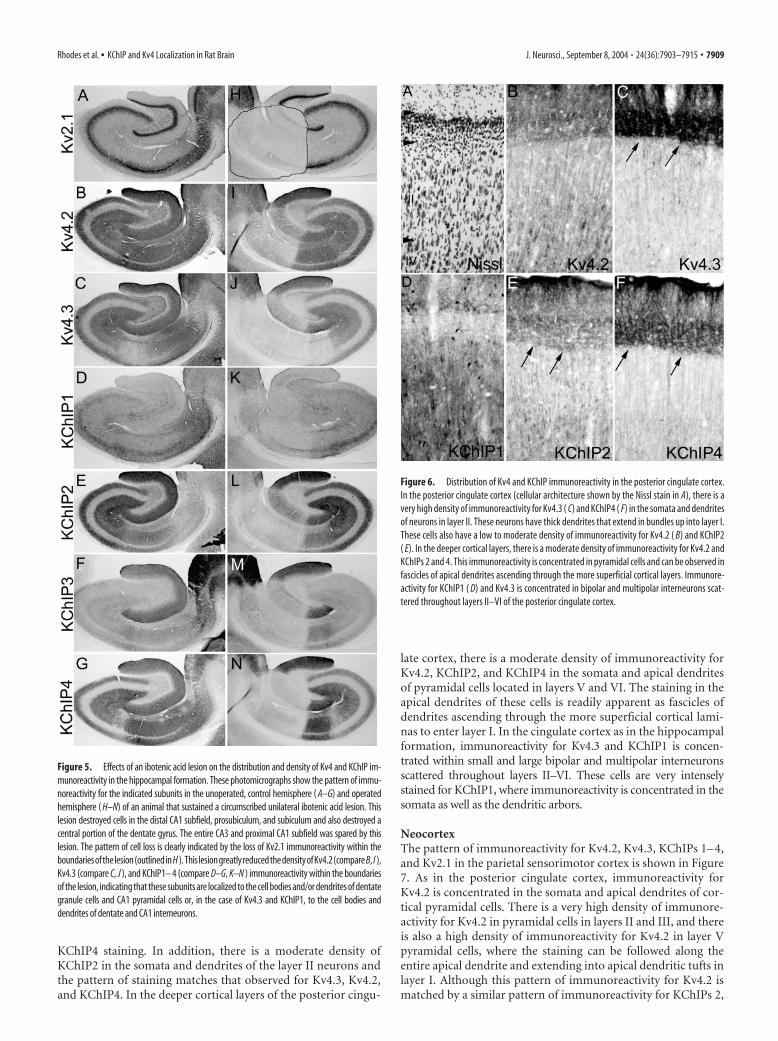
Posterior cingulate cortexPrevious descriptions of the pattern of Kv4.3 mRNA and proteinexpression in brain noted the very high levels Kv4.3 expressionand immunoreactivity in the posterior cingulate cortex (Area 23)(Tsaur et al., 1997). To confirm and extend these observations,we examined the distribution of immunoreactivity for Kv4 �subunits and KChIPs in this brain region. As shown in Figure 6,there is an exceptionally high density of immunoreactivity forKv4.3 in the somata and dendrites of large neurons in layer II ofthe posterior cingulate cortex. Staining in these cells is concen-trated in the apical dendrites that extend in bundles into layer I.Although there is a far lower density of immunoreactivity forKv4.2 compared with Kv4.3 in these cells, the high density ofKv4.3 immunoreactivity is matched by a similarly high density of
Figure 4. Distribution of Kv4 and KChIP immunoreactivity in the CA1 subfield. In the CA1 subfield (cellular architecture shownby the Nissl stain in A), immunoreactivity for Kv4.2 ( B), KChIP2 ( E), and KChIP4 ( G) but not Kv4.3 ( C) is concentrated in the apicaland basal dendrites and fine dendritic branches of pyramidal cells. There is little, if any, observable immunoreactivity for thesesubunits in the somata of CA1 pyramidal cells. Although there is also immunoreactivity for KChIP3 ( F) in CA1 pyramidal celldendrites, the density of KChIP3 immunoreactivity is far lower than KChIPs 2 or 4. As in CA3, there is a far greater density of Kv4.2,KChIP2, and KChIP4 immunoreactivity in the proximal two-thirds of the dendritic fields of CA3 pyramidal cells (stratum radiatum)compared with the distal third (stratum moleculare). These staining patterns contrast sharply with that for Kv2.1 ( H ), which isstrikingly restricted to the proximal one-third of the larger-caliber apical and basal dendritic branches. As in the dentate gyrus andCA3 subfields, immunoreactivity for Kv4.3 and KChIP1 ( D) is concentrated in the somata and dendrites of large, multipolarinterneurons. These Kv4.3- and KChIP1-immunoreactive interneurons are concentrated in the stratum oriens (or), stratum pyra-midale (py), stratum radiatum (rd), and along the junction of stratum radiatum and stratum moleculare. Interestingly, it appearsthat total number of Kv4.3-positive interneurons is a subset of those that are immunoreactive for KChIP1. ml, Stratum lacunosummoleculare.
7908 • J. Neurosci., September 8, 2004 • 24(36):7903–7915 Rhodes et al. • KChIP and Kv4 Localization in Rat Brain
KChIP4 staining. In addition, there is a moderate density ofKChIP2 in the somata and dendrites of the layer II neurons andthe pattern of staining matches that observed for Kv4.3, Kv4.2,and KChIP4. In the deeper cortical layers of the posterior cingu-
late cortex, there is a moderate density of immunoreactivity forKv4.2, KChIP2, and KChIP4 in the somata and apical dendritesof pyramidal cells located in layers V and VI. The staining in theapical dendrites of these cells is readily apparent as fascicles ofdendrites ascending through the more superficial cortical lami-nas to enter layer I. In the cingulate cortex as in the hippocampalformation, immunoreactivity for Kv4.3 and KChIP1 is concen-trated within small and large bipolar and multipolar interneuronsscattered throughout layers II–VI. These cells are very intenselystained for KChIP1, where immunoreactivity is concentrated in thesomata as well as the dendritic arbors.
NeocortexThe pattern of immunoreactivity for Kv4.2, Kv4.3, KChIPs 1– 4,and Kv2.1 in the parietal sensorimotor cortex is shown in Figure7. As in the posterior cingulate cortex, immunoreactivity forKv4.2 is concentrated in the somata and apical dendrites of cor-tical pyramidal cells. There is a very high density of immunore-activity for Kv4.2 in pyramidal cells in layers II and III, and thereis also a high density of immunoreactivity for Kv4.2 in layer Vpyramidal cells, where the staining can be followed along theentire apical dendrite and extending into apical dendritic tufts inlayer I. Although this pattern of immunoreactivity for Kv4.2 ismatched by a similar pattern of immunoreactivity for KChIPs 2,
Figure 5. Effects of an ibotenic acid lesion on the distribution and density of Kv4 and KChIP im-munoreactivity in the hippocampal formation. These photomicrographs show the pattern of immu-noreactivity for the indicated subunits in the unoperated, control hemisphere ( A–G) and operatedhemisphere ( H–N) of an animal that sustained a circumscribed unilateral ibotenic acid lesion. Thislesion destroyed cells in the distal CA1 subfield, prosubiculum, and subiculum and also destroyed acentral portion of the dentate gyrus. The entire CA3 and proximal CA1 subfield was spared by thislesion. The pattern of cell loss is clearly indicated by the loss of Kv2.1 immunoreactivity within theboundariesofthelesion(outlinedinH ).This lesiongreatlyreducedthedensityofKv4.2(compareB, I ),Kv4.3 (compare C, J ), and KChIP1– 4 (compare D–G, K–N ) immunoreactivity within the boundariesof the lesion, indicating that these subunits are localized to the cell bodies and/or dendrites of dentategranule cells and CA1 pyramidal cells or, in the case of Kv4.3 and KChIP1, to the cell bodies anddendrites of dentate and CA1 interneurons.
Figure 6. Distribution of Kv4 and KChIP immunoreactivity in the posterior cingulate cortex.In the posterior cingulate cortex (cellular architecture shown by the Nissl stain in A), there is avery high density of immunoreactivity for Kv4.3 ( C) and KChIP4 ( F) in the somata and dendritesof neurons in layer II. These neurons have thick dendrites that extend in bundles up into layer I.These cells also have a low to moderate density of immunoreactivity for Kv4.2 ( B) and KChIP2( E). In the deeper cortical layers, there is a moderate density of immunoreactivity for Kv4.2 andKChIPs 2 and 4. This immunoreactivity is concentrated in pyramidal cells and can be observed infascicles of apical dendrites ascending through the more superficial cortical layers. Immunore-activity for KChIP1 ( D) and Kv4.3 is concentrated in bipolar and multipolar interneurons scat-tered throughout layers II–VI of the posterior cingulate cortex.
Rhodes et al. • KChIP and Kv4 Localization in Rat Brain J. Neurosci., September 8, 2004 • 24(36):7903–7915 • 7909
3, and 4, it is clear that the pattern ofKChIP3 and KChIP4 immunoreactivitymost closely matches the staining patternfor Kv4.2. For KChIP2, there is a muchgreater density of immunoreactivity in theapical dendrites of layer II–III pyramidalcells compared with the density of stainingin layer V neurons, and there is also a veryhigh density of immunoreactivity forKChIP2 in layer IV, where the staining isconcentrated in small multipolar neuronsand perhaps also in afferent fibers. The im-munoreactivity for Kv4.2 and KChIPs 2, 3,and 4 in fascicles of apical dendrites of corti-cal pyramidal cells stands out in marked con-trast to the staining for Kv2.1. Kv2.1 immu-noreactivity is restricted to the proximalportion of the apical and basal dendrites,whereas the immunostaining for Kv4.2 andthese KChIPs extends along the entire apicaldendrite and into distal dendritic arbors.
As in the hippocampus and other brainregions, immunoreactivity for KChIP1and Kv4.3 is concentrated in the somataand dendritic branches of interneuronsscattered throughout all cortical laminas(Fig. 7). Immunoreactivity was present invirtually all morphological classes of inter-neurons, including bipolar, multipolar,and bi-tufted cells, among others. Stainingfor KChIP1 was concentrated in the pe-rinuclear cytoplasm as well as the dendriticcytoplasm of these interneurons, whereasimmunoreactivity for Kv4.3 was most in-tense in the somatodendritic membrane ofthese cells. Note that in many of these cellsthe Kv4.3 staining on somata was muchless apparent for Kv4.3 than for KChIP1(Fig. 7).
StriatumThe pattern of immunoreactivity for Kv4 � subunits and KChIPsin the caudate/putamen is shown in Figure 8. In this brain region,immunoreactivity for Kv4.2, Kv4.3, KChIP2, KChIP3, andKChIP4 is concentrated in the neuropil, where the staining islikely to be associated with the somata and dendrites of striatalprojection neurons. However, immunoreactivity for Kv4.2 andKChIP2 in the striatal neuropil is so dense that it is difficult toattribute the staining to a specific cell type or subcellular domain.In contrast to this, immunoreactivity for Kv4.3 and KChIP1 isclearly visible in medium to large-sized multipolar striatal inter-neurons. Based on the size, distribution, and morphology of thesecells, they are likely to represent the somatostatin- and/or neu-ropeptide Y-containing interneurons. However, detailed analy-ses using labeling with these specific markers is necessary for aconclusive identification of these immunoreactive cells. Interest-ingly, immunoreactivity of KChIP4 is concentrated in very largemultipolar interneurons (Fig. 8F). Based on their size and den-dritic morphology, these KChIP4-positive cells are likely to belarge cholinergic interneurons that have large A-type currents(Song et al., 1998), although conclusive double-labeling experi-ments were not performed.
Colocalization of Kv4 and KChIP immunoreactivityAs described above, in many cell types and brain regions, there isclear correspondence and overlap in the distribution of Kv4 andKChIP immunoreactivity. To determine whether and where in-dividual Kv4 � subunits and KChIP isoforms are colocalizedwithin the same cell, we performed double-label immunofluores-cence analyses and examined the staining patterns by laser scan-ning confocal microscopy. Although overlap of immunofluores-cence signals indicates that the two target antigens are presentwithin the same cell and subcellular domain, we cannot concludefrom this type of analysis that the antigens are coassociated.Thus, these data should be interpreted as suggestive, indicat-ing sites where it is likely that two channel subunits form achannel complex. Our ability to perform these colocalizationanalyses were limited by the availability of mAbs having com-patible isotypes; however, because we initiated our efforts togenerate Kv4- and KChIP-specific antibodies with the aim ofperforming double-label immunofluorescence studies, wewere able to select antibody reagents that met our strict selec-tivity criteria for immunohistochemistry and also satisfiedour requirement of isotype compatibility for double-labelimmunofluorescence.
Representative confocal images showing colocalization of Kv4and KChIP isoforms are shown in Figure 9. As described above, inmany brain regions, immunoreactivity for KChIP1 and Kv4.3 is
Figure 7. Distribution of Kv4 and KChIP immunoreactivity in the parietotemporal cortex. In neocortical regions such as theparietotemporal cortex (cellular architecture shown by the Nissl stain in A), immunoreactivity for Kv4.2 ( B), Kv4.3 ( C), and KChIPs2– 4 ( D–G) is concentrated along the apical dendrites and in the apical dendritic arbors of cortical pyramidal cells. This distributionis particularly striking for Kv4.2, KChIP2, KChIP3, and KChIP4, where the staining lines the large apical dendrites of layer Vpyramidal cells as they ascend through layer IV and into more superficial layers and contrasts with the staining pattern for Kv2.1( H ), which is limited to the somata and proximal dendritic branches of cortical neurons. As in the hippocampal formation,immunoreactivity for Kv4.3 and KChIP1 is concentrated in the somata and dendrites of cortical interneurons. These interneuronsare multipolar, bipolar and fusiform in shape, and are scattered throughout all cortical layers.
7910 • J. Neurosci., September 8, 2004 • 24(36):7903–7915 Rhodes et al. • KChIP and Kv4 Localization in Rat Brain
observed in interneurons. As shown in Figure 9, A–C, in many,but not all, of these interneurons, Kv4.3 and KChIP1 are colocal-ized. However, in these interneurons, there is a high density ofimmunoreactivity for KChIP1 in the perinuclear cytoplasm, aspredicted from studies in heterologous cells (Shibata et al., 2003),
but no detectable cytoplasmic staining for Kv4.3. Instead, theimmunoreactivity for Kv4.3 is predominantly concentratedalong the somatodendritic membrane, and here there is also ahigh density of staining for KChIP1. It is important to point outthat in the hippocampus, neocortex, and striatum, virtually all ofthe cells in which Kv4.3 and KChIP1 are colocalized are interneu-rons, based on both morphology, density, and location and stain-ing with immunohistochemical markers (supplemental material,available at www.jneurosci.org). In the hippocampus, virtuallyevery Kv4.3-positive cell also expresses KChIP1, and vice versa.Figure 9, D–F, also shows colocalization of Kv4.2 with KChIP4 inthe dendritic arbors of layer II neurons in the posterior cingulatecortex, in the apical and basal dendrites of CA1 pyramidal cells,and in the glomeruli formed by the dendrites of cerebellar gran-ule cells. In these regions, there is very tight overlap in the pat-terns of immunofluorescence for Kv4.2 and KChIP4, and it isimportant to point out that in many of the cells in which colocal-ization of Kv4.2 with KChIP4 (and KChIP2) was observed areexcitatory glutamatergic projection neurons.
Coimmunoprecipitation analysesThe immunohistochemical and immunofluorescence data de-scribed above indicate that Kv4.2, Kv4.3, and the four KChIPisoforms show tight overlap in their regional and subcellular dis-tribution across several brain regions but that there is preferentialcodistribution of Kv4.2 with KChIPs 2, 3, and 4 and preferentialcodistribution of Kv4.3 with KChIP1. To determine that thesechannel subunits are not only codistributed but are also coasso-ciated in brain Kv channel complexes, we performed reciprocalcoimmunoprecipitation analyses on detergent extracts of rat hip-pocampal membranes. Because the molecular mass of individualKChIP isoforms and their mobility on SDS-PAGE gels corre-sponds closely to that for the light chain of IgG, it was not possibleto obtain clean, clear, and interpretable results from immunopre-cipitation analyses when we used the purified mAbs describedabove for both immunoprecipitation and immunoblotting.
Thus, for coimmunoprecipitation studies,we used affinity-purified rabbit polyclonalantibodies for immunoprecipitation andthe purified mAbs to detect associatedproteins via immunoblotting. As such, wewere limited in the repertoire of availablesubtype-specific antibodies for the immu-noprecipitation reactions. Moreover, thepropensity of Kv4.2 to aggregate (Sheng etal., 1993; Shibata et al., 2003) limited thequantitative interpretation of these data.However, as shown in Figure 10, immuno-precipitation reactions performed usingan affinity-purified anti-KChIP antibodythat recognizes all known KChIP isoformsyielded strong coimmunoprecipitation ofKv4.2 but not Kv2.1 and, as expected,yielded strong immunoprecipitation ofeach KChIP (1–3) polypeptide analyzed.Immunoprecipitation reactions per-formed using an affinity-purified antibodyagainst Kv4.2 yielded coimmunoprecipi-tation of each KChIP isoform. Interest-ingly, the anti-Kv4.2 antibody also coim-munoprecipitated Kv4.3 (but not Kv2.1),suggesting that Kv4.2 and Kv4.3 may bepresent in heteromeric Kv4 complexes in
Figure 8. Localization of Kv4 � subunits and KChIPs in the striatum. In the striatal neuropil,there is a very high density of immunoreactivity for Kv4.2 ( A) and KChIP2 ( D) and a lowerdensity of immunoreactivity for Kv4.3 ( B) and KChIPs 3 ( E) and 4 ( F). Immunoreactivity forKv4.3 and KChIP1 ( C) is concentrated in what appear to be medium-sized striatal interneurons,whereas immunoreactivity for KChIP4 can be observed in very large, multipolar interneurons.
Figure 9. Confocal images showing colocalization of Kv4 and KChIP immunoreactivity. These confocal images show that Kv4.3(red) and KChIP1 (green) tend to be colocalized in the somatodendritic membranes of hippocampal ( A), neocortical ( B), andstriatal ( C) presumed inhibitory interneurons, whereas Kv4.2 (red) and KChIP4 (green) tend to be colocalized in the somatoden-dritic membranes of presumed excitatory neurons, in the posterior cingulate cortex ( D), hippocampus ( E), and cerebellar granulecell layer ( F).
Rhodes et al. • KChIP and Kv4 Localization in Rat Brain J. Neurosci., September 8, 2004 • 24(36):7903–7915 • 7911
regions in which Kv4.2/Kv4.3 colocalization is observed (e.g.,hippocampal dentate granule cells and CA3 pyramidal cell den-drites, glomeruli of cerebellar granule neurons). Taken together,these analyses confirm that Kv4.2 and Kv4.3 are coassociatedwith KChIP isoforms in rat brain membranes and that theKChIPs are tightly associated with Kv4 � subunits in rat brain Kvchannel complexes.
DiscussionHere, we generated and characterized mAbs against Kv4.2, Kv4.3,and KChIPs 1– 4 that were used in immunohistochemical andimmunoprecipitation analyses of rat brain Kv4 channel com-plexes. We did not analyze Kv4.1 because specific antibodies werenot available. In general, KChIPs 2, 3, and 4 colocalize with Kv4.2in excitatory neurons including cortical and hippocampal CA1pyramidal cells, and KChIP1 and Kv4.3 colocalize in inhibitoryinterneurons throughout the cortex and hippocampus and instriatal interneurons. However, this general inverse relationshipbetween Kv4.2 and Kv4.3, as noted in previous mRNA analyses(Song et al., 1998; Tkatch et al., 2000; Liss et al., 2001; Lien et al.,2002), does not hold for all cell types, in that hippocampal den-tate granule and CA3 pyramidal cells and cerebellar granule cellsexpress high levels of colocalized Kv4.2 and Kv4.3. Coimmuno-precipitation analysis in detergent-solubilized hippocampalmembranes revealed that all four KChIPs coassociate with Kv4 �
subunits in hippocampal membranes, confirming that theKChIPs are tightly associated with Kv4 � subunits in native brainA-type channel complexes.
Although our data confirm that each of the anti-KChIP mAbsdescribed here are specific for their respective antigen, it is im-portant to note that alternatively spliced isoforms of each KChIPpolypeptide have been reported (An et al., 2000; Bahring et al.,2001; Decher et al., 2001; Liss et al., 2001; Morohashi et al., 2002;Patel et al., 2002; Boland et al., 2003). Most of the alternativesplicing occurs within the variable N termini; a smaller number ofsplice variants have been reported that contain insertions or de-letions within the C-terminal “core” domain. As part of our an-tibody characterization efforts, we expressed N-terminal trunca-tion mutants of each KChIP polypeptide (An et al., 2000;Holmqvist et al., 2002) in COS cells and tested each mAb forreactivity against the core domain by immunofluorescence. Us-ing this approach, we determined that the epitope for each of theanti-KChIP mAbs maps within the conserved 185 amino acidcore domain. Because of this, the staining patterns described herereflect the distribution of all possible N-terminal splice forms foreach individual KChIP family member. This point must be con-sidered in interpreting the immunohistochemical staining de-scribed here because the specific contribution of KChIP expres-sion to the phenotype of Kv4-mediated currents is likely todepend on the specific splice variant that is expressed (Boland etal., 2003; Decher et al., 2004; Patel et al., 2004). This is especiallycritical for KChIP4, for which the “KChIP4a” splice variant pro-foundly slows the kinetics of Kv4 inactivation, but the“KChIP4ap” variant does not (Holmqvist et al., 2002). Althoughimmunohistochemical localization of individual KChIP spliceforms will require generation of variant-specific antibodies, valu-able information about the contribution of individual KChIPsplice variants to Kv4 currents in native cells can also be gainedfrom single-cell analyses of KChIP isoform expression (Liss et al.,2001).
The protein we identified as KChIP3 (An et al., 2000) wasidentified independently as DREAM, a Ca 2�-dependent tran-scriptional repressor (Carrion et al., 1999), and calsenilin, a pre-senilin interacting protein (Buxbaum et al., 1999). Recently,Zaidi et al. (2002) described the distribution of calsenilin immu-noreactivity in mouse brain, using a rabbit polyclonal antibodyraised against a full-length calsenilin/KChIP3 fusion protein,which is likely to cross-react with other KChIP family members.Their immunoblot data strongly support this contention becausein mouse brain membranes their antibody detects several bandsin the 25–35 kDa range, corresponding to the predicted molecu-lar masses of other KChIP family members (Zaidi et al., 2002).Interestingly, neither the polyclonal antibody used by Zaidi et al.(2002) nor our KChIP3-specific mAb exhibit staining in the nu-cleus of neurons or glial cells, which might be expected for aCa 2�-dependent regulator of transcription (Carrion et al., 1999;Cheng et al., 2002). The remarkable correspondence betweenKv4.2 (Sheng et al., 1992; present study) and KChIP3 (Zaidi et al.,2002; present study) localization suggests that the preferred bind-ing partner for KChIP3 in neurons is Kv4.2, raising questions asto whether KChIP3 in native cells is truly the multifunctionalprotein suggested by studies in heterologous expression systems.
Somatodendritic A-currents have been studied most exten-sively by patch clamp in hippocampal pyramidal cells and in thelarge layer V pyramidal neurons in the motor and somatosensorycortex (Wu and Barish, 1992; Hoffman et al., 1997; Hoffman andJohnston, 1998; Golding et al., 1999; Bekkers, 2000a,b; Johnstonet al., 2000; Korngreen and Sakmann, 2000; Storm, 2000). In CA1
Figure 10. Heteromeric Kv4 and KChIP channel complexes in rat hippocampus. Immunopre-cipitation reactions were performed on detergent extracts of adult rat hippocampal membranes(HCM) with the indicated rabbit polyclonal antibodies (anti-KChIP, anti-Kv4.2, or anti-Kv2.1).Aliquots of the HCM detergent extract and of the products of the immunoprecipitation reactionswere size fractionated by 4 –12% (Kv4 blots) or 10% (KChIP blots) SDS-PAGE. The samples werethen transferred to nitrocellulose and probed with mouse mAbs as indicated below each blotpanel. Bound antibody was detected by ECL/autoradiography. The arrows or brackets to left ofthe panels highlight the band resulting from specific detection of the antigen listed below eachpanel. The numbers to left of the panels refer to electrophoretic mobility of molecular weightstandards.
7912 • J. Neurosci., September 8, 2004 • 24(36):7903–7915 Rhodes et al. • KChIP and Kv4 Localization in Rat Brain
pyramidal cells, A-type currents play a critical role in synapticintegration and plasticity by controlling subthreshold excitationand the amplitude of back-propagating action potentials (Hoff-man et al., 1997; Johnston et al., 2000). In these cells, the densityof A-type currents on the trunk of the apical dendrite increasesdramatically with distance from the pyramidal cell soma (Hoff-man et al., 1997). We did not observe a striking gradient in theintensity of Kv4.2, KChIP2, or KChIP4 immunoreactivity acrossthe stratum radiatum of CA1 using immunoperoxidase staining,although a striking gradient was observed across dendrites ofdentate granule cells in the molecular layer of the denate gyrus.Confocal analysis of sections with double immunofluorescencestaining for Kv4.2 and KChIP4 (Fig. 9E) or Kv4.2 and PSD-95(supplemental material, available at www.jneurosci.org) revealedonly a subtle increase in the density of Kv4.2 immunoreactivity inthe superficial third of the stratum radiatum. Unlike in the neo-cortex (see below), the extremely close packing of CA1 pyramidalcells precludes detailed immunocytochemical analyses of indi-vidual apical dendrites required for conclusive correlations withelectrophysiological studies. Moreover, our experiments labelKv4.2 not only on the main trunk of the apical dendrites accessi-ble by patch clamp but also each branch and collateral in theentire, and very complex, intertwining dendritic trees of thedensely packed CA1 pyramidal neurons. We are also visualizingboth intracellular and surface pools of Kv4.2 in our detergent-permeabilized sections. Finally, dynamic modulation of den-dritic Kv4 channel activity by phosphorylation (Hoffman andJohnston, 1998; Varga et al., 2000; Schrader et al., 2002; Frick etal., 2004) may lead to inconsistencies between the density ofchannel protein and functional channels.
Two striking findings in the present study are that in CA1–CA3, there is an abrupt decrease in the density of Kv4.2 (andKChIP2, 3, and 4) at the transition from the stratum radiatum tothe stratum moleculare and that there is little or no Kv4.3 immu-noreactivity in CA1 pyramidal cells but a high density of Kv4.3(with Kv4.2) immunoreactivity in CA3 pyramids. The very lowdensity of Kv4.3 immunoreactivity in CA1 compared with CA3pyramidal cells suggest that in CA1 pyramidal cells, the majorityof A-type channel complexes are homomeric Kv4.2 channels. Toour knowledge, there is little published data exploring the de-tailed biophysical properties of heteromeric channels formed bycoexpression of Kv4.2 and Kv4.3 or concatenated � subunits (butsee Guo et al., 2002). Nevertheless, the dramatic change in Kv4 �subunit expression in these adjacent hippocampal subfields isintriguing. The drop-off of Kv4 and KChIP immunoreactivity inthe stratum moleculare suggests a very low density of Kv4-mediated A-type current in very distal dendrites of CA pyramidalcells.
Neocortical layer V pyramidal cells differ from hippocampalCA1 pyramidal cells in that they do not express a gradient ofA-current along the apical dendrite and in having a much lowerdensity of dendritic A-type current (Hoffman et al., 1997; Bek-kers, 2000a,b; Korngreen and Sakmann, 2000). This suggests thatdendritic A-current does not exert as dominant an influence incortical pyramidal cells as it does in CA1 (Storm, 2000). Here, weobserved that immunoreactivity for Kv4.2 and KChIPs 2, 3, and 4can be followed along the entire apical dendrite of cortical layer Vpyramidal cells and that there is a fairly uniform density of im-munoreactivity along the dendritic shaft. It is important to notethat Kv4.2 and KChIP immunoreactivity can be followed all theway into the most distal dendritic branches and the apical den-
dritic tufts of cortical pyramidal cells. The presence of Kv4 im-munoreactivity in the most distal dendritic regions indicates thatthese channels may modulate excitatory input along the entiredendrite and that in neocortical pyramidal cells, A-type currentsare not as compartmentalized within the dendrite as they are inhippocampal CA pyramids.
Although KChIPs are clearly a component of the Kv4 modu-latory factors encoded in brain mRNA (Nadal et al., 2001), an-other factor that accelerates Kv4 inactivation has been identifiedas the CD26-like dipeptidyl-peptidase dipeptidyl aminopeptidase-like protein (DPPX) (Nadal et al., 2003). Coexpression of Kv4 �subunits with KChIPs and DPPX in heterologous cells gives riseto currents that recapitulate the features of native A-type cur-rents, indicating that native brain Kv4 channels are likely to beformed as complexes of all three proteins. Future studies areneeded to explore the relative contributions of DPPX andKChIPs to the formation of A-type currents in native neurons.
ReferencesAn WF, Bowlby MR, Betty M, Cao J, Ling HP, Mendoza G, Hinson JW,
Mattsson KI, Strassle BW, Trimmer JS, Rhodes KJ (2000) Modulation ofA-type potassium channels by a family of calcium sensors. Nature403:553–556.
Bahring R, Dannenberg J, Peters HC, Leicher T, Pongs O, Isbrandt D (2001)Conserved Kv4 N-terminal domain critical for effects of Kv channel-interacting protein 2.2 on channel expression and gating. J Biol Chem276:23888 –23894.
Baldwin TJ, Tsaur ML, Lopez GA, Jan YN (1991) Characterization of amammalian cDNA for an inactivating voltage-sensitive K� channel. Neu-ron 7:471– 483.
Bekele-Arcuri Z, Matos MF, Manganas L, Strassle BW, Monaghan MM,Rhodes KJ, Trimmer JS (1996) Generation and characterization ofsubtype-specific monoclonal antibodies to K � channel �- and �-subunitpolypeptides. Neuropharmacology 35:851– 865.
Bekkers JM (2000a) Properties of voltage-gated potassium currents in nu-cleated patches from large layer 5 cortical pyramidal neurons of the rat.J Physiol (Lond) 525:593– 609.
Bekkers JM (2000b) Distribution and activation of voltage-gated potassiumchannels in cell-attached and outside-out patches from large layer 5 cor-tical pyramidal neurons of the rat. J Physiol (Lond) 525:611– 620.
Boland LM, Jiang M, Lee SY, Fahrenkrug SC, Harnett MT, O’Grady SM(2003) Functional properties of a brain-specific N-terminally splicedmodulator of Kv4 channels. Am J Physiol Cell Physiol 285:C161–C170.
Buxbaum JD, Choi EK, Luo Y, Lilliehook C, Crowley AC, Merriam DE,Wasco W (1998) Calsenilin: a calcium-binding protein that interactswith the presenilins and regulates the levels of a presenilin fragment. NatMed 4:1177–1181.
Carrion AM, Link WA, Ledo F, Mellstrom B, Naranjo JR (1999) DREAM isa Ca 2�-regulated transcriptional repressor. Nature 398:80 – 84.
Chabala LD, Bakry N, Covarrubias M (1993) Low molecular weightpoly(A)� mRNA species encode factors that modulate gating of a non-Shaker A-type K � channel. J Gen Physiol 102:713–728.
Cheng HY, Pitcher GM, Laviolette SR, Whishaw IQ, Tong KI, Kockeritz LK,Wada T, Joza NA, Crackower M, Goncalves J, Sarosi I, Woodgett JR,Oliveira-dos-Santos AJ, Ikura M, van der Kooy D, Salter MW, PenningerJM (2002) DREAM is a critical transcriptional repressor for pain mod-ulation. Cell 108:31– 43.
Decher N, Uyguner O, Scherer CR, Karaman B, Yuksel-Apak M, Busch AE,Steinmeyer K, Wollnik B (2001) hKChIP2 is a functional modifier ofhKv4.3 potassium channels: cloning and expression of a short hKChIP2splice variant. Cardiovasc Res 52:255–264.
Decher N, Barth AS, Gonzalez T, Steinmeyer K, Sanguinetti MC (2004)Novel KChIP2 isoforms increase functional diversity of transient outwardpotassium currents. J Physiol (Lond) 557:761–772.
Du J, Tao-Cheng JH, Zerfas P, McBain CJ (1998) The K� channel, Kv2.1, isapposed to astrocytic processes and is associated with inhibitory postsyn-aptic membranes in hippocampal and cortical principal neurons and in-hibitory interneurons. Neuroscience 84:37– 48.
Frick A, Magee J, Johnston D (2004) LTP is accompanied by an enhanced
Rhodes et al. • KChIP and Kv4 Localization in Rat Brain J. Neurosci., September 8, 2004 • 24(36):7903–7915 • 7913
local excitability of pyramidal neuron dendrites. Nat Neurosci 7:126 –135.
Golding NL, Jung H, Mickus T, Spruston N (1999) Dendritic calcium spikeinitiation and repolarization are controlled by distinct potassium channelsubtypes in CA1 pyramidal neurons. J Neurosci 19:8789 – 8798.
Guo W, Li H, Aimond F, Johns DC, Rhodes KJ, Trimmer JS, Nerbonne JM(2002) Role of heteromultimers in the generation of myocardial tran-sient outward K� currents. Circ Res 90:586 –593.
Gutman GA, Chandy KG, Adelman JP, Aiyar J, Bayliss DA, Clapham DE,Covarriubias M, Desir GV, Furuichi K, Ganetzky B, Garcia ML, GrissmerS, Jan LY, Karschin A, Kim D, Kuperschmidt S, Kurachi Y, Lazdunski M,Lesage F, Lester HA, et al. (2003) International Union of Pharmacology.XLI. Compendium of voltage-gated ion channels: potassium channels.Pharmacol Rev 55:583–586.
Hoffman DA, Johnston D (1998) Downregulation of transient K � channelsin dendrites of hippocampal CA1 pyramidal neurons by activation ofPKA and PKC. J Neurosci 18:3521–3528.
Hoffman DA, Magee JC, Colbert CM, Johnston D (1997) K � channel reg-ulation of signal propagation in dendrites of hippocampal pyramidal neu-rons. Nature 387:869 – 875.
Holmqvist MH, Cao J, Knoppers MH, Jurman ME, Distefano PS, Rhodes KJ,Xie Y, An WF (2001) Kinetic modulation of Kv4-mediated A-current byarachidonic acid is dependent on potassium channel interacting proteins.J Neurosci 21:4154 – 4161.
Holmqvist MH, Cao J, Hernandez-Pineda R, Jacobson MD, Carroll KI, SungMA, Betty M, Ge P, Gilbride KJ, Brown ME, Jurman ME, Lawson D,Silos-Santiago I, Xie Y, Covarrubias M, Rhodes KJ, Distefano PS, An WF(2002) Elimination of fast inactivation in Kv4 A-type potassium chan-nels by an auxiliary subunit domain. Proc Natl Acad Sci USA99:1035–1040.
Johnston D, Hoffman DA, Magee JC, Poolos NP, Watanabe S, Colbert CM,Migliore M (2000) Dendritic potassium channels in hippocampal pyra-midal neurons. J Physiol (Lond) 525:75– 81.
Johnston D, Christie BR, Frick A, Gray R, Hoffman DA, Schexnayder LK,Watanabe S, Yuan LL (2003) Active dendrites, potassium channels andsynaptic plasticity. Philos Trans R Soc Lond B Biol Sci 358:667– 674.
Kim LA, Furst J, Butler MH, Xu S, Grigorieff N, Goldstein SA (2004a) Itochannels are octomeric complexes with four subunits of each Kv4.2 andK� channel-interacting protein 2. J Biol Chem 279:5549 –5554.
Kim LA, Furst J, Gutierrez D, Butler MH, Xu S, Goldstein SA, Grigorieff N(2004b) Three-dimensional structure of I(to); Kv4.2-KChIP2 ion chan-nels by electron microscopy at 21 Angstrom resolution. Neuron41:513–519.
Korngreen A, Sakmann B (2000) Voltage-gated K � channels in layer 5 neo-cortical pyramidal neurones from young rats: subtypes and gradients.J Physiol (Lond) 525:621– 639.
Lien CC, Martina M, Schultz JH, Ehmke H, Jonas P (2002) Gating, modu-lation and subunit composition of voltage-gated K(�) channels in den-dritic inhibitory interneurons of rat hippocampus. J Neurophysiol538:405– 409.
Liss B, Franz O, Sewing S, Bruns R, Neuhoff H, Roeper J (2001) Tuningpacemaker frequency of individual dopaminergic neurons by Kv4.3L andKChip3.1 transcription. EMBO J 20:5715–5724.
Maletic-Savatic M, Lenn NJ, Trimmer JS (1995) Differential spatiotemporalexpression of K � channel polypeptides in rat hippocampal neurons de-veloping in situ and in vitro. J Neurosci 15:3840 –3851.
Monaghan MM, Trimmer JS, Rhodes KJ (2001) Experimental localizationof Kv1 family voltage-gated K � channel � and � subunits in rat hip-pocampal formation. J Neurosci 21:5973–5983.
Morohashi Y, Hatano N, Ohya S, Takikawa R, Watabiki T, Takasugi N,Imaizumi Y, Tomita T, Iwatsubo T (2002) Molecular cloning and char-acterization of CALP/KChIP4, a novel EF-hand protein interacting withpresenilin 2 and voltage-gated potassium channel subunit Kv4. J BiolChem 277:14965–14975.
Nadal MS, Amarillo Y, Vega-Saenz de Miera E, Rudy B (2001) Evidence forthe presence of a novel Kv4-mediated A-type K(�) channel-modifyingfactor. J Physiol (Lond) 537:801– 809.
Nadal MS, Ozaita A, Amarillo Y, de Miera EV, Ma Y, Mo W, Goldberg EM,Misumi Y, Ikehara Y, Neubert TA, Rudy B (2003) The CD26-relateddipeptidyl aminopeptidase-like protein DPPX is a critical component ofneuronal A-type K � channels. Neuron 37:449 – 461.
Nakamura TY, Coetzee WA, Vega-Saenz De Miera E, Artman M, Rudy B
(1997) Modulation of Kv4 channels, key components of rat ventric-ular transient outward K � current, by PKC. Am J Physiol273:H1775–H1786.
Patel SP, Campbell DL, Morales MJ, Strauss HC (2002) Heterogeneous ex-pression of KChIP2 isoforms in the ferret heart. J Physiol (Lond)15:649 – 656.
Rhodes KJ, Keilbaugh SA, Barrezueta NX, Lopez KL, Trimmer JS (1995)Association and colocalization of K � channel �- and �-subunit polypep-tides in rat brain. J Neurosci 15:5360 –5371.
Rhodes KJ, Monaghan MM, Barrezueta NX, Nawoschik S, Bekele-Arcuri Z,Matos M, Nakahira K, Schechter LE, Trimmer JS (1996) Voltage-gatedK � channel �-subunits: expression and distribution of Kv�1 and Kv�2 inadult rat brain. J Neurosci 16:4846 – 4860.
Rhodes KJ, Strassle BW, Monaghan MM, Bekele-Arcuri Z, Matos MF, Trim-mer JS (1997) Association and colocalization of Kv�1 and Kv�2 withKv1 �-subunits in mammalian brain K � channel complexes. J Neurosci17:8246 – 8258.
Rudy B, Hoger JH, Lester HA, Davidson N (1988) At least two mRNA spe-cies contribute to the properties of rat brain A-type potassium channelsexpressed in Xenopus oocytes. Neuron 1:649 – 658.
Scannevin RH, Murakoshi H, Rhodes KJ, Trimmer JS (1996) Identificationof a cytoplasmic domain important in the polarized expression and clus-tering of the Kv2.1 K� channel. J Cell Biol 135:1619 –1632.
Scannevin RH, Wang K, Jow F, Megules J, Kopsco DC, Edris W, Carroll KC,Lu Q, Xu W, Xu Z, Katz AH, Olland S, Lin L, Taylor M, Stahl M, MalakianK, Somers W, Mosyak L, Bowlby MR, Chanda P, Rhodes KJ (2004) TwoN-terminal domains of Kv4 K(�) channels regulate binding to and mod-ulation by KChIP1. Neuron 19:587–598.
Schrader LA, Anderson AE, Mayne A, Pfaffinger PJ, Sweatt JD (2002) PKAmodulation of Kv4.2-encoded A-type potassium channels requires for-mation of a supramolecular complex. J Neurosci 22:10123–10133.
Serodio P, Rudy B (1998) Differential expression of Kv4 K � channel sub-units mediating subthreshold transient K � (A-type) currents in rat brain.J Neurophysiol 79:1081–1091.
Serodio P, Kentros C, Rudy B (1994) Identification of molecular compo-nents of A-type channels activating at subthreshold potentials. J Neuro-physiol 72:1516 –1529.
Serodio P, Vega-Saenz de Miera E, Rudy B (1996) Cloning of a novelcomponent of A-type K � channels operating at subthreshold poten-tials with unique expression in heart and brain. J Neurophysiol75:2174 –2179.
Sheng M, Tsaur ML, Jan YN, Jan LY (1992) Subcellular segregation of twoA-type K � channel proteins in rat central neurons. Neuron 9:271–284.
Sheng M, Liao YJ, Jan YN, Jan LY (1993) Presynaptic A-current based onheteromultimeric K� channels detected in vivo. Nature 365:72–75.
Shibata R, Misonou H, Campomanes CR, Anderson AE, Schrader LA,Doliveira LC, Carroll KI, Sweatt JD, Rhodes KJ, Trimmer JS (2003) Afundamental role for KChIPs in determining the molecular propertiesand trafficking of Kv4.2 potassium channels. J Biol Chem 278:36445–36454.
Song WJ, Tkatch T, Baranauskas G, Ichinohe N, Kitai ST, Surmeier DJ(1998) Somatodendritic depolarization-activated potassium currents inrat neostriatal cholinergic interneurons are predominantly of the A typeand attributable to coexpression of Kv4.2 and Kv4.1 subunits. J Neurosci18:3124 –3137.
Storm JF (2000) K( �) channels and their distribution in large cortical pyra-midal neurones. J Physiol (Lond) 525:565–566.
Tago H, Kimura H, Maeda T (1986) Visualization of detailed acetylcho-linesterase fiber and neuron staining in rat brain by a sensitive histochem-ical procedure. J Histochem Cytochem 34:1431–1438.
Tekirian TL, Merriam DE, Marshansky V, Miller J, Crowley AC, Chan H,Ausiello D, Brown D, Buxbaum JD, Xia W, Wasco W (2001) Subcellularlocalization of presenilin 2 endoproteolytic C-terminal fragments. BrainRes Mol Brain Res 96:14 –20.
Tkatch T, Baranauskas G, Surmeier DJ (2000) Kv4.2 mRNA abundance andA-type K(�) current amplitude are linearly related in basal ganglia andbasal forebrain neurons. J Neurosci 20:579 –588.
7914 • J. Neurosci., September 8, 2004 • 24(36):7903–7915 Rhodes et al. • KChIP and Kv4 Localization in Rat Brain
Trimmer JS (1991) Immunological identification and characterization of adelayed rectifier K � channel polypeptide in rat brain. Proc Natl Acad SciUSA 88:10764 –10768.
Trimmer JS, Rhodes KJ (2004) Localization of voltage-gated ion channels inmammalian brain. Annu Rev Physiol 66:477–519.
Trimmer JS, Trowbridge IS, Vacquier VD (1985) Monoclonal antibody to amembrane glycoprotein inhibits the acrosome reaction and associatedCa 2� and H � fluxes of sea urchin sperm. Cell 40:697–703.
Tsaur ML, Chou CC, Shih YH, Wang HL (1997) Cloning, expression andCNS distribution of Kv4.3, an A-type K� channel alpha subunit. FEBSLett 400:215–220.
specific immunolocalization of differentially phosphorylated Kv4.2 in themouse brain. Learn Mem 7:321–332.
Wu RL, Barish ME (1992) Two pharmacologically and kinetically distincttransient potassium currents in cultured embryonic mouse hippocampalneurons. J Neurosci 12:2235–2246.
Zaidi NF, Berezovska O, Choi EK, Miller JS, Chan H, Lilliehook C, HymanBT, Buxbaum JD, Wasco W (2002) Biochemical and immunocyto-chemical characterization of calsenilin in mouse brain. Neuroscience114:247–263.
Zhou W, Qian Y, Kunjilwar K, Pfaffinger PJ, Choe S (2004) Structural in-sights into the functional interaction of KChIP1 with Shal-type K(�)channels. Neuron 41:573–586.
Rhodes et al. • KChIP and Kv4 Localization in Rat Brain J. Neurosci., September 8, 2004 • 24(36):7903–7915 • 7915