20
THE CELL WALL
| Date post: | 14-Jul-2015 |
| Category: |
Documents |
| Upload: | genevia-vincent |
| View: | 57 times |
| Download: | 0 times |
THE CELL WALL
•The plant cell wall is a remarkable structure. It
provides the most significant difference between
plant cells and other eukaryotic cells.
•The cell wall is rigid (up to many micrometers in
thickness) and gives plant cells a very defined
shape.
•While most cells have a outer membrane, none is
comparable in strength to the plant cell wall. The
cell wall is the reason for the difference between
plant and animal cell functions. Because the plant
has evolved this rigid structure.
INTRODUCTION
•On the basis of chemical composition of cell wall there are
three types of cell wall:
1) Green Plant Cell Wall : which is made up of Cellulose.
2) Cell Wall of Fungi: made up of Chitin.
3) Bacteria Cell Wall: made up of Mucopeptide and Muramic
Acid.
•The cell wall is composed of Cellulose, fibres,
polysaccharides and proteins i.e Living Protoplast.
•It consist of the following:
Middle lamella
Primary Cell Wall
Secondary Cell Wall
Tertiary Cell Wall

•It is present between two adjacent cells.
•It is situated outside primary cell wall and is made up
of calcium and magnesium pectate.
•It acts as cement which holds the adjacent cells
together.
2. PRIMARY CELL WALL
1. MIDDLE LAMELLA
•It is present beneath Middle lamella.
•It is made up of Cellulose, Hemi-cellulose, pectic
substances, lipids, proteins, minerals, elements
and water.

3. SECONDARY CELL WALL•It is made up of Cellulose, Hemi-cellulose
and polysaccharides.
•Secondary Cell wall is deposited Lignin. It is
present beneath Primary Cell wall.
4. TERTIARY CELL WALL•Tertiary Cell wall is deposited in few cells.
•It is considered to be dry residue of
protoplast.
•Besides Cellulose and Hemi-cellulose, Xylan
is also present.

•On the whole, each cell's cell wall
interacts with its neighbours to
form a tightly bound plant
structure. Despite the rigidity of
the cell wall, chemical signals and
cellular excretions are allowed to
pass between cells.

The primary wall of cells are capable of expansion. The middle
lamella is formed during cell division and grows coordinately during
cell expansion. Contact between certain cells is maintained by the
middle lamella , and the cell corners are often filled with pectin rich
polysaccharides. In older cells the material in the cell corners is
sometimes degraded and an air space forms.
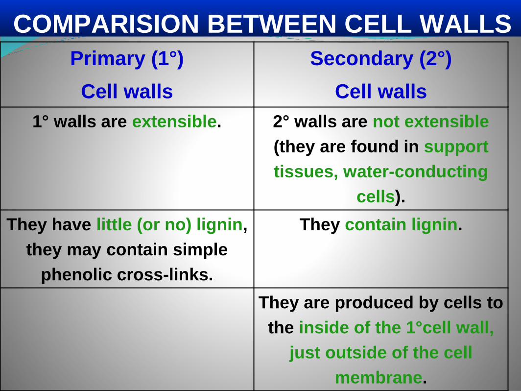
Primary (1°)
Cell walls
Secondary (2°)
Cell walls
1° walls are extensible. 2° walls are not extensible
(they are found in support
tissues, water-conducting
cells).
They have little (or no) lignin,
they may contain simple
phenolic cross-links.
They contain lignin.
They are produced by cells to
the inside of the 1°cell wall,
just outside of the cell
membrane.
COMPARISION BETWEEN CELL WALLS

Cell Wall components
1.Cellulose
2.Other carbohydrates
3.Lignin (other polyphenolics)
4.Proteins
The main components of Cell wall are as follows:

Carbohydrates
•Classified by solubilities
•Pectins – complex carbohydrates extracted in
water using Calcium chelators
oPolyuronic acids
oArabinans
oGlactans
•Hemicelluloses – soluble in 4M KOH
oXylans - common
oMannans – abundant in conifers
oArabinoglactans
•Microfibrillar components
oCellulose
oBeta 1,4 mannans – algae,Beta 1,3 xylans - algae
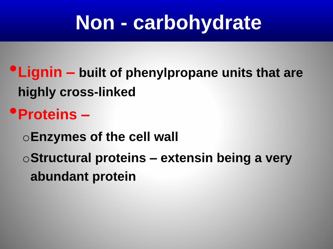
Non - carbohydrate
•Lignin – built of phenylpropane units that are
highly cross-linked
•Proteins –
oEnzymes of the cell wall
oStructural proteins – extensin being a very
abundant protein

The Cell wall is made of two or three structurally
independent networks
These networks interlace.
•Cellulose and cross-linking glycans
•Pectic polysaccharides
•Structural proteins and/or phenylpropanoid
network
.
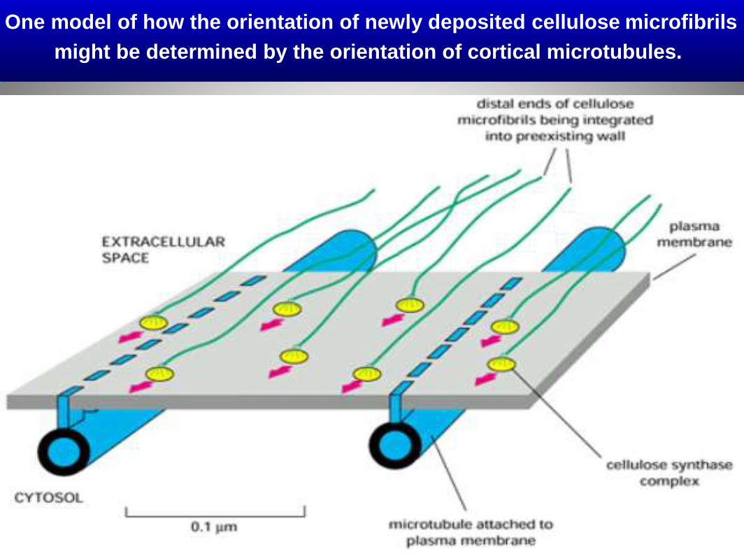
One model of how the orientation of newly deposited cellulose microfibrils
might be determined by the orientation of cortical microtubules.

The large cellulose synthase complexes are integral membrane proteins that continuously synthesize cellulose microfibrils on the outer face of the plasma membrane. The distal ends of the stiff microfibrilsbecome integrated into the texture of the wall, and their elongation at the proximal end pushes the synthase complex along in the plane of the membrane. Because the cortical array of microtubules is attached to the plasma membrane in a way that confines this complex to defined membrane channels, the microtubule orientation determines the axis along which the microfibrils are laid down.
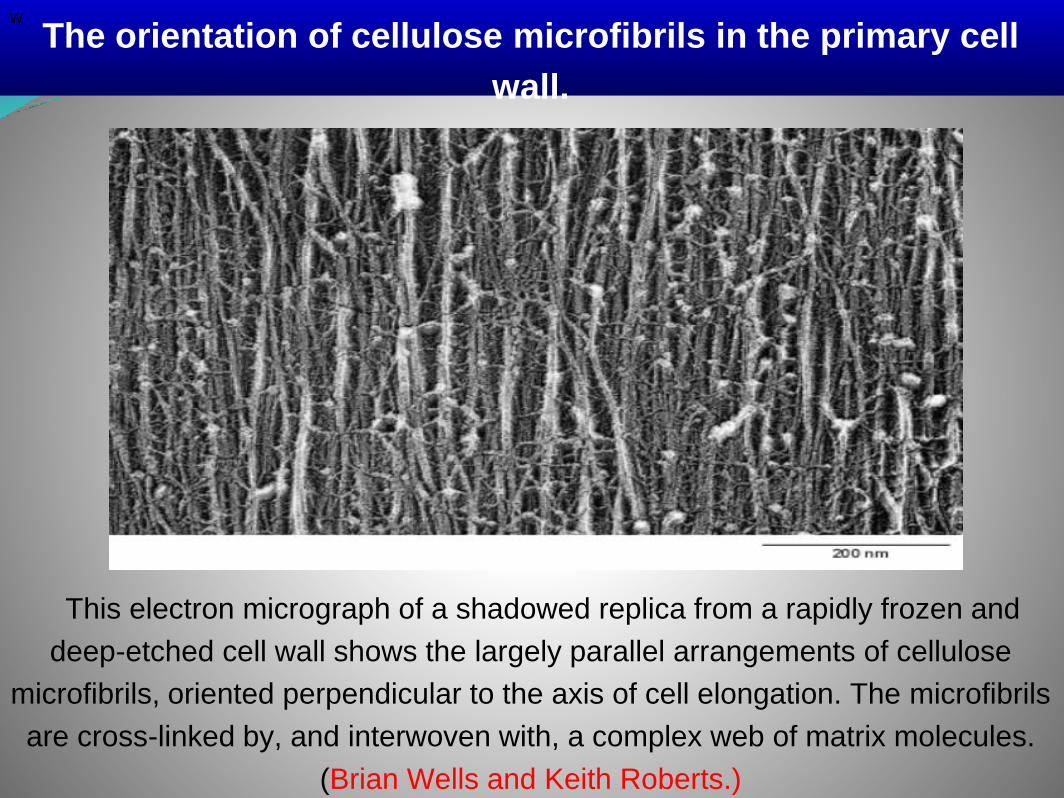
This electron micrograph of a shadowed replica from a rapidly frozen and
deep-etched cell wall shows the largely parallel arrangements of cellulose
microfibrils, oriented perpendicular to the axis of cell elongation. The microfibrils
are cross-linked by, and interwoven with, a complex web of matrix molecules.
(Brian Wells and Keith Roberts.)
w
The orientation of cellulose microfibrils in the primary cell
wall.
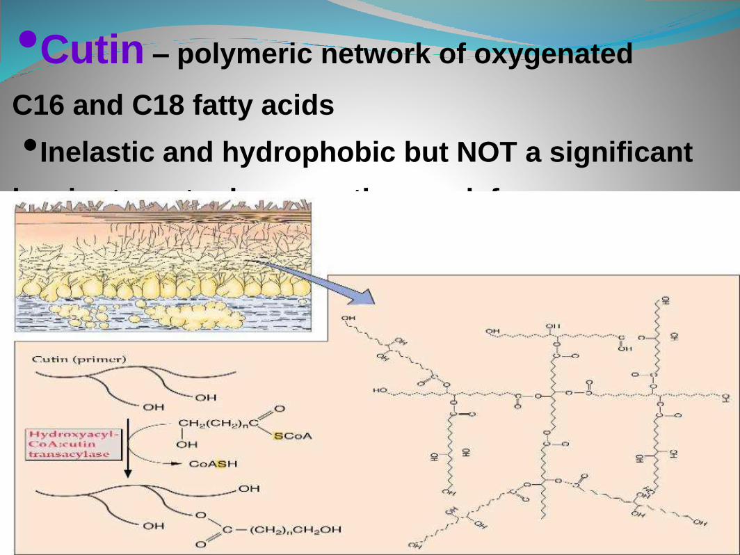
•Cutin – polymeric network of oxygenated
C16 and C18 fatty acids
•Inelastic and hydrophobic but NOT a significant
barrier to water loss – pathogen defense – yes

Suberin – similar but longer fatty acids, less oxygenated
And linked to phenolics – more hydrophobic than cutin
Aerial surfaces covered with waxes – extremely long
Chain fatty acids – prevents water loss

Lignin- very resistant to solubilization
40% HCl used
Made of linked phenylpropane units.
An example of
a “lignan”
component

•Is an important source of strength (rigidity) for plant cells
and, as such, supports the shape of plant cells, tissues and
organs. It is also an important barrier to pathogens and
insects.
•They generally try to breach that barrier by producing and
secreting cell wall-degrading enzymes.
•However, because the barrier is made of sugars and amino
acids, the wall itself is also food for insects and pathogens.
•Because of its composition, the wall is also a potentially
important feedstock for production of biofuels
…particularly if we can learn to operate like insects and
pathogens and take it apart efficiently.
Functions of plant cell wall

Cell walls
1.Plant cell walls provide protection against
abrasion, osmotic stress, and pathogens.
2.Microfibrils of cellulose form the fibrous
component of the cell wall.
C. The matrix of cell wall contains hemicellulose,
pectins, and hydroxyproline-rich, proline-rich, and
glycine-rich structural proteins.