Central mucoepidermoid carcinoma with an atypicalradiographic appearanceBrad Johnson, DMD,a and Ines Velez, DDS, MS,b Fort Lauderdale, FloridaCOLLEGE OF DENTAL MEDICINE, NOVA SOUTHEASTERN UNIVERSITY
Central mucoepidermoid carcinoma (CMC) is a rare osseous neoplasm occurring most often in the fourth toseventh decades of life. Its usual presentation is one of a locally destructive radiolucent lesion found primarily in themandible, in the premolar region. No reported case to date has shown a mixed radiolucent-radiopaque expression ofthis tumor. The objective of this article is to report such a case: a CMC that presented an atypical radiographic
appearance of a mixed lesion. (Oral Surg Oral Med Oral Pathol Oral Radiol Endod 2008;106:e51-e53)
Mucoepidermoid carcinoma is a malignant neoplasm ofsalivary gland origin. It accounts for about 15% ofsalivary gland tumors, with most of them arising withinthe parotid gland. Central osseous origin of this tumoris exceeding rare, representing only about 2%-4% of allmucoepidermoid carcinomas.1 These central mucoepi-dermoid carcinomas (CMC) have a 2:1 predilection forthe mandible, and one-half are associated with im-pacted teeth.2
Although the exact pathogenesis of this lesion isunknown, several current theories of its origin do exist:
1) Ectopic salivary gland tissue: embryonic salivarygland remnants included within the mandible; en-trapment of mucous glands within the bone.
2) Transformation of mucous cells found in odonto-genic cysts.
3) Maxillary sinus or submucosal mucous glands withintraosseous extension.
The clinical presentation of this tumor is varied. Symp-toms of CMC may include pain, swelling, movement ofteeth, and even altered sensation of the inferior alveolarnerve for a long-standing lesion. Most of the time,however, CMC is asymptomatic and an incidental find-ing on a dental radiograph.
The radiographic presentation of this tumor usu-ally reveals a well defined unilocular, lobulated, ormultilocular radiolucency. Root resorption and dis-placement of the teeth are not uncommon. In long-standing cases, this tumor may also exhibit corticalperforation.3
In an extensive review of the English-language lit-erature, we found no reported cases to date of a mixedradiolucent/radiopaque presentation of CMC. To theauthors’ knowledge, all of the reported cases of CMChave presented a radiolucent image.
To be classified as a CMC, the following criteriahave been suggested to establish the diagnosis:
1) Intact cortical bone.2) Radiographic evidence of bony destruction.3) Uninvolved mucous membrane overlying the lesion.4) Absence of primary salivary gland or odontogenic
tumors.5) Histopathologic evidence of CMC.4
Once diagnosed, most CMCs are of low or intermediategrade.5 Current surgical treatment of choice is blockresection. Brookstone and Huvos reported 40% recur-rence after enucleation and marginal resection and 4%recurrence after segmental resection.6
CASE REPORTA 40-year-old African-American woman presented with a
chief complaint of severe pain in the anterior maxilla associ-ated with teeth #8–#11. The patient denied any history oftrauma or recent signs of infection. The symptoms had anabrupt onset and had increased over time. The patient’s fullmedical and surgical history was reviewed and proved to beunremarkable.
Upon full clinical examination, no gross abnormalitieswere identified. All of the maxillary anterior teeth testedcompletely vital. The area was covered by normal mucosaand exhibited no osseous expansion. There was no notedlymphadenopathy. The only positive finding was pain topercussion to teeth #9–#11.
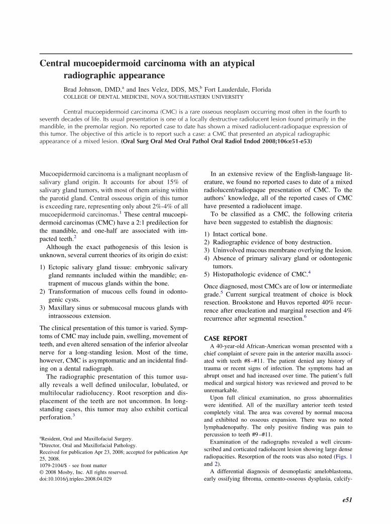
Examination of the radiographs revealed a well circum-scribed and corticated radiolucent lesion showing large denseradiopacities. Resorption of the roots was also noted (Figs. 1and 2).
A differential diagnosis of desmoplastic ameloblastoma,
early ossifying fibroma, cemento-osseous dysplasia, calcify-
e51
OOOOEe52 Johnson and Velez October 2008
ing epithelial odontogenic tumor, and calcifying odontogeniccyst was considered, owing to the radiographic expression ofthis lesion.
At the next appointment an incisional biopsy was per-formed and sent for microscopic examination.
Pathologic examinationThe biopsy was performed in a clinical setting. A palatal
flap was raised and a bony window procured with a #4 roundbur and drill. There was no cortical perforation of the bone aswitnessed by direct visualization.
Gross examination of the biopsy specimen revealed it to bea small lesion that was found at the apex of teeth #9–#10. Itappeared as an irregular soft tissue mass interspersed with alarge amount of hard tissue.
The hematoxylin-eosin slides exhibited very dense scle-rotic bone with reversal lines. It was intermixed within a softtissue matrix composed of solid and cystic isles of epidermoidepithelium, intermediate cells, and mucous cells. A diagnosisof CMC was rendered (Figs. 3 and 4). Owing to the atypicalpresentation, confirmation of the diagnosis was obtained fromthe Armed Forces Institute of Pathology.
DISCUSSIONAberrant salivary gland tissue may be found in
Fig. 1. Well circumscribed and corticated radiolucent lesionwith dense radiopacities and resorption of the roots.
numerous regions of the body to include lymph
nodes, ear, thyroglossal duct, and pituitary gland.However, central salivary gland tumors are ex-
Fig. 2. Well circumscribed and corticated radiolucent lesionwith dense radiopacities and resorption of the roots.
Fig. 3. Dense sclerotic bone with islands of tumor. Hema-toxylin and eosin, �10.
tremely rare.
OOOOEVolume 106, Number 4 Johnson and Velez e53
Mucoepidermoid carcinomas comprise between 4%and 8% of all salivary gland tumors and present con-siderable variation between cases. Central mucoepider-moid carcinoma constitutes less than 2%-4% of allmucoepidermoid tumors.7 They are composed of epi-dermoid, intermediate, and mucous cells. These inter-mediate cells form the primary dividing cell in theseneoplasms and appear to be able to differentiate intoboth mucous and epidermoid cells. At the ultrastruc-tural level, it has been suggested that this tumor holdsthe capability for differentiation into a wide variety ofcell types.8 Perhaps it is this capability that allows forthe potential to form hard tissue as well.
Central mucoepidermoid carcinoma usually exhibitsa rapid clinical onset. Osseous expansion, destructionof local structures, and pain may be expressed in aslittle as 1 year. It generally is only locally aggressive,but metastases can occur and are usually limited to the
Fig. 4. Sclerotic vital bone showing fragments of mucoepi-dermoid carcinoma. Hematoxylin and eosin, �40.
pression of CMC consistently illustrates destruction ofbone with a multilocular or cystic-like radiolucency.We now know that there is also a potential for thistumor to form hard tissue and be expressed as a mixedlesion as well.
In conclusion, CMC is a rare but serious pathologicentity which is capable of variable radiographic expres-sion. We should now consider this neoplasm as a pos-sibility when diagnosing mixed radiolucent/radiopaquelesions.
REFERENCES1. Pires FR, Paes de Almeida O, Lopes MA, Elias Da Cruz Perez D.
Central mucoepidermoid carcinoma of the mandible: report offour cases. Int J Oral Maxillofac Surg 2003;32:378-82.
2. Eversole LR. Mucoepidermoid carcinoma: review of 815 reportedcases. Oral Surg Oral Med Oral Pathol Oral Radiol Endod1970;28:490-5.
3. Freje JE, Campbell BH, Yousif WJ, Clowry LJ. Central muco-epidermoid carcinoma of the mandible. Otolaryngol Head NeckSurg 1995;112:453-6.
4. Alexander RW, Dupuis RH, Holton H. Central mucoepidermoidtumor (carcinoma) of the mandible. J Oral Surg 1974;32:541-7.
5. Browand BC, Waldron CA. Central mucoepidermoid tumors ofthe jaws. Oral Surg Oral Med Oral Pathol Oral Radiol Endod1975;40:631-43.
6. Brookstone MS, Huvos AG. Central salivary gland tumors ofmaxilla and mandible: report of 11 cases and analysis of theliterature. J Oral Maxillofac Surg 1992;50:229-36.
7. Gingell JC, Beckerman T, Levy BA, Snider LA. Central muco-epidermoid carcinoma: review of the literature and report of a caseassociated with an apical periodontal cyst. Oral Surg Oral MedOral Pathol Oral Radiol Endod 1984;57:436-40.
8. Dardick I. Salivary gland tumor pathology. New York: Igaku-Shoin; 1996. p. 163-76.
Reprint requests:
Brad Johnson, DMDOral and Maxillofacial SurgeryCollege of Dental MedicineNova Southeastern University3200 S. University Dr.Fort Lauderdale, FL 33328