24
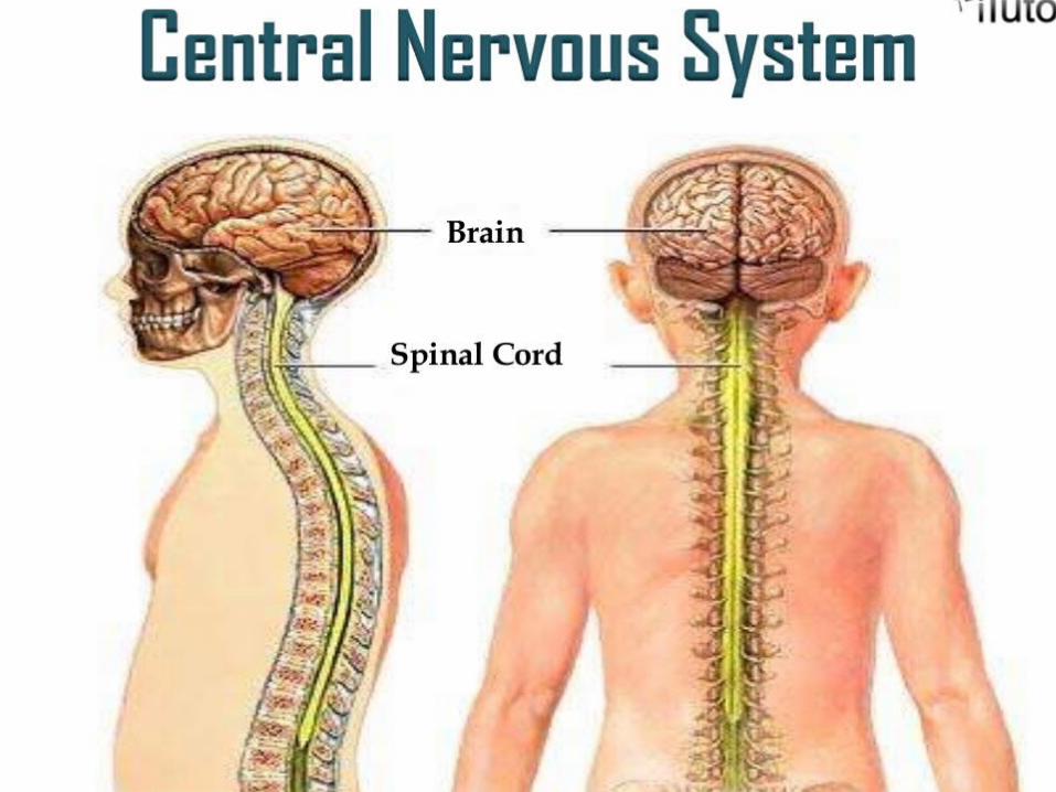
CENTRAL NERVOUS SYSTEM Sarita Sharma Assistant professor of pharmacology Mumbai
| Date post: | 11-Apr-2017 |
| Category: |
Science |
| Upload: | sarita1916 |
| View: | 46 times |
| Download: | 0 times |

CENTRAL NERVOUS SYSTEM
Sarita Sharma Assistant professor of pharmacology Mumbai

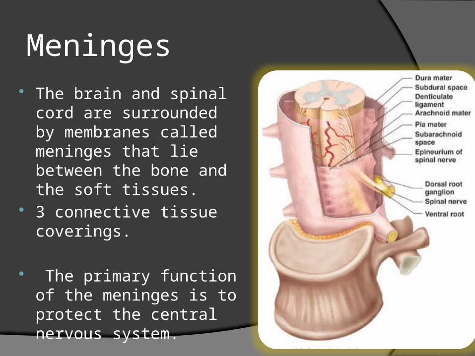
Meninges The brain and spinal cord
are surrounded by membranes called meninges that lie between the bone and the soft tissues.
3 connective tissue coverings.
The primary function of the meninges is to protect the central nervous system.
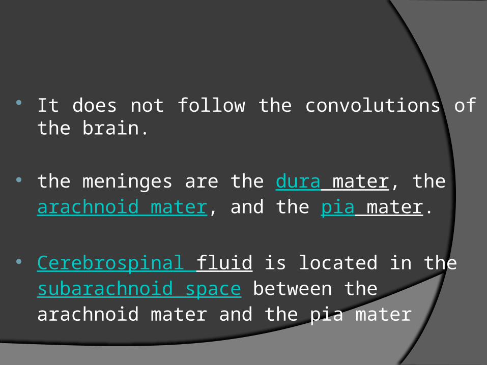
It does not follow the convolutions of the brain. the meninges are the dura mater, the
arachnoid mater, and the pia mater.
Cerebrospinal fluid is located in the subarachnoid space between the arachnoid mater and the pia mater


1.Dura mater Thick, outermost fibrous membrane, Lined the
interior of the skull Composed of dense, irregular connective tissues.
It forms a sec from the level of the foramen magnum in the occipital bone, where it continuous with the duramater of brain, to the 2nd secral vertebra.
The spinal cord is also protected by a cushion of fat & connective tissue located into the epidural spaces(a space between the duramater & the wall of the vertibral canal)
2. Arachnoid mater Middle transparent layer Lies between the duramater & piamater
Between the arachnoid and pia maters is a subarachnoid space containing cerebrospinal fluid(CSF).

3. Pia mater
The innermost layer Closely covers the brain & spinal cord It also dips into the fissures of the brain Contain the minute blood vessels which
supply the brain & spinal cord
Spinal cord Runs through the vertebral canal The spinal cord begins at the base of the brain (medulla
oblongata)and extends as a slender cord to the level of superior border of 2nd lumbar vertebrae.
Adult spinal cord is about 42-45 cm long. Regions
Cervical Thoracic LumbarSacralCoccygeal
CSF presant in central canal of spinal cord & in the subarachnoid space

Has 2 enlargements which are seen on external observations
Cervical enlargement
Extenting from 4th cervical vertibra to 1st thoracic vertibra
corresponds with the attachments of the large nerves which supply the
upper limbs.
Lumber enlargement
Extenting from 9th thoracic vertibra to 12th thoracic vertibra
gives attachment to the nerves which supply the lower limbs.


Internal anatomy of spinal cord
The spinal cord can be anatomically divided into 31 spinal segments based on the origins of the spinal nerves.
Each segment of the spinal cord is associated with a pair of ganglia, called dorsal root ganglia, which are situated just outside the spinal cord. These ganglia contain cell bodies of sensory neurons. Axons of these sensory neurons travel into the spinal cord via the dorsal roots.
Ventral roots consist of axons from motor neurons, which bring information to the periphery from cell bodies within the CNS. Dorsal roots and ventral roots come together and exit the intervertebral foramina as they become spinal nerves.
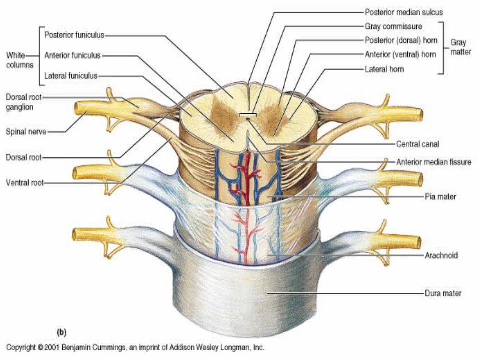


(a) Gray mater The grey column, in the center of the cord, is shaped
like a butterfly and consists of cell bodies of interneurons, motor neurons, neuroglia cells and unmyelinated axons.
The anterior and posterior grey column present as projections of the grey matter and are also known as the horns of the spinal cord form the "grey H.“
In the center of the gray commissure is a small space called “center canal”, it extends the entire length of spinal cord & filled with CSF.
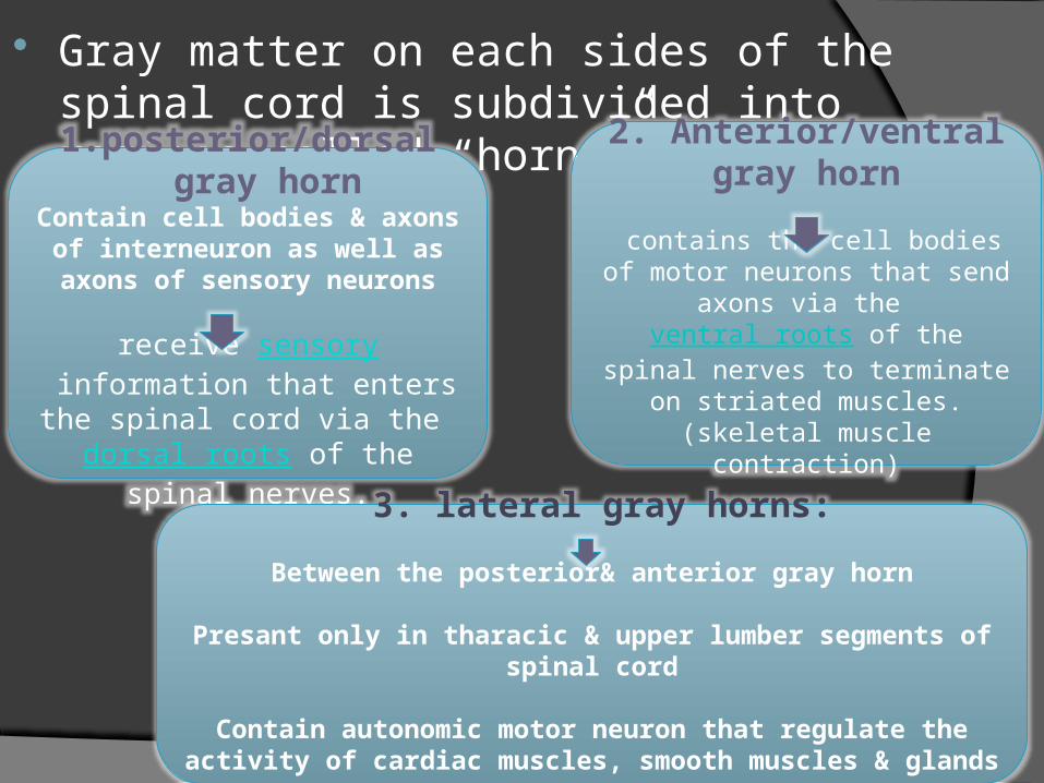
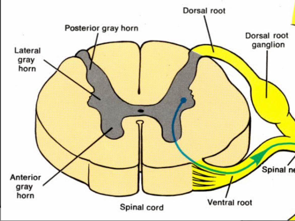
Gray matter on each sides of the spinal cord is subdivided into regions called “horns”. 2 types
1. posterior/dorsal gray horn
Contain cell bodies & axons of interneuron as well as axons of
sensory neurons
receive sensory information that enters the spinal cord via the
dorsal roots of the spinal nerves.
2. Anterior/ventral gray horn
contains the cell bodies of motor neurons that send axons via the
ventral roots of the spinal nerves to terminate on striated muscles. (skeletal muscle contraction)
3. lateral gray horns:
Between the posterior& anterior gray horn
Presant only in tharacic & upper lumber segments of spinal cord
Contain autonomic motor neuron that regulate the activity of cardiac muscles, smooth muscles & glands
(b)White mater The white matter is located outside of the grey matter and
consists almost totally of myelinated motor and sensory axons. "Columns" of white matter carry information either up or down the spinal cord.(white color due to myelin)
Like gray mater it is also divided in to 3 parts 1. Anterior/ventral white columns 2. posterior/dorsal white columns 3. lateral white columns Each column contain different bundles of axons
having common origin or destination & carrying similar information

Processing of sensory input and motor output by the spinal cord.

Reflex action/ reflex arc

Spinal nerves: Spinal nerves are the path of communication
between spinal cord & specific regions of the body
31 pairs of spinal nerves are presant & they connected with spinal cord by “Roots”.
2 types of rootsPosterior or dorsal roots(sensory roots)
Contain sensory nerve fibres
Conduct nerve impulse from periphery to spinal
cord
Anterior or ventral roots(Motor roots)
Contain motor nerons
Conduct nerve impulse from spinal cord to periphery

spinal nerve is a mixed nerve, which carries motor, sensory, and autonomic signals between the spinal cord and the body.
In the human body there are 31 pairs of spinal nerves, one on each side of the vertebral column. These are grouped into the corresponding cervical, thoracic, lumbar, sacral and coccygeal regions of the spine.
(1) cervical nerves = 8 pairs (2) thoracic nerves = 12 pairs (3) lumbar nerves = 5 pairs (4) sacral nerves= 5 pairs (5) coccygeal nerves = 1 pairs
Naming of spinal nerves are according
to the segment in which they are located

Functions of the Spinal Cord The spinal cord has two major functions: to
transmit impulses to and from the brain, and to house spinal reflexes.
Tracts carrying sensory information to the brain are called ascending tracts; descending tracts carry motor information from the brain.
The names that identify nerve tracts identify the origin and termination of the fibers in the tract.
Many spinal reflexes also pass through the spinal cord.


