Cancer Investigation, 30:404–414, 2012 ISSN: 0735-7907 print / 1532-4192 online Copyright C 2012 Informa Healthcare USA, Inc. DOI: 10.3109/07357907.2012.672844 CELLULAR AND MOLECULAR BIOLOGY Centroblastic Diffuse Large B Cell Lymphoma Displays Distinct Expression Pattern and Prognostic Role of Apoptosis Resistance Related Proteins Roberta Soares Faccion, 1–3 Lidia Maria Magalh ˜ aes Rezende, 4 S´ ergio de Oliveira Romano, 4 Ricardo de S ´ a Bigni, 5 Gelcio Luiz Quintella Mendes, 6 and Raquel Ciuvalschi Maia 1,5, ∗ Laborat´ orio de Hemato-Oncologia Celular e Molecular, Programa de Pesquisa em Hemato-Oncologia Molecular, Coordenac ¸˜ ao Geral T´ ecnico-Cient´ ıfica, Instituto Nacional de Cˆ ancer (INCA), Rio de Janeiro, Brazil, 1 Programa de P´ os-graduac ¸˜ ao em Ciˆ encias Morfol´ ogicas, Instituto de Ciˆ encias Biom´ edicas, Centro de Ciˆ encias da Sa´ ude, Universidade Federal do Rio de Janeiro, Rio de Janeiro, Brazil, 2 Programa de P´ os-Graduac ¸˜ ao em Ciˆ encias Fisiol´ ogicas, Instituto de Biof´ ısica Carlos Chagas Filho, Centro de Ciˆ encias da Sa´ ude, Universidade Federal do Rio de Janeiro, Rio de Janeiro, Brazil, 3 Divis˜ ao de Anatomia Patol´ ogica, Coordenac ¸˜ ao Geral de Gest˜ ao Assistencial, INCA, Rio de Janeiro, Brazil, 4 Servic ¸o de Hematologia, Hospital do Cˆ ancer I, Coordenac ¸˜ ao Geral de Gest˜ ao Assistencial, INCA, Rio de Janeiro, Brazil, 5 Servic ¸o de Oncologia, Hospital do Cˆ ancer I, Coordenac ¸˜ ao Geral de Gest˜ ao Assistencial, INCA, Rio de Janeiro, Brazil 6 Centroblastic diffuse large B cell lymphoma (DLBCL) samples were analyzed by immunohistochemistry to evaluate the expression of p53, Bcl-2, Survivin, XIAP, and Ki-67. Survivin was the only protein which expression exhibited a trend for impact in progression-free (p = .077) and overall survival (p = .054). In the Mann–Whitney test, Survivin expression correlated with a negative overall survival (p = .045). These results appeared to be intimately related to Survivin cytoplasmic localization. Moreover, the anti-apoptotic proteins Bcl-2 and Survivin were less frequent in centroblastic DLBCL. Our results indicate that centroblastic DLBCL may be a disease with characteristic biology and clinical course and, therefore, specific prognostic factors. Keywords: Centroblastic diffuse large B cell lymphoma; p53; Bcl-2; Survivin; XIAP; Ki-67 co-expression; Prognosis; Immunohistochemistry INTRODUCTION Diffuse large B-cell lymphomas (DLBCL) are the most com- mon type of high-grade non-Hodgkin lymphoma (NHL), accounting for 30% of NHL. It comprises an aggressive and heterogeneous group of B-cell NHL, which differ in terms of morphology, gene expression profile, and treatment outcome. The most common morphological entities that can be distinguished among DLBCL are the centroblastic and immunoblastic subtypes, which are currently equally treated (1). However, centroblastic DLBCL is not only more common than immunoblastic DLBCL, but is also more Correspondence to: Raquel C. Maia, MD, PhD, Laborat´ orio de Hemato-Oncologia Celular e Molecular, Programa de Pesquisa em Hemato-Oncologia Molecular, Coordenac ¸˜ ao Geral T´ ecnico-Cient´ ıfica, Instituto Nacional de Cˆ ancer, Prac ¸a da Cruz Vermelha 23, 6 ◦ andar, Centro, Rio de Janeiro, RJ, 20230-130 Brazil. email: [email protected]susceptible to treatment (2–4). Moreover, centroblastic DLBCL is thought to arise from centroblasts—proliferating germinal center B lymphocytes—and hence, is denoted as centroblastic DLBCL. On the other hand, immunoblastic DLBCL is thought to arise from immunoblasts—positive- selected post-germinal center B lymphocytes—and hence, is termed as immunoblastic DLBCL (5, 6). Owing to the sub- types heterogeneity in terms of clinical course and molecular features, there is a debate on whether these variants should be regarded as different diseases, with adjusted treatment and independent research attention for each subtype. One of the limitations that has led to the inclusion of both the subtypes in the same category is that the distinction be- tween them is morphological, and therefore, can be subjec- tive. However, perhaps, the main limitation is the absence of conclusive studies on the differences between them with regard to treatment response (7). On the other hand, nowa- days, immunohistochemistry represents an important tool in routine services to aid in the discrimination of DLBCL sub- types when the morphology is doubtful (8, 9). Furthermore, several long-term follow-up studies have indeed shown that immunoblastic DLBCL has a worse prognosis than centrob- lastic DLBCL, although during the first two years after diag- nosis, this difference might not be obvious (2–4, 10). Recently, a correlation between the ABC microar- ray/immunohistochemistry classification and immunoblas- tic morphology has been identified (11, 12). Furthermore, at this stage of maturation, the lymphocyte is associated with a prosurvival expression profile (5, 6). However, it has not been possible to correlate the centroblastic subtype to the Cancer Invest Downloaded from informahealthcare.com by University of Sussex Library on 02/23/13 For personal use only.
Centroblastic Diffuse Large B Cell Lymphoma Displays DistinctExpression Pattern and Prognostic Role of Apoptosis Resistance RelatedProteins
Roberta Soares Faccion,1–3 Lidia Maria Magalhaes Rezende,4 Sergio de Oliveira Romano,4
Ricardo de Sa Bigni,5 Gelcio Luiz Quintella Mendes,6 and Raquel Ciuvalschi Maia1,5,∗
Laboratorio de Hemato-Oncologia Celular e Molecular, Programa de Pesquisa em Hemato-Oncologia Molecular, CoordenacaoGeral Tecnico-Cientıfica, Instituto Nacional de Cancer (INCA), Rio de Janeiro, Brazil,1 Programa de Pos-graduacao em CienciasMorfologicas, Instituto de Ciencias Biomedicas, Centro de Ciencias da Saude, Universidade Federal do Rio de Janeiro, Rio deJaneiro, Brazil,2 Programa de Pos-Graduacao em Ciencias Fisiologicas, Instituto de Biofısica Carlos Chagas Filho, Centro deCiencias da Saude, Universidade Federal do Rio de Janeiro, Rio de Janeiro, Brazil,3 Divisao de Anatomia Patologica,Coordenacao Geral de Gestao Assistencial, INCA, Rio de Janeiro, Brazil,4 Servico de Hematologia, Hospital do Cancer I,Coordenacao Geral de Gestao Assistencial, INCA, Rio de Janeiro, Brazil,5 Servico de Oncologia, Hospital do Cancer I,Coordenacao Geral de Gestao Assistencial, INCA, Rio de Janeiro, Brazil6
Centroblastic diffuse large B cell lymphoma (DLBCL) sampleswere analyzed by immunohistochemistry to evaluate theexpression of p53, Bcl-2, Survivin, XIAP, and Ki-67. Survivin wasthe only protein which expression exhibited a trend for impactin progression-free (p = .077) and overall survival (p = .054). Inthe Mann–Whitney test, Survivin expression correlated with anegative overall survival (p = .045). These results appeared tobe intimately related to Survivin cytoplasmic localization.Moreover, the anti-apoptotic proteins Bcl-2 and Survivin wereless frequent in centroblastic DLBCL. Our results indicate thatcentroblastic DLBCL may be a disease with characteristicbiology and clinical course and, therefore, specific prognosticfactors.
Keywords: Centroblastic diffuse large B cell lymphoma; p53;Bcl-2; Survivin; XIAP; Ki-67 co-expression; Prognosis;Immunohistochemistry
INTRODUCTION
Diffuse large B-cell lymphomas (DLBCL) are the most com-mon type of high-grade non-Hodgkin lymphoma (NHL),accounting for 30% of NHL. It comprises an aggressiveand heterogeneous group of B-cell NHL, which differ interms of morphology, gene expression profile, and treatmentoutcome. The most common morphological entities thatcan be distinguished among DLBCL are the centroblasticand immunoblastic subtypes, which are currently equallytreated (1). However, centroblastic DLBCL is not only morecommon than immunoblastic DLBCL, but is also more
Correspondence to: Raquel C. Maia, MD, PhD, Laboratorio de Hemato-Oncologia Celular e Molecular, Programa de Pesquisa emHemato-Oncologia Molecular, Coordenacao Geral Tecnico-Cientıfica, Instituto Nacional de Cancer, Praca da Cruz Vermelha 23, 6◦ andar,Centro, Rio de Janeiro, RJ, 20230-130 Brazil. email: [email protected]
susceptible to treatment (2–4). Moreover, centroblasticDLBCL is thought to arise from centroblasts—proliferatinggerminal center B lymphocytes—and hence, is denoted ascentroblastic DLBCL. On the other hand, immunoblasticDLBCL is thought to arise from immunoblasts—positive-selected post-germinal center B lymphocytes—and hence, istermed as immunoblastic DLBCL (5, 6). Owing to the sub-types heterogeneity in terms of clinical course and molecularfeatures, there is a debate on whether these variants shouldbe regarded as different diseases, with adjusted treatmentand independent research attention for each subtype.
One of the limitations that has led to the inclusion of boththe subtypes in the same category is that the distinction be-tween them is morphological, and therefore, can be subjec-tive. However, perhaps, the main limitation is the absenceof conclusive studies on the differences between them withregard to treatment response (7). On the other hand, nowa-days, immunohistochemistry represents an important tool inroutine services to aid in the discrimination of DLBCL sub-types when the morphology is doubtful (8, 9). Furthermore,several long-term follow-up studies have indeed shown thatimmunoblastic DLBCL has a worse prognosis than centrob-lastic DLBCL, although during the first two years after diag-nosis, this difference might not be obvious (2–4, 10).
Recently, a correlation between the ABC microar-ray/immunohistochemistry classification and immunoblas-tic morphology has been identified (11, 12). Furthermore, atthis stage of maturation, the lymphocyte is associated witha prosurvival expression profile (5, 6). However, it has notbeen possible to correlate the centroblastic subtype to the
Can
cer
Inve
st D
ownl
oade
d fr
om in
form
ahea
lthca
re.c
om b
y U
nive
rsity
of
Suss
ex L
ibra
ry o
n 02
/23/
13Fo
r pe
rson
al u
se o
nly.
Apoptosis Related Proteins in Centroblastic DLBCL
GCB microarray/immunohistochemistry classification (11,12). In addition, the centroblastic subtype has been charac-terized by a proliferative gene expression profile (5, 6). Onthe other hand, it has not been well characterized accordingto survival-related expression profile. We therefore decidedto investigate the expression of apoptosis-related proteins inthis subtype of proteins that not only are involved in apop-tosis, but have also been reported to impact DLBCL biol-ogy, such as Survivin and XIAP; caspase inhibitors, IAPs(Inhibitor of Apoptosis Proteins) (13), Bcl-2, mitochondrialmembrane depolarization inhibitor, p53, cell cycle controller,and apoptosis inductor; and ki-67, a proliferation-associatedantigen (14).
Moreover, as the immunoblast has a prosurvival profile,the expression of some of these apoptosis resistance relatedproteins might be associated with this DLBCL variant, whichhas a worse prognosis than the centroblastic variant. There-fore, when including both the variants in a DLBCL prognos-tic factor quest, a worse prognosis value of a given apoptosisresistance related protein might actually merely reflect theworse prognosis value of the immunoblastic subtype. Cur-rently, the only parameter used to access DLBCL prognosis isthe International Prognostic Index (IPI) (15). Furthermore,therapy provides long-term overall survival of as much as halfof the patients (1, 16, 17). Therefore, there is a need to identifyand establish biological prognostic factors that can help im-prove risk stratification and function as targets for new thera-pies. In the past decade, the expression of several proteins hasbeen studied as possible prognostic biomarkers for DLBCL.The classical candidates are proteins that play crucial roles inthe cell cycle (e.g., the cell cycle guardian p53 and the prolif-eration antigen ki-67 (14)) and apoptotic pathways (e.g., theantiapoptotic members of the Bcl-2 family, such as Bcl-2 itself(14) and the caspase inhibitors, XIAP, and Survivin (13)).
Because of the limited knowledge regarding the expres-sion of apoptosis resistance related proteins in centroblasticDLBCL, we addressed this issue in a group of 81 centroblasticDLBCL cases with the aim of understanding p53, Ki-67, Bcl-2, XIAP, and Survivin expression and features, analyzed usingimmunohistochemistry, and also evaluated their prognosticvalue in centroblastic DLBCL.
MATERIAL AND METHODS
Patients’ selectionFrom the 191 adult lymphoma cases registered at the Na-tional Cancer Institute (Rio de Janeiro, Brazil) between 1989and 1993, a total of 81 adult centroblastic DLBCL patientsand 6 adult immunoblastic DLBCL patients were enrolledfor the present study. They were selected to enter the analysisbased on having DLBCL with confirmed morphology, avail-able paraffin-embedded tumor sample(s), and adequatelong-term follow-up information. The demographic andclinical data were obtained from the patients’ records. Outof the 81 centroblastic DLBCL patients and of the 6 im-munoblastic DLBCL patients, 69 and 5, respectively, receivedanthracycline-based chemotherapy protocols, e.g., CHOP orCNOP. Progression-free survival (PFS) and overall survival
(OS) were calculated based on the clinical information re-trieved from their medical charts. The PFS and OS were notevaluated in the other 13 patients because their treatmentwas heterogeneous. The local Institutional Ethic Committeeapproved this study, which was conducted in accordance withthe recommendations of the Helsinki Declaration.
ImmunohistochemistryTumor samples were collected at diagnostic biopsies. Thecentroblastic DLBCL diagnosis was confirmed by twopathologists (L.M. Rezende and S.O. Romano) indepen-dently and based on standard morphological parameters, fol-lowing the current WHO classification parameters (18). Theimmunohistochemical analysis was adopted from a previ-ous work of our group (19). Briefly, 4 µm of formalin-fixedparaffin-embedded sample sections were deparaffinized inxylene and rehydrated in ethanol baths. Bcl-2, XIAP, and Sur-vivin antigenic retrieval was performed in a steamer with acitrate buffer of pH 6.0 for 30 min at 98◦C. p53 and Ki-67 anti-genic retrieval was performed with the same buffer, but in apressure cooker for 3 min. Endogenous peroxidase and non-specific antibody labeling were blocked with 3% hydrogenperoxide and a blocking solution, respectively. Tumor slideswere incubated overnight at 4◦C with anti-p53 (clone DO-7Dako), anti-Ki-67 (MIB Dako), anti-Bcl-2 (clone 124 Dako),anti-XIAP (Sigma-Aldrich X4503), or anti-Survivin antibod-ies (Sigma-Aldrich S8191). As the detection system, a labeledstreptavidin biotin method with a coupled HRP-peroxidase(LSAB2-Dako) was employed. After 3,3’ diaminobenzidinetetrahydrochloride (DAB) staining, Harris hematoxylin wasused for a slight counterstaining. The positive control forXIAP and Survivin expression was a normal stomach mu-cosa, and for p53, Bcl-2, and Ki-67 expression, previouslydetermined positive tumor samples were utilized. As a nega-tive control, the primary antibody was omitted. When morethan one sample was available for a given patient, immuno-histochemistry was performed in both the samples to eval-uate possible discrepancies between samples from the sametumor.
Immunostaining results were analyzed by two indepen-dent observers and registered in an Eclipse E200 Nikon mi-croscope connected to a Digital Sight System. For p53, Bcl-2,XIAP and Survivin, cases with fewer than 5% of positive tu-mor cells were considered negative, and cases with 5% ormore positive tumor cells were considered positive. For ki-67antigen, the cutoff of positivity was defined as 60% or moreof positive tumor cells. Scoring analysis was performed in atleast 10 fields in a 40 × magnification. Subcellular localiza-tion of XIAP and Survivin was evaluated as nuclear and/orcytoplasmic in all positive samples.
Statistical analysisStatistical analysis was performed in the SPSS 17.0 soft-ware. PFS was evaluated as the time between the diagno-sis and the progression of the disease. The events regardingPFS were considered progression of the disease (for the non-responsive patients), relapse (for patients who achieved com-plete remission), or disease-related death. The remaining
cases were censored at the last follow-up. OS was evaluated asthe time between the diagnosis and the end of the study. Theevent regarding OS was considered as disease-related death.The remaining cases were censored at the last follow-up.Co-expression of the proteins was evaluated through thePearson χ 2 test with continuity correction when appro-priated. Survival curves were plotted by the Kaplan–Meiermethod. The correlation between treatment response andproteins expression was examined by the log-rank test. For a95% confidence interval, the difference between the analyzedgroups was considered significant when p < .05.
RESULTS
Clinical, demographic, and treatment response dataof centroblastic DLBCL patientsMedian age at diagnosis was 58 years (range: 23–85 years).From the 81 patients included in this study, 12 were nottreated with anthracycline-based protocols and were there-fore excluded from the treatment response analysis. Theremaining 69 patients were treated with anthracycline-basedregimens and were therefore eligible for entering the prog-nostic analysis. PFS and OS ranged from 0 to 166 monthswith the median period being 4 months for PFS and 76months for OS. Although PFS of the centroblastic DLBCLpatients did not significantly differ from immunoblasticDLBCL patients, centroblastic DLBCL patients had betterOS than immunoblastic DLBCL patients (SupplementaryFigure 1). From the clinical and demographic featuresanalyzed, the only characteristic that had an impact on PFSwas age, with patients older than 60 years having a worsePFS than those younger than 60 years (p = .002). Althoughit was not significant, patients with low IPI tended to have abetter PFS (p = .098). Regarding OS, low IPI (p = .042), andnormal LDH level (.046) were favorable prognostic factors.A summary of patients’ characteristics is listed in Table 1.
Apoptosis resistance related proteins expressionin centroblastic DLBCLFor p53, Bcl-2, Ki-67, XIAP, and Survivin expression analysis,it was not possible to access p53 expression in 4 cases, Bcl-2expression in 6 cases, Ki-67 in 15 cases, and XIAP expressionin 10 cases. p53 expression was observed in 14 cases (18.2%),Bcl-2 expression was observed in 10 cases (13.3%), Ki-67 wasobserved in 41 cases (62.1%), XIAP expression was observedin 23 cases (32.4%), and Survivin expression was observed in19 cases (23.5%). In Table 2, the positivity frequencies foundin centroblastic DLBCL cases in our study, in contrast to thefindings in DLBCL cases from other studies, are shown.
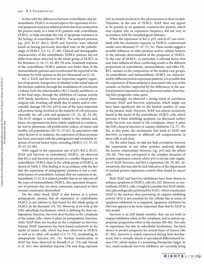
Another feature shown in Table 2 is the subcellular local-ization of the analyzed proteins. In this regard, the expres-sion of p53 and Ki-67 antigen was detected in the nuclei oftumor cells and the expression of Bcl-2 was observed in thecytoplasm of tumor cells in all positive cases. With regard tothe expression of XIAP, it was observed almost always in thenuclei of tumor cells (Figure 1(a)), whereas the expressionof Survivin was detected almost always in the cytoplasm oftumor cells (Figure 1(b)) in all positive cases. In the XIAP
positive cases, we observed that the positive cells usually hada larger and undifferentiated morphology than the negativeones even in the same tissue (Figure 1(a)).
Apoptosis resistance related proteins co-expressionin centroblastic DLBCLAs these proteins have close relations in normal cell biology—e.g., p53 can inhibit the expression of Bcl-2 (20–23), XIAP(24, 25), and Survivin (26, 27), and has a role in cell cycle con-trol (22, 28), whereas Ki-67 is also found throughout the cellcycle in cycling cells (29–31)—we investigated whether p53,Bcl-2, Ki-67, XIAP, and Survivin expressions were associatedwith one another in centroblastic DLBCL. However, althoughthere was a trend for XIAP and Survivin expressions to havea direct correlation (p = .059), there was no significant corre-lation between the expression of any of the analyzed proteins(p > .05) (data not shown).
Proteins expression and treatment outcome of centroblasticDLBCL patientsAs p53, Bcl-2, Ki-67, XIAP, and Survivin expressions havebeen correlated to a worse survival for DLBCL patients; wetested whether this was true for the centroblastic DLBCL sub-set as well. However, there was no correlation between p53,Bcl-2, Ki-67, and XIAP expression and PFS (p = .968, p =.941, p = .737, and p = .742, respectively) or OS (p = .494,p = .939, p = .816, and p = .936, respectively) (Table 1 andFigure 2).
On the other hand, there was a trend for Survivinexpression to confer a poor PFS (p = .077) and OS (p =.054). Indeed, this tendency persisted when we performedthe Mann–Whitney test to evaluate the correlation betweenthe PFS of the Survivin-positive versus the Survivin-negative patients group (Table 3) (p = .087). In fact, in theMann–Whitney test, Survivin expression correlated to anegative OS (p = .045). In particular, the Mann–Whitneytest also corroborated the log-rank results for p53, Bcl-2,Ki-67, and XIAP expression (Table 3).
DISCUSSION
DLBCL is an aggressive form of NHL and exhibits a greatheterogeneity in terms of molecular features and treat-ment response (1, 14, 32, 33). In the past decade, a greatimprovement has taken place in the treatment of DLBCLpatients with the inclusion of the antibody, anti-CD20, inthe treatment (rituximab) (34–36). However, it is a veryexpensive therapy and currently not all public health careservices are able to include rituximab in their standard NHLtreatment protocols. Anthracycline-based therapy is themost common treatment protocol, irrespective of whetherit includes rituximab or not. Overall, it provides as muchas 50% cure rate for DLBCL patients (1, 16, 17). In terms ofmorphology, this rate is higher for centroblastic DLBCL andlower for immunoblastic DLBCL (2–4), and represents dis-tinct variants among the DLBCL. Although the morphologyis often a subjective classification with the risk of variance
Cancer Investigation
Can
cer
Inve
st D
ownl
oade
d fr
om in
form
ahea
lthca
re.c
om b
y U
nive
rsity
of
Suss
ex L
ibra
ry o
n 02
/23/
13Fo
r pe
rson
al u
se o
nly.
Apoptosis Related Proteins in Centroblastic DLBCL
Table 1. Clinical and Demographic Data of the Centroblastic DLBCL Patients
Feature
All Included Patients –Absolute and RelativeFrequencies (%)
Patients Included in thePrognosis Analysis –Absolute and RelativeFrequencies (%)
PFS: Progression-free survival. OS: overall survival. SE: standard error. Associations were investigated in the Kaplan-Meier plot by the log-rank test. ∗p < .05 was consideredsignificant. N.D.: not determined. N.A.: not applicable.
Table 2. Positivity Frequencies of p53, Bcl-2, ki-67, XIAP and Survivin Expression in Centroblastic DLBCL Found in the Present Study andPositivity Frequencies of These Proteins in DLBCL Found by Other Groups
Positivity Frequecy
SubcellularLocalization
Number of Positive Cases/TotalNumber of Analyzed Cases
CentroblasticDLBCL DLBCL∗ References n∗∗
p53 Nucleus 14/77 18.2% 13–38% (39, 41, 43, 44, 95) 55–372Bcl-2 Cytoplasm 10/75 13.3% 26–61% (39, 41, 43, 44, 95, 96) 39–372ki67 Nucleus 41/66 62.1% 18–100% (37, 43, 45, 95) 55–405XIAP Nucleus 23/71 32.4% 26–67% (13, 70, 96) 38–73Survivin Cytoplasm 19/81 23.5% 52–82% (48, 96–99) 27–222∗Expression frequencies in DLBCL cases as reported by other groups. ∗∗Number of cases analyses in the cited studies.
Figure 1. Imunohistochemical detection of XIAP and Survivin in centroblastic DLBCL cells. XIAP localized in the nucleus (a) while Survivinlocalized in the cytoplasm (b) of the tumoral cells. Positive expression controls were normal stomach mucosa tumor sections both for XIAP (c) andSurvivin (d). Negative tumor cells for both XIAP (e) and Survivin (f) exhibit the same color pattern as negative control (omission of the primaryantibody) (g).
among the observers, the fundamental basis of the classifi-cation criteria lies in the subtypes’ greatest discrepancies: thecentroblastic subtype is composed of large undifferentiatedcells with modest cytoplasm and prominent nucleoli, whilethe immunoblastic subtype is composed of small roundeddifferentiated cells with little cytoplasm and dense nuclei(18, 32). More recently, a less subjective and, thus, morereliable classification foundation has become available asthe immunoblastic subtype was shown to correlate with thenon-GCB microarray/immunophenotypic profiling (11, 12).However, the centroblastic subtype has not been signifi-cantly correlated to the GCB microarray/immunophenotypicprofiling, which left many questions regarding the issueof molecular alterations in the morphological subtypesunsolved.
Table 3. Mann-Whitney Test of Correlation Between p53, Bcl-2,ki-67, XIAP and Survivin Expression in Centroblastic DLBCL andTreatment Outcome (PFS and OS) of the Patients in the Study
Associations were investigated by the Mann-Whitney test. ∗p < .05 was consideredsignificant. In italics, note the trend for Survivin expression to be associated with PFS.
Cancer Investigation
Can
cer
Inve
st D
ownl
oade
d fr
om in
form
ahea
lthca
re.c
om b
y U
nive
rsity
of
Suss
ex L
ibra
ry o
n 02
/23/
13Fo
r pe
rson
al u
se o
nly.
Apoptosis Related Proteins in Centroblastic DLBCL
Figure 2. Kaplan–Meier survival curves showing the probability of survival (P.S.) in relation to the expression of the analyzed proteins. Progression-free survival (PFS) and overall survival (OS) of the centroblastic DLBCL patients do not seem to be affected by the expression of p53, Bcl-2, Ki-67,and XIAP. For each protein, straight curve represents the group of patients with negative tumor expression and dotted curve represents the groupof patients with positive tumor expresion. Survivin was the only protein among the proposed prognostic factor that had borderline impact in theoutcome of centroblastic DLBCL patients (bottom graphics).
In line with the differences between centroblastic and im-munoblastic DLBCL in several aspects the expression of sev-eral apoptosis resistance related proteins has been analyzed inthe present study, in a total of 81 patients with centroblasticDLBCL, to help elucidate the role of apoptosis resistance inthe biology of centroblastic DLBCL. The analyzed proteins,e.g., p53, Ki-67, Bcl-2, XIAP, and Survivin, were selectedbased on having previously described roles in the pathobi-ology of DLBCL (13, 14, 37–48). Clinical and demographiccharacteristics of the centroblastic DLBCL patients did notdiffer from those observed in the whole group of DLBCL inthe literature (1, 16, 17, 43, 49). Of note, treatment responseof the centroblastic DLBCL patients was better than of theimmunoblastic patients and corroborated the reported in theliterature for both variants in the pre-Rituximab era (2–4).
Bcl-2, XIAP, and Survivin are important negative regula-tors of apoptosis. Irrespective of whether in the initial steps ofthe intrinsic pathway, through the modulation of cytochromec release from the mitochondria (Bcl-2 family members), orin the final steps, through the inhibition of caspase activity(IAP family members), these proteins play a crucial physi-ological role, avoiding cell death due to minor and/or over-comable damage (50–53). p53 is one of the most importantcell proteins being involved in the majority of cell processes,especially the cell cycle and apoptosis (21, 23, 28, 54–56).The ki-67 antigen is intimately related to the mitosis and,hence, its expression has been widely employed as a molecu-lar tool to determine cell proliferation indices of tumor andhealthy cell populations (29–31, 57–61). In association withother factors or in isolation, the expression of these proteinshas been associated with the pathogenesis and treatment re-sponse of several tumor types, including DLBCL (17, 37, 43,45–47, 62–69).
With regard to the expression rate of p53, Bcl-2, Ki-67,XIAP, and Survivin in centroblastic DLBCL, we observedthat Bcl-2 and Survivin are present in a smaller frequency incentroblastic DLBCL than in the whole group of DLBCL, asshown in Table 2. This finding is in accordance with the factthat the expression of antiapoptotic proteins is not a com-mon feature of centroblasts. Instead, they are common in im-munoblasts (5, 6). It is indeed possible that as we selected offthe cases of immunoblastic DLBCL, the expression frequen-cies of proteins that are more commonly expressed in theirnormal counterpart diminished.
On the other hand, XIAP is also known as a potentantiapoptotic protein, but its expression in centroblasticDLBCL is not inferior to that found for the whole group ofDLBCL in the literature (13, 70). However, if we look at theIAPs subcellular localization, which is defining for their an-tiapoptotic function, Survivin does localize in the cytoplasmof the tumor cells, where it plays its antiapoptotic function,while XIAP does not localize in the cytoplasm as Survivin.Instead, XIAP expression has been found exclusively in thenuclei of tumor cells, which has been observed in DLBCLas well as in other cell models (13, 71–73), modulating itsantiapoptotic function (65, 74). Nuclear translocation ofXIAP has been observed by Russell et al. (74) and Nowaket al. (65) who identified hypoxia (74) and drug exposure
(65) as stimuli involved in this phenomenon in their models.Therefore, in the case of DLBCL, XIAP does not appearto be present as an apoptosis counteractive protein, whichmay explain why its expression frequency did not vary inaccordance with the morphological subtypes.
When the expression of Bcl-2, p53, and ki-67 was corre-lated with the treatment response in DLBCL, controversialresults were obtained (37–47, 75, 76). These results suggest apossible influence of other proteins and/or cellular featuresin the ultimate determination of the prognosis of DLBCL.In the case of DLBCL, in particular, a relevant factor thatmay have influenced these conflicting results is the differentproportions of centroblastic, immunoblastic, and other DL-BCL variants in the composition of the studied case groups.As centroblastic and immunoblastic DLBCL are character-ized by different protein expression patterns, it is possible thatthe expression of these proteins is associated with one of thevariants, as further supported by the differences in the ana-lyzed proteins expression rates as discussed earlier, thus bias-ing their prognostic value quest.
Interestingly, we observed a strong trend of correlationbetween XIAP and Survivin expression, which might nothave been significant due to the limited number of casesin the present study. However, XIAP was almost exclusivelyfound in the nuclei of the centroblastic DLBCL cells, whichprevents it from inhibiting apoptosis (as discussed earlier)while Survivin was found in the cytoplasm, where it playsthe IAPs classical function of caspase inhibition (72, 77, 78).Yet, at this point, the mechanism that leads to XIAP andSurvivin co-expression in different cell compartments inthese cells is not clear.
On the other hand, we did not find correlation betweenthe expressions of any other proteins analyzed, despitethe known relationships between, e.g., p53 and the others(20–28). This may correspond to the complex dynamics ofprotein expression control, where p53 is not the only regula-tor of XIAP, Survivin, and Bcl-2 expression (20, 79, 80). Al-ternatively, this may also be and indicative of the impairmentof normal protein expression control often found in cancercells.
Both XIAP and Survivin inhibitions have been shown toenhance apoptosis in DLBCL cells (81, 82). However, in cen-troblastic DLBCL cells, it might be possible that XIAP inhibi-tion (physiologically performed by XAF1, which translocatesXIAP to the nucleus, thus preventing its caspase-inhibitingactivity (83)) is not essential for the cellular fate in terms ofapoptosis inhibition or is impaired. Apoptosis inhibition bySurvivin appears to be more important than that by XIAP inthis model.
Survivin is an IAP family member that can act both incaspase inhibition when in the cytoplasm, and in mitosis ap-propriate progression when in the nucleus (84–86). Not onlyits expression, but also its subcellular localization, has beenshown to predict prognosis for several types of tumors (68,87, 88)). Survivin is widely expressed during fetal develop-ment, but is virtually absent in specialized differentiated tis-sues (78), which makes it a promising therapeutic target. Infact, small-molecule Survivin inhibitors are currently being
Cancer Investigation
Can
cer
Inve
st D
ownl
oade
d fr
om in
form
ahea
lthca
re.c
om b
y U
nive
rsity
of
Suss
ex L
ibra
ry o
n 02
/23/
13Fo
r pe
rson
al u
se o
nly.
Apoptosis Related Proteins in Centroblastic DLBCL
tested and the first clinical trials have demonstrated its safety(89, 90). Currently, Survivin inhibitors are being evaluatedfor DLBCL treatment (91).
Furthermore, there might be an apoptosis-prone pathwayactivated in these cells, leading to XIAP nuclear translo-cation, but a more crucial survival pathway might besurrogating the apoptotic signaling and maintaining sur-vival through cytoplasmic Survivin antiapoptotic function.Indeed, in the centroblastic DLBCL patients analyzed,Survivin expression had a strong tendency of being a poorprognostic factor both for PFS and OS, as observed byboth log-rank and Mann–Whitney tests. However, XIAPexpression was not a PFS or OS prognostic factor in eithertests, which reinforces the concept that nuclear XIAP doesnot exhibit an antiapoptotic function, and thus, does notstand as a determinant of the cellular fate and, ultimately, ofthe chemotherapy outcome.
On the other hand, the proposed role for cytoplasmicSurvivin concerning apoptosis inhibition is a “passive” func-tion as an XIAP ligand that stabilizes XIAP against differentdegradation stimuli, thus allowing XIAP to directly inhibitcaspase activity (92, 93). In fact, the proapoptotic XIAP-associated factor 1 (XAF1)/XIAP complex has been shownto enhance Survivin degradation (94). However, in centrob-lastic DLBCL, XIAP nuclear localization, although may bemediated by XAF1, does not support this model; instead,these results support a more “active” role in which Survivinis able to inhibit apoptosis in the absence of XIAP in thecytoplasm.
In conclusion, taken together, our results point Survivinas an interesting treatment target for centroblastic DLBCL.Moreover, subcellular localization analysis of XIAP and Sur-vivin highlights the importance of this feature for an IAP toexhibit its antiapoptotic role. Last, the differential expressionof Bcl-2 and Survivin and the lack of prognostic value of p53,ki-67, Bcl-2, and XIAP in centroblastic DLBCL strongly sup-port that DLBCL should be regarded as a group of differentlymphomas with respect to molecular, morphological, andprognostic features. Therefore, each entity should be studiedand treated considering its specificities.
ACKNOWLEDGMENT
The authors thank MSc Mauricio G. S. Costa for the aid inthe artwork formatting and M.D. Diogo G. Luque for the as-sistance in collecting patients chart data. This work was sup-ported by The Swissbridge Foundation, CNPq, FAPERJ andINCT para Controle do Cancer/CNPq.
DECLARATION OF INTERESTThe authors declare no conflicting interests. The authorsalone are responsible for the content and writing of the paper.
REFERENCES
1. Ng AK. Diffuse large B-cell lymphoma. Semin Radiat Oncol2007;17:169–175.
2. Baars JW, de Jong D, Willemse EM, Gras L, Dalesio O, v Heerde P,Huygens PC, vd Lelie H, Kr vd Borne AE. Diffuse large B-cell non-Hodgkin lymphomas: the clinical relevance of histological sub-classification. Br J Cancer 1999;79:1770–1776.
3. Diebold J, Anderson JR, Armitage JO, Connors JM, MaclennanKA, Muller-Hermelink HK, Nathwani BN, Ullrich F, Weisen-burger DD. Diffuse large B-cell lymphoma: a clinicopathologicanalysis of 444 cases classified according to the updated Kiel clas-sification. Leuk Lymphoma 2002;43:97–104.
4. Engelhard M, Brittinger G, Huhn D, Gerhartz HH, Meusers P,Siegert W, Thiel E, Wilmanns W, Aydemir U, Bierwolf S, GriesserH, Tiemann M, Lennert K. Subclassification of diffuse large B-cell lymphomas according to the Kiel classification: distinction ofcentroblastic and immunoblastic lymphomas is a significant prog-nostic risk factor. Blood 1997;89:2291–2297.
5. Guzman-Rojas L, Sims-Mourtada JC, Rangel R, Martinez-ValdezH. Life and death within germinal centres: a double-edged sword.Immunology 2002;107:167–175.
6. Klein U, Dalla-Favera R. Germinal centres: role in B-cellphysiology and malignancy. Nat Rev Immunol 2008;8:22–33.
7. Harris NL, Jaffe ES, Diebold J, Flandrin G, Muller-Hermelink HK,Vardiman J, Lister TA, Bloomfield CD. The World Health Orga-nization classification of neoplastic diseases of the hematopoieticand lymphoid tissues. Report of the Clinical Advisory Commit-tee meeting, Airlie House, Virginia, November, 1997. Ann Oncol1999;10:1419–1432.
8. de Leval L, Harris NL. Variability in immunophenotype in diffuselarge B-cell lymphoma and its clinical relevance. Histopathology2003;43:509–528.
9. Pileri SA, Dirnhofer S, Went P, Ascani S, Sabattini E, Marafi-oti T, Tzankov A, Leoncini L, Falini B, Zinzani PL. Diffuselarge B-cell lymphoma: one or more entities? Present controver-sies and possible tools for its subclassification. Histopathology2002;41:482–509.
10. Salar A, Fernandez de Sevilla A, Romagosa V, Domingo-Claros A,Gonzalez-Barca E, Pera J, Climent J, Granena A. Diffuse large B-cell lymphoma: is morphologic subdivision useful in clinical man-agement? Eur J Haematol 1998;60:202–208.
11. Camara DA, Stefanoff CG, Pires AR, Soares F, Biasoli I, Zal-cberg I, Spector N, Lopes VS, Morais JC. Immunoblastic mor-phology in diffuse large B-cell lymphoma is associated with anongerminal center immunophenotypic profile. Leuk Lymphoma2007;48:892–896.
12. Bernd HW, Ziepert M, Thorns C, Klapper W, Wacker HH, Hum-mel M, Stein H, Hansmann ML, Ott G, Rosenwald A, Muller-Hermelink HK, Barth TF, Moller P, Cogliatti SB, PfreundschuhM, Schmitz N, Trumper L, Holler S, Loffler M, Feller AC. Loss ofHLA-DR expression and immunoblastic morphology predict ad-verse outcome in diffuse large B-cell lymphoma - analyses of casesfrom two prospective randomized clinical trials. Haematologica2009;94:1569–1580.
13. Akyurek N, Ren Y, Rassidakis GZ, Schlette EJ, Medeiros LJ.Expression of inhibitor of apoptosis proteins in B-cell non-Hodgkin and Hodgkin lymphomas. Cancer 2006;107:1844–1851.
14. Bai M, Skyrlas A, Agnantis NJ, Kamina S, Papoudou-Bai A,Kitsoulis P, Kanavaros P. B-cell differentiation, apoptosis andproliferation in diffuse large B-cell lymphomas. Anticancer Res2005;25:347–362.
15. A predictive model for aggressive non-Hodgkin’s lymphoma.The International Non-Hodgkin’s Lymphoma Prognostic FactorsProject. N Engl J Med 1993;329:987–994.
18. Swerdlow SH, International Agency for Research on Cancer.WHO Classification of Tumours of Haematopoietic and Lym-phoid Tissues. Lyon, France: International Agency for Researchon Cancer, 2008.
19. Faccion RS, Ferreira RM, Grabois MF, Fonseca TC, de Oliveira JA,Maia RC. Lack of prognostic significance of survivin in pediatricmedulloblastoma. Pathol Oncol Res 2011;17:899–908.
20. Phillips K, Luisi B. The virtuoso of versatility: POU proteins thatflex to fit. J Mol Biol 2000;302:1023–1039.
21. Meulmeester E, Jochemsen AG. p53: a guide to apoptosis. CurrCancer Drug Targets 2008;8:87–97.
22. Vousden KH, Lane DP. p53 in health and disease. Nat Rev MolCell Biol 2007;8:275–283.
23. Riley T, Sontag E, Chen P, Levine A. Transcriptional control of hu-man p53-regulated genes. Nat Rev Mol Cell Biol 2008;9:402–412.
24. Tun C, Guo W, Nguyen H, Yun B, Libby RT, Morrison RS, Gar-den GA. Activation of the extrinsic caspase pathway in culturedcortical neurons requires p53-mediated down-regulation of theX-linked inhibitor of apoptosis protein to induce apoptosis. J Neu-rochem 2007;102:1206–1219.
25. Plesnila N, von Baumgarten L, Retiounskaia M, Engel D,Ardeshiri A, Zimmermann R, Hoffmann F, Landshamer S, Wag-ner E, Culmsee C. Delayed neuronal death after brain trauma in-volves p53-dependent inhibition of NF-kappaB transcriptional ac-tivity. Cell Death Differ 2007;14:1529–1541.
26. Mirza A, McGuirk M, Hockenberry TN, Wu Q, Ashar H, Black S,Wen SF, Wang L, Kirschmeier P, Bishop WR, Nielsen LL, PickettCB, Liu S. Human survivin is negatively regulated by wild-typep53 and participates in p53-dependent apoptotic pathway. Onco-gene 2002;21:2613–2622.
27. Wang Z, Fukuda S, Pelus LM. Survivin regulates the p53 tumorsuppressor gene family. Oncogene 2004;23:8146–8153.
28. Efeyan A, Serrano M. p53: guardian of the genome and policemanof the oncogenes. Cell Cycle 2007;6:1006–1010.
29. Gerdes J, Lemke H, Baisch H, Wacker HH, Schwab U, Stein H.Cell cycle analysis of a cell proliferation-associated human nuclearantigen defined by the monoclonal antibody Ki-67. J Immunol1984;133:1710–1715.
30. Schluter C, Duchrow M, Wohlenberg C, Becker MH, Key G, FladHD, Gerdes J. The cell proliferation-associated antigen of antibodyKi-67: a very large, ubiquitous nuclear protein with numerous re-peated elements, representing a new kind of cell cycle-maintainingproteins. J Cell Biol 1993;123:513–522.
31. Endl E, Gerdes J. The Ki-67 protein: fascinating forms and an un-known function. Exp Cell Res 2000;257:231–237.
32. Stein H, Hummel M. Histopathology in the light of molecular pro-filing. Ann Oncol 2006;17(Suppl 4):iv5–iv7.
33. Alizadeh AA, Eisen MB, Davis RE, Ma C, Lossos IS, Rosenwald A,Boldrick JC, Sabet H, Tran T, Yu X, Powell JI, Yang L, Marti GE,Moore T, Hudson J, Lu L, Lewis DB, Tibshirani R, Sherlock G,Chan WC, Greiner TC, Weisenburger DD, Armitage JO, WarnkeR, Levy R, Wilson W, Grever MR, Byrd JC, Botstein D, BrownPO, Staudt LM. Distinct types of diffuse large B-cell lymphomaidentified by gene expression profiling. Nature 2000;403:503–511.
34. Flowers CR, Sinha R, Vose JM. Improving outcomes for patientswith diffuse large B-cell lymphoma. CA Cancer J Clin 60:393–408.
35. Cabanillas F. Front-line management of diffuse large B cell lym-phoma. Curr Opin Oncol 2010;22:642–645.
36. Niitsu N. Current treatment strategy of diffuse large B-cell lym-phomas. Int J Hematol 92:231–237.
37. Grogan TM, Lippman SM, Spier CM, Slymen DJ, Rybski JA,Rangel CS, Richter LC, Miller TP. Independent prognostic signif-icance of a nuclear proliferation antigen in diffuse large cell lym-phomas as determined by the monoclonal antibody Ki-67. Blood1988;71:1157–1160.
38. Piris MA, Villuendas R, Martinez JC, Sanchez-Beato M, OrradreJL, Mateo MS, Martinez P. p53 expression in non-Hodgkin’slymphomas: a marker of p53 inactivation? Leuk Lymphoma1995;17:35–42.
39. Kramer MH, Hermans J, Parker J, Krol AD, Kluin-Nelemans JC,Haak HL, van Groningen K, van Krieken JH, de Jong D, KluinPM. Clinical significance of bcl2 and p53 protein expression indiffuse large B-cell lymphoma: a population-based study. J ClinOncol 1996;14:2131–2138.
40. Kramer MH, Hermans J, Wijburg E, Philippo K, Geelen E, vanKrieken JH, de Jong D, Maartense E, Schuuring E, Kluin PM. Clin-ical relevance of BCL2, BCL6, and MYC rearrangements in diffuselarge B-cell lymphoma. Blood 1998;92:3152–3162.
41. Maartense E, Kramer MH, le Cessie S, Kluin-Nelemans JC, KluinPM, Snijder S, Noordijk EM. Lack of prognostic significance ofBCL2 and p53 protein overexpression in elderly patients withdiffuse large B-cell non-Hodgkin’s lymphoma: results from apopulation-based non-Hodgkin’s lymphoma registry. Leuk Lym-phoma 2004;45:101–107.
42. Monni O, Franssila K, Joensuu H, Knuutila S. BCL2 overex-pression in diffuse large B-cell lymphoma. Leuk Lymphoma1999;34:45–52.
43. Llanos M, Alvarez-Arguelles H, Aleman R, Oramas J, Diaz-FloresL, Batista N. Prognostic significance of Ki-67 nuclear proliferativeantigen, bcl-2 protein, and p53 expression in follicular and diffuselarge B-cell lymphoma. Med Oncol 2001;18:15–22.
44. Sohn SK, Jung JT, Kim DH, Kim JG, Kwak EK, Park T, ShinDG, Sohn KR, Lee KB. Prognostic significance of bcl-2, bax, andp53 expression in diffuse large B-cell lymphoma. Am J Hematol2003;73:101–107.
45. Jerkeman M, Anderson H, Dictor M, Kvaloy S, Akerman M,Cavallin-Stahl E. Assessment of biological prognostic factors pro-vides clinically relevant information in patients with diffuse largeB-cell lymphoma–a Nordic Lymphoma Group study. Ann Hema-tol 2004;83:414–419.
46. Pennanen H, Kuittinen O, Soini Y, Turpeenniemi-Hujanen T.Prognostic significance of p53 and matrix metalloproteinase-9 expression in follicular lymphoma. Eur J Haematol2008;81:289–297.
47. Young KH, Leroy K, Moller MB, Colleoni GW, Sanchez-BeatoM, Kerbauy FR, Haioun C, Eickhoff JC, Young AH, GaulardP, Piris MA, Oberley TD, Rehrauer WM, Kahl BS, Malter JS,Campo E, Delabie J, Gascoyne RD, Rosenwald A, Rimsza L,Huang J, Braziel RM, Jaffe ES, Wilson WH, Staudt LM, VoseJM, Chan WC, Weisenburger DD, Greiner TC. Structural pro-files of TP53 gene mutations predict clinical outcome in diffuselarge B-cell lymphoma: an international collaborative study. Blood2008;112:3088–3098.
48. Adida C, Haioun C, Gaulard P, Lepage E, Morel P, Briere J, Dom-bret H, Reyes F, Diebold J, Gisselbrecht C, Salles G, Altieri DC,Molina TJ. Prognostic significance of survivin expression in dif-fuse large B-cell lymphomas. Blood 2000;96:1921–1925.
49. de Leval L, Hasserjian RP. Diffuse large B-cell lymphomasand burkitt lymphoma. Hematol Oncol Clin North Am2009;23:791–827.
50. Elmore S. Apoptosis: a review of programmed cell death. ToxicolPathol 2007;35:495–516.
51. Ameisen JC. On the origin, evolution, and nature of programmedcell death: a timeline of four billion years. Cell Death Differ2002;9:367–393.
52. Opferman JT, Korsmeyer SJ. Apoptosis in the developmentand maintenance of the immune system. Nat Immunol2003;4:410–415.
53. Vermeulen K, Van Bockstaele DR, Berneman ZN. Apop-tosis: mechanisms and relevance in cancer. Ann Hematol2005;84:627–639.
54. Finlay CA, Hinds PW, Levine AJ. The p53 proto-oncogenecan act as a suppressor of transformation. Cell 1989;57:1083–1093.
55. Bensaad K, Vousden KH. p53: new roles in metabolism. TrendsCell Biol 2007;17:286–291.
56. Liu B, Chen Y, St Clair DK. ROS and p53: a versatile partnership.Free Radic Biol Med 2008;44:1529–1535.
Cancer Investigation
Can
cer
Inve
st D
ownl
oade
d fr
om in
form
ahea
lthca
re.c
om b
y U
nive
rsity
of
Suss
ex L
ibra
ry o
n 02
/23/
13Fo
r pe
rson
al u
se o
nly.
Apoptosis Related Proteins in Centroblastic DLBCL
57. Gerdes J, Schwab U, Lemke H, Stein H. Production of a mousemonoclonal antibody reactive with a human nuclear antigen as-sociated with cell proliferation. Int J Cancer 1983;31:13–20.
58. Gerdes J, Li L, Schlueter C, Duchrow M, Wohlenberg C, GerlachC, Stahmer I, Kloth S, Brandt E, Flad HD. Immunobiochemicaland molecular biologic characterization of the cell proliferation-associated nuclear antigen that is defined by monoclonal antibodyKi-67. Am J Pathol 1991;138:867–873.
59. Scholzen T, Gerdes J. The Ki-67 protein: from the known and theunknown. J Cell Physiol 2000;182:311–322.
60. Kausch I, Lingnau A, Endl E, Sellmann K, Deinert I, Ratliff TL,Jocham D, Sczakiel G, Gerdes J, Bohle A. Antisense treatmentagainst Ki-67 mRNA inhibits proliferation and tumor growth invitro and in vivo. Int J Cancer 2003;105:710–716.
61. Wang R, Luo D, Ma X, Yang W, Chen R, Liu Y, Meng L, ZhouJ, Xu G, Lu YP, Wang S, Ma D. Antisense Ki-67 cDNA transfec-tion reverses the tumorigenicity and induces apoptosis in humanbreast cancer cells. Cancer Invest 2008;26:830–835.
62. Eliyahu D, Michalovitz D, Eliyahu S, Pinhasi-Kimhi O, Oren M.Wild-type p53 can inhibit oncogene-mediated focus formation.Proc Natl Acad Sci USA 1989;86:8763–8767.
63. Datta R, Oki E, Endo K, Biedermann V, Ren J, Kufe D. XIAP reg-ulates DNA damage-induced apoptosis downstream of caspase-9cleavage. J Biol Chem 2000;275:31733–31738.
64. Notarbartolo M, Cervello M, Dusonchet L, Cusimano A,D’Alessandro N. Resistance to diverse apoptotic triggers inmultidrug resistant HL60 cells and its possible relationship tothe expression of P-glycoprotein, Fas and of the novel anti-apoptosis factors IAP (inhibitory of apoptosis proteins). CancerLett 2002;180:91–101.
65. Nowak D, Boehrer S, Brieger A, Kim SZ, Schaaf S, Hoelzer D,Mitrou PS, Weidmann E, Chow KU. Upon drug-induced apop-tosis in lymphoma cells X-linked inhibitor of apoptosis (XIAP)translocates from the cytosol to the nucleus. Leuk Lymphoma2004;45:1429–1436.
66. Zaffaroni N, Pennati M, Daidone MG. Survivin as a target for newanticancer interventions. J Cell Mol Med 2005;9:360–372.
67. Kuo MT. Roles of multidrug resistance genes in breast cancerchemoresistance. Adv Exp Med Biol 2007;608:23–30.
68. Pennati M, Folini M, Zaffaroni N. Targeting survivin in cancertherapy: fulfilled promises and open questions. Carcinogenesis2007;28:1133–1139.
69. Pennati M, Folini M, Zaffaroni N. Targeting survivin in cancertherapy. Expert Opin Ther Targets 2008;12:463–476.
70. Muris JJ, Cillessen SA, Vos W, van Houdt IS, Kummer JA, vanKrieken JH, Jiwa NM, Jansen PM, Kluin-Nelemans HC, Os-senkoppele GJ, Gundy C, Meijer CJLM, Oudejans JJ. Immunohis-tochemical profiling of caspase signaling pathways predicts clini-cal response to chemotherapy in primary nodal diffuse large B-celllymphomas. Blood 2005;105:2916–2923.
71. Vischioni B, van der Valk P, Span SW, Kruyt FA, Rodriguez JA,Giaccone G. Expression and localization of inhibitor of apoptosisproteins in normal human tissues. Hum Pathol 2006;37:78–86.
72. Srinivasula SM, Ashwell JD. IAPs: what’s in a name? Mol Cell2008;30:123–135.
74. Russell JC, Whiting H, Szuflita N, Hossain MA. Nuclear translo-cation of X-linked inhibitor of apoptosis (XIAP) determinescell fate after hypoxia ischemia in neonatal brain. J Neurochem2008;106:1357–1370.
75. Monni O, Joensuu H, Franssila K, Klefstrom J, Alitalo K, KnuutilaS. BCL2 overexpression associated with chromosomal amplifica-tion in diffuse large B-cell lymphoma. Blood 1997;90:1168–1174.
76. Barrans SL, Evans PA, O’Connor SJ, Kendall SJ, Owen RG, HaynesAP, Morgan GJ, Jack AS. The t(14;18) is associated with germinalcenter-derived diffuse large B-cell lymphoma and is a strong pre-dictor of outcome. Clin Cancer Res 2003;9:2133–2139.
77. Ambrosini G, Adida C, Sirugo G, Altieri DC. Induction of apop-tosis and inhibition of cell proliferation by survivin gene targeting.J Biol Chem 1998;273:11177–11182.
78. Ambrosini G, Adida C, Altieri DC. A novel anti-apoptosisgene, survivin, expressed in cancer and lymphoma. Nat Med1997;3:917–921.
79. Quillard T, Devalliere J, Chatelais M, Coulon F, Seveno C, Ro-magnoli M, Barille Nion S, Charreau B. Notch2 signaling sensi-tizes endothelial cells to apoptosis by negatively regulating the keyprotective molecule survivin. PLoS One 2009;4:e8244.
80. Riley A, Jordan LE, Holcik M. Distinct 5’ UTRs regulate XIAPexpression under normal growth conditions and during cellularstress. Nucleic Acids Res 2010;38:4665–4674.
81. Hussain AR, Uddin S, Ahmed M, Bu R, Ahmed SO, AbubakerJ, Sultana M, Ajarim D, Al-Dayel F, Bavi PP, Al-Kuraya KS.Prognostic significance of XIAP expression in DLBCL and ef-fect of its inhibition on AKT signalling. J Pathol 2010;222:180–190.
82. Kita A, Nakahara T, Yamanaka K, Nakano K, Nakata M, Mori M,Kaneko N, Koutoku H, Izumisawa N, Sasamata M. Antitumor ef-fects of YM155, a novel survivin suppressant, against human ag-gressive non-Hodgkin lymphoma. Leuk Res 2011;35:787–792.
83. Liston P, Fong WG, Kelly NL, Toji S, Miyazaki T, Conte D, TamaiK, Craig CG, McBurney MW, Korneluk RG. Identification ofXAF1 as an antagonist of XIAP anti-Caspase activity. Nat Cell Biol2001;3:128–133.
84. Tamm I, Wang Y, Sausville E, Scudiero DA, Vigna N, Oltersdorf T,Reed JC. IAP-family protein survivin inhibits caspase activity andapoptosis induced by Fas (CD95), Bax, caspases, and anticancerdrugs. Cancer Res 1998;58:5315–5320.
85. Li F, Ambrosini G, Chu EY, Plescia J, Tognin S, Marchisio PC,Altieri DC. Control of apoptosis and mitotic spindle checkpointby survivin. Nature 1998;396:580–584.
86. Honda R, Korner R, Nigg EA. Exploring the functional interac-tions between Aurora B, INCENP, and survivin in mitosis. MolBiol Cell 2003;14:3325–3341.
87. Stauber RH, Mann W, Knauer SK. Nuclear and cytoplasmic sur-vivin: molecular mechanism, prognostic, and therapeutic poten-tial. Cancer Res 2007;67:5999–6002.
88. Mita AC, Mita MM, Nawrocki ST, Giles FJ. Survivin: key regulatorof mitosis and apoptosis and novel target for cancer therapeutics.Clin Cancer Res 2008;14:5000–5005.
89. Satoh T, Okamoto I, Miyazaki M, Morinaga R, Tsuya A, HasegawaY, Terashima M, Ueda S, Fukuoka M, Ariyoshi Y, Saito T, MasudaN, Watanabe H, Taguchi T, Kakihara T, Aoyama Y, HashimotoY, Nakagawa K. Phase I study of YM155, a novel survivin sup-pressant, in patients with advanced solid tumors. Clin Cancer Res2009;15:3872–3880.
90. Tolcher AW, Mita A, Lewis LD, Garrett CR, Till E, Daud AI,Patnaik A, Papadopoulos K, Takimoto C, Bartels P, Keating A,Antonia S. Phase I and pharmacokinetic study of YM155, asmall-molecule inhibitor of survivin. J Clin Oncol 2008;26:5198–5203.
91. Leonard JP, Martin P, Barrientos J, Elstrom R. Targeted treatmentand new agents in diffuse large B-cell lymphoma. Semin Hematol2008;45:S11–S16.
93. Hu D, Liu S, Shi L, Li C, Wu L, Fan Z. Cleavage of survivinby Granzyme M triggers degradation of the survivin-X-linkedinhibitor of apoptosis protein (XIAP) complex to free caspaseactivity leading to cytolysis of target tumor cells. J Biol Chem285:18326–18335.
94. Arora V, Cheung HH, Plenchette S, Micali OC, Liston P, Kor-neluk RG. Degradation of survivin by the X-linked inhibitor ofapoptosis (XIAP)-XAF1 complex. J Biol Chem 2007;282:26202–26209.
95. Sanchez E, Chacon I, Plaza MM, Munoz E, Cruz MA, MartinezB, Lopez L, Martinez-Montero JC, Orradre JL, Saez AI, Garcia JF,Piris MA. Clinical outcome in diffuse large B-cell lymphoma isdependent on the relationship between different cell-cycle regula-tor proteins. J Clin Oncol 1998;16:1931–1939.
96. Markovic O, Marisavljevic D, Cemerikic V, Perunicic M, SavicS, Filipovic B, Mihaljevic B. Clinical and Prognostic Significanceof Apoptotic Profile in Patients with Newly Diagnosed NodalDiffuse Large B Cell Lymphoma (Dlbcl). Eur J Haematol 2011;86:246–255.
97. Kuttler F, Valnet-Rabier MB, Angonin R, Ferrand C, DeconinckE, Mougin C, Cahn JY, Fest T. Relationship between expression of
genes involved in cell cycle control and apoptosis in diffuse largeB cell lymphoma: a preferential survivin-cyclin B link. Leukemia2002;16:726–735.
98. Watanuki-Miyauchi R, Kojima Y, Tsurumi H, Hara T, GotoN, Kasahara S, Saio M, Moriwaki H, Takami T. Expressionof survivin and of antigen detected by a novel monoclonalantibody, T332, is associated with outcome of diffuse largeB-cell lymphoma and its subtypes. Pathol Int 2005;55:324–330.
99. Liu L, Zhang M, Zou P. Expression of PLK1 and survivin indiffuse large B-cell lymphoma. Leuk Lymphoma 2007;48:2179–2183.