F195 Archives of Disease in Childhood 1994; 70: F195-F200 Cerebral palsy and neonatal encephalopathy Geraldine Gaffney, Valerie Flavell, Ann Johnson, Marian Squier, Susan Sellers Abstract A retrospective cohort study was carried out to test the hypothesis that children born at term with cerebral palsy with signs of neurological dysfunction preceded by depression at birth (termed neonatal encephalopathy) differ from those without such signs in the frequency of antenatal and perinatal factors, and in the severity and characteristics of their impairment and disability. The study was carried out in the area covered by Oxford Regional Health Authority. Antenatal, intrapartum, neo- natal factors, and the later clinical status of the two groups of children were used as the main outcome measures. Although most maternal and antenatal characteristics were similar in the two groups, the mothers of children with a history of neonatal encephalopathy were more likely to be primigravidae (odds ratio (OR) 2-0; 95% confidence interval (CI) 1P0 to 4-3) and to have a pregnancy of greater than 41 weeks' gestation (OR 3 5; 95% CI 1*0 to 12-1). Intrapartum compli- cations were more frequent in the neo- natal encephalopathy group: meconium staining of the amniotic fluid (OR 3*5; 95% CI 1-5 to 7.8), an ominous first stage cardiotocograph (OR 10-2; 95% CI 2-9 to 36.4), with a longer median duration of abnormality (200 v 48 minutes). At 5 years of age those with neonatal encephalopathy were more likely to have developed spastic quadriplegia (OR 4*8; 95% CI 2-2 to 10.5), to have visual impairment (OR 3-0; 95% CI 1-0 to 8.6), and to be non-walking (OR 4-0; 95% CI 1-8 to 8.8) than those without neonatal encephalopathy. Children with cerebral palsy who were born at term and have neonatal encepha- lopathy are more likely to have had signs of intrapartum asphyxia and are more likely to have a more severe form of cerebral palsy than those without a history of neonatal encephalopathy. Although this group represents only one in 10 of all cases of cerebral palsy, some of these may be obstetrically preventable. (Arch Dis Child 1994; 70: F195-F200) Cerebral palsy is used to refer to a spectrum of motor disorders of differing aetiology and clinical manifestation. A number of predispos- ing antepartum, intrapartum, and postnatal factors have been identified.' In children with cerebral palsy born after 37 weeks' gestation and who have no congenital anomaly, the neurological deficit is often attributed to an acute intrapartum hypoxic episode, possibly obstetrically preventable.2 In some term infants who later develop cerebral palsy, how- ever, there are pre-existing genetic or develop- mental factors which could have resulted in damage to the developing brain in the antenatal period, or which may have altered the vulnerability of the fetus to the normal stress of labour.3 4 The challenge is to identify those infants who show the effects of an acute intrapartum hypoxic-ischaemic injury as opposed to an earlier antenatal insult. Neuropathological study of infants who die may help to dis- tinguish the two groups by identifying the age and characteristics of the lesions in the brain. In term infants acute asphyxia typically produces damage in the hippocampus, thalamus, brain stem, and anterior horn cells of the spinal cord.5-9 Earlier antenatal ischaemic injury affecting the less mature brain tends to produce more severe damage in the white matter of the cerebral hemispheres with relative sparing of the cerebral cortex.'0 1 1 In life, this distinction may not be made so clearly. Neuroimaging, such as ultrasound, may not detect mild diffuse lesions of antenatal origin and may also be unreliable soon after an acute hypoxic injury.'2 Newer techniques such as magnetic resonance imaging'3 14 and computed tomography,'5-17 though more discriminating, have not been available for routine use until recently. We therefore have to turn to clinical obser- vations to distinguish when brain injury might have occurred. For many years it has been assumed that intrapartum clinical signs of fetal distress such as changes on a cardiotoco- graph or meconium staining of amniotic fluid indicate acute hypoxic stress. It has become clear, however, that these signs may also reflect earlier antenatal ischaemic damage and this non-specificity limits their usefulness as markers of recent hypoxic events.'8 It has been suggested that signs of neonatal neuro- logical abnormality in the first hours after birth are the most reliable indicator of a recent period of hypoxia.'920 This view has been adopted to the extent that the term 'hypoxic- ischaemic encephalopathy' has been used to describe infants with abnormal neonatal neurological signs. Over the last few years a consensus has evolved that, to attribute cerebral palsy to intrapartum hypoxia, the infant must be both depressed at birth and have signs of neonatal neurological abnor- mality.2' It is likely that not all infants with neurological signs have had intrapartum hypoxia and a more general term such as neonatal encephalopathy is preferable.22 In this study we have used the term neonatal encephalopathy to describe infants who have neonatal neurological dysfunction and National Perinatal Epidemiology Unit, Radcliffe Infirmary, Oxford OX2 6HE G GafEney V Flavell A Johnson John Radcliffe Maternity Hospital, Oxford S Sellers Department of Neuropathology, Radcliffe Infirmary, Oxford M V Squier Correspondence to: Dr Johnson. Accepted 11 February 1994 on November 22, 2021 by guest. Protected by copyright. http://fn.bmj.com/ Arch Dis Child Fetal Neonatal Ed: first published as 10.1136/fn.70.3.F195 on 1 May 1994. Downloaded from
Transcript
F195Archives of Disease in Childhood 1994; 70: F195-F200
Cerebral palsy and neonatal encephalopathy
Geraldine Gaffney, Valerie Flavell, Ann Johnson, Marian Squier, Susan Sellers
AbstractA retrospective cohort study was carriedout to test the hypothesis that childrenborn at term with cerebral palsy with signsof neurological dysfunction preceded bydepression at birth (termed neonatalencephalopathy) differ from those withoutsuch signs in the frequency of antenataland perinatal factors, and in the severityand characteristics of their impairmentand disability.The study was carried out in the area
covered by Oxford Regional HealthAuthority. Antenatal, intrapartum, neo-natal factors, and the later clinical statusof the two groups of children were used asthe main outcome measures.Although most maternal and antenatal
characteristics were similar in the twogroups, the mothers of children with ahistory of neonatal encephalopathy weremore likely to be primigravidae (oddsratio (OR) 2-0; 95% confidence interval(CI) 1P0 to 4-3) and to have a pregnancy ofgreater than 41 weeks' gestation (OR 3 5;95% CI 1*0 to 12-1). Intrapartum compli-cations were more frequent in the neo-natal encephalopathy group: meconiumstaining ofthe amniotic fluid (OR 3*5; 95%CI 1-5 to 7.8), an ominous first stagecardiotocograph (OR 10-2; 95% CI 2-9 to36.4), with a longer median duration ofabnormality (200 v 48 minutes). At 5 yearsofage those with neonatal encephalopathywere more likely to have developed spasticquadriplegia (OR 4*8; 95% CI 2-2 to 10.5),to have visual impairment (OR 3-0; 95%CI 1-0 to 8.6), and to be non-walking (OR4-0; 95% CI 1-8 to 8.8) than those withoutneonatal encephalopathy.
Children with cerebral palsy who wereborn at term and have neonatal encepha-lopathy are more likely to have had signsof intrapartum asphyxia and are morelikely to have a more severe form ofcerebral palsy than those without a historyofneonatal encephalopathy. Although thisgroup represents only one in 10 of all casesof cerebral palsy, some of these may beobstetrically preventable.(Arch Dis Child 1994; 70: F195-F200)
Cerebral palsy is used to refer to a spectrum ofmotor disorders of differing aetiology andclinical manifestation. A number of predispos-ing antepartum, intrapartum, and postnatalfactors have been identified.' In children withcerebral palsy born after 37 weeks' gestationand who have no congenital anomaly, theneurological deficit is often attributed to anacute intrapartum hypoxic episode, possibly
obstetrically preventable.2 In some terminfants who later develop cerebral palsy, how-ever, there are pre-existing genetic or develop-mental factors which could have resulted indamage to the developing brain in theantenatal period, or which may have alteredthe vulnerability of the fetus to the normalstress of labour.3 4The challenge is to identify those infants
who show the effects of an acute intrapartumhypoxic-ischaemic injury as opposed to anearlier antenatal insult. Neuropathologicalstudy of infants who die may help to dis-tinguish the two groups by identifying the ageand characteristics of the lesions in the brain.In term infants acute asphyxia typicallyproduces damage in the hippocampus,thalamus, brain stem, and anterior horn cellsof the spinal cord.5-9 Earlier antenatalischaemic injury affecting the less mature braintends to produce more severe damage in thewhite matter of the cerebral hemispheres withrelative sparing of the cerebral cortex.'0 1 1
In life, this distinction may not be made soclearly. Neuroimaging, such as ultrasound,may not detect mild diffuse lesions of antenatalorigin and may also be unreliable soon after anacute hypoxic injury.'2 Newer techniquessuch as magnetic resonance imaging'3 14and computed tomography,'5-17 though morediscriminating, have not been available forroutine use until recently.We therefore have to turn to clinical obser-
vations to distinguish when brain injury mighthave occurred. For many years it has beenassumed that intrapartum clinical signs offetal distress such as changes on a cardiotoco-graph or meconium staining of amniotic fluidindicate acute hypoxic stress. It has becomeclear, however, that these signs may alsoreflect earlier antenatal ischaemic damage andthis non-specificity limits their usefulness asmarkers of recent hypoxic events.'8 It hasbeen suggested that signs of neonatal neuro-logical abnormality in the first hours afterbirth are the most reliable indicator of a recentperiod of hypoxia.'920 This view has beenadopted to the extent that the term 'hypoxic-ischaemic encephalopathy' has been used todescribe infants with abnormal neonatalneurological signs. Over the last few years aconsensus has evolved that, to attributecerebral palsy to intrapartum hypoxia, theinfant must be both depressed at birth andhave signs of neonatal neurological abnor-mality.2' It is likely that not all infants withneurological signs have had intrapartumhypoxia and a more general term such asneonatal encephalopathy is preferable.22 Inthis study we have used the term neonatalencephalopathy to describe infants whohave neonatal neurological dysfunction and
National PerinatalEpidemiology Unit,Radcliffe Infirmary,Oxford OX2 6HEG GafEneyV FlavellA Johnson
John RadcliffeMaternity Hospital,OxfordS Sellers
Department ofNeuropathology,Radcliffe Infirmary,OxfordM V Squier
Correspondence to:Dr Johnson.Accepted 11 February 1994
depression at birth. We assume that this groupof infants includes those most likely to havehad a recent hypoxic episode.
If this is so, children with cerebral palsywithout neonatal encephalopathy might beexpected to differ in a number of ways fromthose with encephalopathy. For example, thosewithout neonatal encephalopathy would have:(a) a higher frequency of adverse maternal andantenatal factors (as they are more likely tohave cerebral palsy of antenatal origin); (b) alower frequency of acute events in labour suchas haemorrhage and less severe signs of fetaldistress (as measured by the frequency andduration of abnormalities on a cardiotoco-graph); and (c) a different pattern and distri-bution of motor and associated sensory andintellectual deficits. Those without neonatalencephalopathy (cerebral palsy assumed to beof antenatal origin) are more likely to havesigns reflecting white matter damage, whichpredominantly affects motor tracts. Those withneonatal encephalopathy (cerebral palsyassumed to be of acute intrapartum origin)would be more likely to have evidence ofextensive deep grey matter damage in additionto the white matter being affected, manifest assevere motor deficits with associated intellec-tual and sensory impairment.We tested these hypotheses by comparing
the antenatal histories, intrapartum events, theduration of stress in the fetus, and the clinicalstatus of the surviving children between twogroups derived from a total birth population.These two groups are: (1) children withcerebral palsy born of a singleton pregnancyafter 37 completed weeks' gestation withoutcongenital anomaly and who did not haveneonatal encephalopathy (no neonatalencephalopathy group); and (2) children withcerebral palsy born of a singleton pregnancyafter 37 completed weeks' gestation withoutcongenital anomaly and who did have neonatalencephalopathy (neonatal encephalopathygroup).
MethodsCHILDREN WITH CEREBRAL PALSYChildren with cerebral palsy born between1984 and 1987 who were singleton deliveriesafter 37 completed weeks' gestation wereidentified from the Oxford regional register ofearly childhood impairment. This registerincludes children of mothers who are residentwithin the Oxford health region at the time ofdelivery. Multiple sources of ascertainment areused to compile the register and the status ofchildren is determined at 3 and 5 years. Weexcluded children in whom a major congenitalanomaly was diagnosed. This comprised amixed aetiological group; some had wellrecognised syndromes such as the 'prune belly'syndrome, others had evidence of intrauterineinfection such as congenital varicella, andsome a genetically determined disorder suchas X linked spastic paraplegia. In addition,children in whom there was a definite posmatalcause for cerebral palsy, such as neonatalmeningitis or trauma, were excluded.
The children with cerebral palsy weredivided into those with signs of neonatalencephalopathy and those without. This wasbased on information recorded in the neonatalcase notes. We defined neonatal encephalo-pathy as depression at birth, based on a oneminute Apgar score of less than or equal to six,followed by evidence of neonatal neurologicalabnormality such as lethargy, coma, impairedrespiration, seizures, and/or tone changes.Infants with transient jitteriness wereexcluded. We did not attempt to grade orallocate a level of encephalopathy. Not allinfants with seizures were allocated to the neo-natal encephalopathy group. Infants who hadseizures but who appeared neurologicallynormal between seizures (that is not lethargicor hypotonic) and who were not depressed atbirth were not included in the neonatalencephalopathy group.The Oxford regional register uses a standard
system for describing children with centralmotor deficit.23 On the basis of this, the clinicalcharacteristics of the children in the study weredescribed in terms of the distribution of tonechanges, as walking or non-walking, and withor without intellectual deficit, vision loss,seizures, involuntary movement, or bulbarsigns such as difficulty in swallowing.
INFORMATION ON ANTENATAL ANDINTRAPARTUM EVENTSThe obstetric notes of mothers included in thestudy were obtained. All information about theoutcome of the infant was masked by aresearcher who did not participate in thereview of the notes. Information wasabstracted about the antenatal period andevents during labour, delivery, and immedi-ately after birth. Information was collectedabout the antenatal and intrapartum character-istics blind to the condition of the neonate andlater development.
Gestation was estimated from accuratemenstrual data or by ultrasound assessmentbefore 20 weeks' gestation. If there was a dis-crepancy of greater than 14 days between thetwo, the ultrasound estimate was used.
FETAL HEART RATE MONITORINGAt the time of birth of the subjects, continuouselectronic fetal heart rate monitoring was widelyused in all 10 obstetric units in the region. Someof the mothers in the study who did not havecontinuous electronic fetal heart rate monitor-ing had an admission cardiotocograTh withintermittent monitoring during labour; theremainder had intermittent auscultation. It wasaccepted practice that if there were signs of fetalheart rate abnormality on intermittent ausculta-tion, continuous electronic fetal heart ratemonitoring was started.The original traces were reviewed for the
study and the presence and duration ofominous changes were noted. The terminologyused to describe and classify the cardioto-cograph was that used in the Dublin trial ofcontinuous fetal heart rate monitoring.24
Table 1 Characteristics of mothers of children with cerebral palsy with and withoutneonatal encephalopathy (NE)
No (%) No (%)without NE with NE Odds ratio
Maternal characteristic (n= 100) (n=41) (95% CI)
Unmarried 11 (11) 4 (10) 0 9 (0-3 to 29)Maternal disease 12 (12) 3 (7) 0-6 (0-2 to 2 2)Primigravida 34 (34) 21 (51) 2-0 (1-0 to 4 3)Recurrent abortion 2 (2) 1 (2) 1-2 (0-1 to 13-9)Poor obstetric history 5 (5) 3 (7) 1-5 (0-3 to 6-6)Previous preterm labour 2 (2) 2 (5) 2-5 (0 4 to 18-5)Maternal smoking 25 (25) 7 (17) 0-6 (0-2 to 1-6)Mean age (years) 26-5 26-5 0 (-1-83 to 1.83)*Mean length of menstrual cycle (days) 30-8 29-1 1-8 (-1 1 to 4-7)*
*Difference of means (95% confidence intervals).
Table 2 Antenatalfactors in mothers of children with cerebral palsy with and withoutneonatal encephalopathy (NE)
No (Oo) No (%/6)without NE with NE Odds ratio
Antenatalfactor (n= 100) (n=41) (95% CI)
Antenatal infection 4 (4) 2 (5) 1-2 (0-2 to 7 0)Premature rupture of membranes 1 (1) 2 (5) 5-1 (0-5 to 57 6)Pre-eclampsia 33 (33) 13 (32) 0 9 (0 4 to 2-1)Severe pre-eclampsia 8 (8) 3 (7) 0 9 (0-2 to 3 6)Antepartum haemorrhage 3 (3) 1 (2) 0-8 (0-1 to 8 0)Previous infertility 10 (10) 3 (7) 0-7 (0-2 to 2 7)Induced conception 4 (4) 2 (5) 1-2 (0-2 to 6-9)Raised maternal serum a fetoprotein 3/54 (6) 1/23 (4) 0-8 (0 1 to 8 0)Polyhydramnios 2 (2) 1 (2) 1-2 (0 1 to 13-9)Oligohydramnios 3 (3) 1 (2) 0-8 (0 1 to 8 0)Reduced fetal movement 11 (11) 4 (10) 0 9 (0-3 to 2-9)Complicated antenatal course 51 (51) 20 (49) 0 9 (0-4 to 1-9)
Table 3 Intrapartum factors in mothers of children with and without neonatalencephalopathy (NE)
No (%) No (%/6)without NE with NE Odds ratio
Intrapartum factor (n= 100) (n=41) (95% CI)
Breech presentation 4 (4) 1 (2) 0-6 (0 1 to 5-5)Pregnancy duration ¢42 weeks' gestation 5 (5) 6 (15) 3-5 (1 0 to 12-1)Induction of labour 24 (24) 17 (42) 2-2 (1 0 to 4 9)Induction of labour m primigravidae 9/34 (27) 12/21 (57) 3-7 (1-2 to 11-7)Augmentation of labour 13 (13) 6 (15) 1-2 (0 4 to 3-3)First stage >12 hours 13/84 (16) 7/35 (20) 1-4 (0 5 to 3-8)Second stage >2 hours 6/82 (7) 8/31 (26) 4-4 (1-4 to 14-0)Meconium stained amniotic fluid 17 (17) 17 (42) 3-5 (1-5 to 7-8)Haemorrhage in labour 2 (2) 1 (2) 1 2 (0 1 to 13-9)Spontaneous vaginal delivery 66 (66) 10 (24) 0-2 (0 1 to 0 4)Forceps delivery 16 (16) 16 (39) 3-4 (1-5 to 7 7)Breech delivery 1 (1) 1 (2) 2-5 (0-2 to 40 5)All caesarean sections 16 (16) 14 (34) 2-7 (1-2 to 6-3)All emergency caesarean sections 9 (9) 14 (34) 5-2 (2-1 to 13-4)All emergency caesarean sections in labour 4 (4) 10 (24) 7-7 (2-3 to 26-4)
ETHICS APPROVAL
Approval for the study was obtained from theethics committees of all eight health districts inthe Oxford region.
STATISTICAL ANALYSIS
Analysis of categorical data was performed bycomparison of characteristics of those withneonatal encephalopathy and those withoutneonatal encephalopathy expressed as oddsratios and their 95%/o confidence intervals.Continuous data were analysed comparing thedifference between means and 95% confidenceintervals for data with a normal distributionand the Mann-Whitney test for non-normallydistributed data.
ResultsThere were 339 children with cerebral palsy onthe regional register who were born between1984 and 1987 to mothers resident in theOxford region. Of these 27 (8%) had a
postnatal cause for their disability and 123(36%) were born before 37 completed weeks'gestation. Of the remainder, 42 (22%) had evi-dence of major congenital anomaly and twowere term multiple births. The remaining 145children were eligible for inclusion in thestudy. There was insufficient information inthe case notes of four, three of these being bornoutside the region, and these were excluded.Of the 141 children with cerebral palsy in
the study, 100 (7 1%) had no sign of neonatalencephalopathy and 41 (29%) had evidence ofneonatal encephalopathy.
MATERNAL AND ANTENATAL CHARACTERISTICSExisting maternal disease occurred more oftenin the group without neonatal encephalopathyand previous preterm labour more often in theneonatal encephalopathy group; numbers weresmall, however, and confidence intervals wide(table 1). The only maternal factor which dif-fered at the 5% level between the two groupswas primigravidity, which was more frequentin the neonatal encephalopathy group (oddsratio (OR) 2-0; 95% confidence interval (CI)10 to 43).
Overall, half of the mothers of infants with-out neonatal encephalopathy (51/100) andmothers of infants with neonatal encephalo-pathy (20/41) had one or more complicatingantenatal factors (table 2). The frequency ofother individual complicating antenatal factorsdid not differ at the 5% level between the twogroups.
INTRAPARTUM CHARACTERISTICSPregnancy which continued after 41 com-pleted weeks' was more frequent in the neo-natal encephalopathy group (OR 3-5; 95% CIIP0 to 1 2@1) (table 3), particularly in primi-gravidae (OR II-0; 95% CI 1.2 to 102-5).
Induction of labour (OR 2-2; 95% CI 1-0 to4.9), particularly in primigravidae (OR 3-7;95% CI 1 2 to 11.7), was more frequent in theneonatal encephalopathy group. A secondstage of labour exceeding two hours (OR 4-4;95% CI 1-4 to 14.0) and meconium staining ofthe amniotic fluid were all more frequent in theneonatal encephalopathy group (OR 3-5; 95%/oCI 1.5 to 7 8).
Seven infants without neonatal encephalo-pathy were delivered by elective caesareansection compared with none in the neonatalencephalopathy group; the indication toperform five of these seven was a previouscaesarean section. Delivery by forceps oremergency caesarean section was morefrequent in the neonatal encephalopathy group(OR 3-4; 950/o CI 1.5 to 7 7), (OR 7-7; 950/o CI2-3 to 26-4) (table 3).A total of48% of mothers of infants without
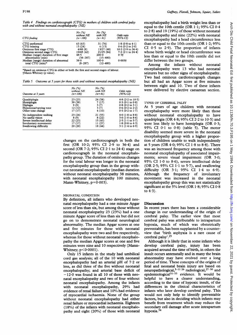
neonatal encephalopathy and 66% of mothersof infants with neonatal encephalopathy had acardiotocograph available for analysis (table4). Eight per cent of the group without neo-natal encephalopathy and 48% of the neonatalencephalopathy group had signs of fetal dis-tress with an increased likelihood of ominous
*Based on ominous CTG in either or both the first and second stages of labour.tMann-Whitney (p value).
Table 5 Outcome at 5 years for those with and without neonatal encephalopathy (NE)
No (%/6) No (%o)without NE with NE Odds ratio
Outcome at S years (n= 100) (n=41) (95% CI)
Quadriplegia 23 (23) 24 (58) 4-8 (2-2 to 10-5)Hemiplegia 39 (39) 7 (17) 0 3 (0-1 to 0 8)Diplegia 9 (9) 3 (7) 0-8 (0-2 to 3-1)Hypotonia/varying tone 12 (12) 3 (7) 0-6 (0-2 to 2 2)Other tone change 17 (17) 4 (10) 0-5 (0-2 to 1-7)No independent walking 23 (24) 21 (55) 4 0 (1-8 to 8 8)No usefulvision 8 (9) 8 (22) 3 0 (1-0 to 8 6)Severe intellectual delay 22 (23) 15 (43) 2-5 (1-1 to 5 7)Involuntary movements 17 (17) 11 (27) 1-8 (0-8 to 4 3)Swallowing difficulty 20 (20) 18 (44) 3-1 (1-4 to 6-9)
changes on the cardiotocograph in both thefirst (OR 10-2; 95% CI 2-9 to 36 4) andsecond (OR 7-2; 95% CI 2 1 to 24 4) stage oncardiotocograph in the neonatal encephalo-pathy group. The duration of ominous changesfor the total labour was longer in the neonatalencephalopathy group than in the group with-out neonatal encephalopathy (median durationwithout neonatal encephalopathy 38 minutes,with neonatal encephalopathy 100 minutes;Mann-Whitney, p=0 003).
NEONATAL CONDITIONBy definition, all infants who developed neo-natal encephalopathy had a one minute Apgarscore of less than six, but among those withoutneonatal encephalopathy 23 (23%) had a one
minute Apgar score of less than six but did notgo on to demonstrate neonatal neurologicalabnormality. The median Apgar scores at oneand five minutes for those with neonatalencephalopathy were two and five respectively,whereas for those without neonatal encephalo-pathy the median Apgar scores at one and fiveminutes were nine and 10 respectively (Mann-Whitney; p<0-0001).
Only 15 infants in the study had umbilicalcord gas analysis; all of the 10 with neonatalencephalopathy had an arterial pH of 7-2 or
less, as did three of the five without neonatalencephalopathy; and arterial base deficit of- 12-0 was found in all 10 of those with neo-
natal encephalopathy and two of four withoutneonatal encephalopathy. Among the infantswith neonatal encephalopathy, 29% hadevidence of renal failure and 10% had evidenceof myocardial ischaemia. None of the groupwithout neonatal encephalopathy had eitherrenal failure or myocardial ischaemia. Eighteen(18%) of the infants with neonatal encephalo-pathy and eight (20%) of those with neonatal
encephalopathy had a birth weight less than orequal to the 10th centile (OR 1 -1; 95% CI 0 4to 2 8) and 19 (19%) ofthose without neonatalencephalopathy and nine (22%) with neonatalencephalopathy had a head circumference lessthan or equal to the 10th centile (OR 1-2; 95%CI 0 5 to 2 9). The proportion of infantswhose birth weight or head circumference wasless than or equal to the 10th centile did notdiffer between the two groups.Among the infants without neonatal
encephalopathy were 13 who had neonatalseizures but no other signs of encephalopathy.Two had ominous cardiotocograph changesbut all had an Apgar score at five minutesbetween eight and 10. Two of these infantswere delivered by elective caesarean section.
TYPES OF CEREBRAL PALSYAt 5 years of age children with neonatalencephalopathy were more likely than thosewithout neonatal encephalopathy to havequadriplegia (OR 4-8; 95°/O CI 2.2 to 10-5) andwere less likely to have hemiplegia (OR 0 3;95% CI 041 to 0 8) (table 5). The motordisability seemed more severe in the neonatalencephalopathy group with a higher propor-tion of children unable to walk independentlyat 5 years (OR 4-0; 95% CI 1.8 to 8-8). Therewas an increased frequency among those withneonatal encephalopathy of associated impair-ments; severe visual impairment (OR 3 0;95% CI 1-0 to 8&6), severe intellectual delay(OR 2-5; 95% CI 1 1 to 5.7), and swallowingdifficulty (OR 341; 95% CI 1V4 to 6-9).Although the frequency of involuntarymovement was increased in the neonatalencephalopathy group this was not statisticallysignificant at the 5% level (OR 1 8; 95% CI 0-8to 4.3).
DiscussionIn recent years there has been a considerablechange in our understanding of the origin ofcerebral palsy. The earlier view that mostcerebral palsy was attributable to intrapartumhypoxia, much of which was obstetricallypreventable, has been supplanted by a counter-view that 'birth asphyxia is a rare cause ofcerebral palsy'.25
Although it is likely that in some infants whodevelop cerebral palsy, injury has beenacquired around the time of birth, in others theinsult occurs antenatally and in many the brainabnormality may have evolved over a longperiod oftime. These concepts ofthe origins offetal and neonatal brain injury are based onneuropathological,5-11 26 radiological,27-30 andepidemiological3l-33 evidence. It would behelpful to have a clearer understanding,according to the time of hypoxic insult, of thedifferences in the clinical characteristics ofinfants who later develop cerebral palsy. Thiswould not only help to identify preventablefactors, but also in deciding which infants maybenefit from treatment which may reduce thesecondary cell damage after acute intrapartumhypoxia.34
We used the clinical finding of depression atbirth followed by neonatal neurological abnor-mality as the most reliable marker of an acuteepisode of intrapartum hypoxia. This findingwas used as the basis for differentiating thosechildren with a predominantly antenatal originof cerebral palsy and those with a primarilyintrapartum origin. We recognised, however,that this division may be rather crude. It ispossible, for example, that within the group ofchildren with cerebral palsy with neonatalencephalopathy there might be some whoseencephalopathic signs reflected a longstandingneurological disorder. Further, it is possiblethat a more critical evaluation of neurologicalstates in the first few days of life might help toidentify different aetiological subgroups. Forexample, we were aware of a group of infantswho had neonatal seizures but did not showany other signs of encephalopathy and whowere not depressed at birth. This group ofinfants appeared to be aetiologically distinctfrom those with changes in consciousness andmuscle tone between seizures. It was also ofinterest that in nine of the 13 infants in thisgroup seizures began before the age of 48hours. In the past it has been suggested thatseizures which start in the first 48 hours of lifeare those most likely to reflect intrapartumhypoxia.35 A large systematic study of infantswith clinical signs of neurological dysfunctionin the neonatal period together withneuroimaging would clarify these issues.Many infants who later have signs of
cerebral palsy do not appear to have abnormalneurological signs in the neonatal period. Ifneonatal encephalopathy is considered to be acondition of term infants, then only 12%(41/339) of all children with cerebral palsyborn to residents of the area in the four birthyears studied had evidence of neonatalencephalopathy. This once again emphasisesthe need to look at aetiological factors whichmay influence earlier intrauterine cerebraldevelopment, not just events related to labour,in the majority of children with cerebral palsy.The search for clinical markers of adverse
antenatal factors in populations of childrenwith cerebral palsy, however, is fairlyunrewarding. Although previous work hasshown an increased risk of pre-eclampsia andintrauterine growth retardation amongmothers of term infants with cerebral palsycompared with a control population,36 this riskdoes not appear to be greater in the groupwithout neonatal encephalopathy as we hadhypothesised. Apart from a lower frequency ofprimigravidae, we were unable to find ante-natal characteristics in the group withoutneonatal encephalopathy which differentiatedthese infants from the group with neonatalencephalopathy. It is likely that more sophisti-cated fetal neuroimaging techniques areneeded to detect signs of antenatal ischaemicdamage, whether focal lesions or the more dif-fuse white matter lesions described withintrauterine hypoxia.'0
It was clear, however, that the clinical intra-partum course was different in the infants withand without neonatal encephalopathy. There
was an increased frequency of induction oflabour and of pregnancies of greater than 41weeks' gestation among the mothers of infantswho went on to develop neonatal encephalo-pathy. In addition, prolonged labour and signsof prolonged severe distress were more fre-quent in the neonatal encephalopathy group.This cluster of adverse intrapartum eventswere also described by Minchom et al in infantswho developed early neonatal seizures.37 Suchassociations do not, of course, imply a causallink.
This group (neonatal encephalopathy),however, includes those cases of cerebral palsywhich are most likely to be obstetricallypreventable. For example, it could be arguedthat reduction in the duration of fetal distressby expediting delivery might have altered theoutcome. The greater frequency of fetaldistress among those with neonatal encephalo-pathy, however, could equally reflect long-standing neurological dysfunction which isnot, at present, obstetrically preventable.The challenge is to find reliable ways of
identifying the infant who is neurologicallyabnormal before or during labour. Severalpotential methods are being examined; forexample, the use of near infrared spectroscopyin labour and antenatal biophysical assess-ment.3842 Using the latter, abnormal in uterobehavioural states can be shown in someinfants who later show abnormal neonatalneurological function. Neonatal evidence ofdamage to other organs may help to identify arecent acute hypoxic event. Ischaemic damageto the kidneys or myocardium in an infant withneonatal encephalopathy who subsequentlyhas cerebral palsy provides supportingevidence of an intrapartum origin to the laterneurological deficit.43-45
It has been previously suggested that clinicalsubgroups of cerebral palsy may have differentaetiologies.46 Our hypothesis that childrenwith cerebral palsy who have a history ofneonatal encephalopathy have more extensivemotor impairment and associated sensory andintellectual disability was sustained. Furtherexploration of the relation between the neuro-pathology of the injury, the later clinical mani-festations, and the appearance of the brain onneuroimaging may be useful in understandingthe time of injury and hence in identifyingthose insults which are most likely to bepreventable.
In conclusion, we consider that childrenwith cerebral palsy and a history of neonatalencephalopathy (depression at birth followedby neonatal neurological dysfunction) aremore likely than those without neonatalencephalopathy to have: (a) evidence ofprolonged fetal distress as measured by theduration of cardiotocograph changes; (b)evidence of other organ damage; and (c) asevere and extensive form of motor deficit withintellectual and sensory involvement.We suggest that this cluster of clinical signs
indicates that the aetiology is likely to be anacute intrapartum insult, preventable or non-preventable. It follows that in the absence ofthis cluster of signs, intrapartum asphyxia is
unlikely to be the cause of a subsequent neuro-logical deficit.We are grateful to Rosemary King, administrative coordinatorof the Oxford regional register of early childhood impairment;to the regional obstetricians and midwives for their assistance;to Georgina Berridge for computing support; and to colleaguesat the National Perinatal Epidemiology Unit for commentingon drafts of the paper. The Oxford regional register of earlychildhood impairment is funded by Oxford Regional HealthAuthority. Geraldine Gaffney was funded by the MedicalResearch Council.
1 Paneth N. The causes of cerebral palsy. Recent evidence.Clin Invest Med 1993; 16: 95-102.
2 Paneth N, Fox HE. The relationship of Apgar score toneurological handicap: a survey of clinicians. ObstetGynecol 1983; 61: 547-50.
3 Stanley F, Blair E. Why have we failed to reduce thefrequency of cerebral palsy? Med3Aust 1991; 154: 623-6.4 Hill A. Current concepts of hypoxic-ischemic injury in the
term newborn. Pediatr Neurol 1991; 7: 317-25.5 Pasternak JF, Predey TA, Mikhael MA. Neonatal asphyxia:
vulnerability of basal ganglia, thalamus, and brainstem.Pediatr Neurol 1991; 7:147-9.
6 Roland EH, Hill A. Selective brainstem injury in anasphyxiated newborn. Ann Neurol 1988; 23: 89-92.
7 Leech RW, Alvord EC. Anoxic-ischaemic encephalopathyin the human neonatal period. The significance of brainstem involvement. Arch Neurol 1977; 34: 109-13.
8 Dambska M, Laure-Kamionowska M, Liebhart M.Brainstem lesions in the course of chronic fetal asphyxia.ClinNeuropathol1987;6: 110-5.
12 Hope PL, Gould SJ, Howard S, Hamilton PA, CostelloAMdelL, Reynolds EOR. Precision of ultrasound diagnosisof pathologically verified lesions in the brains of verypreterm infants. Dev Med Child Neurol 1988; 30: 457-71.
13 BarkovitchJ. MR and CT evaluation of profound neonataland infantile asphyxia. Am .7 Neuroradiol 1992; 13:959-72.
14 Krageloh-MannI, Hagberg B, Petersen D, Riethmuller J,Gut E, Michaelis R. Bilateral spastic cerebral palsy- patho-genetic aspects from MRI. Neuropediatrics 1992; 23: 46-8.
15 Fitzhardinge PM, Flodmark 0, Fitz CR, Ashby S. Theprognostic value of computed tomography as an adjunctto assessment of the term infant with postasphyxialencephalopathy. Pediatrics 1981; 99: 777-81.
16 Adsett DB, Fitz CR, Hill A. Hypoxic-ischaemic cerebralinjury in the term newborn: correlation of CT findingswith neurological outcome. Dev Med Child Neurol 1985;27: 155-60.
17 Lipp-Zwahlen AE, Deonna T, Micheli JL, Calame A,Chrzanowski R, Cetre E. Prognostic value of neonatal CTscans in asphyxiated term babies: low density scorecompared with neonatal neurological signs.Neuropediatncs 1985; 16: 209-17.
18 Gaffney G, Squier M, Johnson A, Flavell V, Sellers S.Clinical associations of prenatal ischaemic white matterinjury. Arch Dis Child 1994; 70: F101-6.
20 Levene MI, Kornber J, Williams THC. The incidence andseverity of post-asphyxial encephalopathy in full-terminfants. Early Hum Dev 1985; 11: 21-6.
21 Hall DMB. Birth asphyxia and cerebral palsy. BM7 1989;299: 279-82.
22 Nelson K, Leviton A. How much of neonatal encephalo-pathy is due to birth asphyxia? Am 7DisChild 1991; 145:1325-31.
23 Evans P, Johnson A, Mutch L, Alberman E. A standardform for recording clinical findings in children with amotor deficit of central origin. Dev Med Child Neruol1989; 31: 119-27.
24 Macdonald D, Grant A, Sheridan-Pereira M, Boylan P,Chalmers I. The Dublin randomized controlled trial ofintrapartum fetal heart rate monitoring. Aml 7 ObstetGynecol 1985; 152: 524-39.
25 Bryce R, Stanley F, Blair E. The effects of intrapartum careon the risk of impairments in childhood. In: Chalmers I,Enkin M, Keirse MSNC, Eds. Effective care in pregnancyand childbirth. Oxford: Oxford University Press, 1989:1313-21.
26 Myers RE. Two patterns of perinatal brain damage andtheir conditions of occurrence. A]n Obstet Gynecol 1972;116: 246-76.
27 Bejar P, Wozniak P, Allard M, et al. Antenatal origin of neu-rologic damage in newborn infants. Am.Y Obstet Gynecol1988; 159: 357-63.
28 Scher MS, Belfar H, Martin J, Painter MJ. Destructivebrain lesions of presumed fetal onset: antepartum causesof cerebral palsy. Pediatrics 1991; 88: 898-906.
29 Johnson MA, Pennock JM, Bydder GM, Dubowitz LMS,Thomas DJ, Young IR. Serial MR imaging in neonatalcerebral injury. AmjiYRadiol 1987; 8: 83-92.
31 Nelson KB, Ellenberg JH. Antecedents of cerebral palsy. NEnglYMed 1986; 8: 81-6.
32 Blair E, Stanley FJ. Intrapartum asphyxia: a rare cause ofcerebral palsy. JPediatr 1988; 112: 515-9.
33 Nelson KB. Relationship of intrapartum and delivery roomevents to longterm neurologic outcome. Clin Perinatol1989; 16: 995-1007.
34 Levene MI. Role of excitatory amino acid antagonists in themanagement of birth asphyxia. Biol Neonate 1992; 62:248-51.
35 Dennis J, Chalmers 1. Very early neonatal seizure rate: apossible epidemiological indicator of the quality of peri-natal care. Br] Obstet Gyniaecol 1982; 89: 418-26.
36 Gaffney G, SellersS, Flavell V, Squier M, Johnson A. Case-control study of intrapartum care, cerebral palsy, anddeath. BM.7 1994; 308: 743-50.
37 Minchom P, Niswander K, Chalmers I, et al. Antecedentsand outcome of very early neonatal seizures in infantsborn at or after term. Br 7 Obstet Gvnaecol 1987; 94:431-9.
38 Wyatt JS. Near infrared spectroscopy. Investigation andassessment of perinatal brain injury. Biol Neonate 1992;62: 290-4.
39 Peebles DM, Edwards AD, Wyatt JS, et al. Changes inhuman fetal cerebral haemoglobin concentration andoxygenation during labor measured by near-infraredspectroscopy. AmY Obstet Gynecol 1992; 166: 1369-73.
40 Nijhuis JG, Prechtl HFR, Martin CB, Bots RSGM. Arethere behavioural states in the human fetus? Early HumDev 1982; 6:177-95.
41 Manning FA, Baskett TF, Morrison I, Lange IR. Fetalbiophysical profile scoring: a prospective study in 1184high-risk patients. Ami _7 Obstet Gynecol 1981; 140:289-94.
42 Horimoto N, Koyanagi T, Macda H, et al. Can brainimpairment be detected by in-utero behavioural patterns.Arch Dis Child 1993; 69: 3-8.
43 Perlman J, Tack E, Martin T, Shackleford G, Amon E.Acute systemic organ injury in term infants after asphyxia.Amn Dis Child 1989; 143: 617-20.
44 Dauber I, Krauss A, Symchych P, Auld P. Renal failurefollowing perinatal anoxia. 7 Pediatr 1976; 88: 851-5.
45 Ruth VJ. Prognostic value of creatine kinase-BB isoenzymein high risk newborn infants. Arch Dis Child 1989; 64:563-8.
46 Stanley FJ, Blair E, Hockey A, Petterson B, Watson L.Spastic quadriplegia in Western Australia: a geneticepidemiological study. 1: Case population and perinatalrisk factors. Dev Med Child Neuirol 1993; 35: 191-201.