1 Cerebrospinal fluid (CSF) The CSF examination Andrea Tipold TiHo Hannover Review IVIS Cerebrospinal fluid - history Presence of fluid in the cavities: known to the ancients first report 17th century B.C. Hippocrates 4th century B.C. Galen 2nd century A.D.: description of ventricular cavities first examinations in animals: 1825 by Magendie Cerebrospinal fluid - history 2 1891 Quincke: diagnostic and therapeutic aid 1900 cytology 2011: nearly the same techniques: CYTOLOGY, PROTEIN-CONTENT
Transcript
1
Cerebrospinal fluid (CSF)
The CSF examination
Andrea TipoldTiHo Hannover
Review IVIS
Cerebrospinal fluid - history
Presence of fluid in the cavities: knownto the ancients
first report 17th century B.C. Hippocrates 4th century B.C. Galen 2nd century A.D.: description of
ventricular cavities first examinations in animals: 1825 by
Magendie
Cerebrospinal fluid - history 2
1891 Quincke: diagnostic andtherapeutic aid
1900 cytology
2011: nearly the same techniques:
CYTOLOGY, PROTEIN-CONTENT
2
CSF – what do you know about the physiology ?
Cerebrospinal fluid - anatomy
CSF: ventricular system, subarachnoid space
cranial cavity – closed space, continuous adjustment of the intracranial pressure
pressure: brain parenchyma, CSF, blood
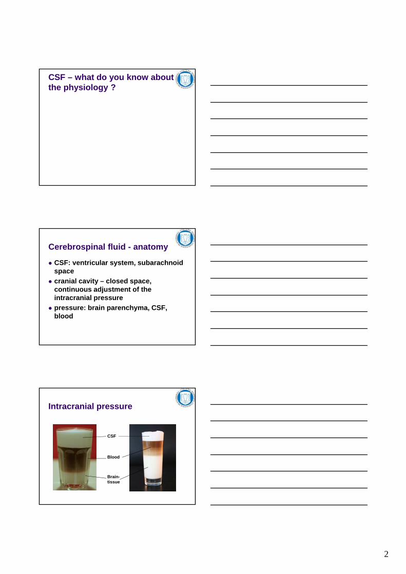
Intracranial pressure
Brain-tissue
Blood
CSF
3
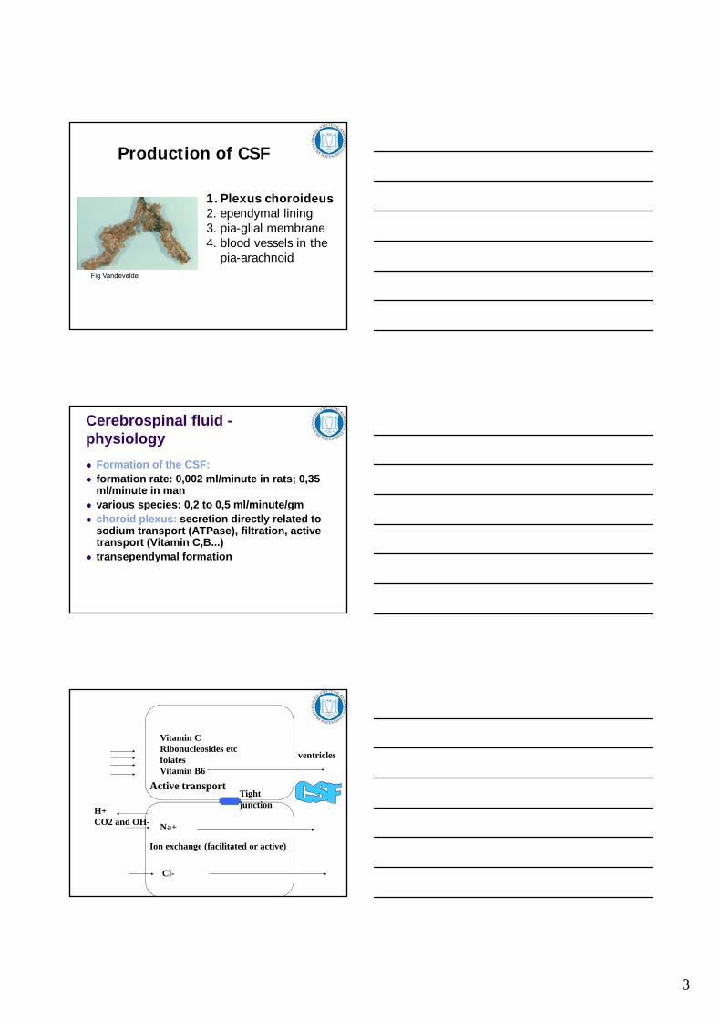
1. Plexus choroideus2. ependymal lining3. pia-glial membrane4. blood vessels in the
pia-arachnoid
Production of CSF
Fig Vandevelde
Cerebrospinal fluid -physiology
Formation of the CSF: formation rate: 0,002 ml/minute in rats; 0,35
ml/minute in man various species: 0,2 to 0,5 ml/minute/gm choroid plexus: secretion directly related to
sodium transport (ATPase), filtration, active transport (Vitamin C,B...)
transependymal formation
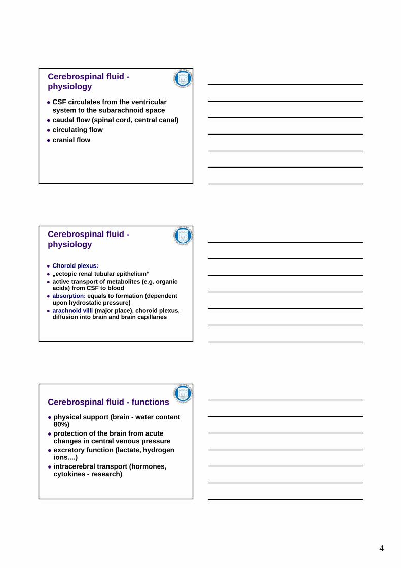
Tight junction
ventricles
Active transport
Vitamin CRibonucleosides etcfolatesVitamin B6
Ion exchange (facilitated or active)
Na+
Cl-
H+CO2 and OH-
4
Cerebrospinal fluid -physiology
CSF circulates from the ventricular system to the subarachnoid space
caudal flow (spinal cord, central canal)
circulating flow
cranial flow
Cerebrospinal fluid -physiology
Choroid plexus: „ectopic renal tubular epithelium“ active transport of metabolites (e.g. organic
acids) from CSF to blood absorption: equals to formation (dependent
upon hydrostatic pressure) arachnoid villi (major place), choroid plexus,
diffusion into brain and brain capillaries
Cerebrospinal fluid - functions
physical support (brain - water content 80%)
protection of the brain from acute changes in central venous pressure
excretory function (lactate, hydrogen ions....)
intracerebral transport (hormones, cytokines - research)
5
Cerebrospinal fluid -composition
watery solution (99% water)
ions (different concentration than plasma)
nutrients, neuroendocrine substances and neurotransmitter
Osmolality: same as plasma (289 mOsm/L)
glucose 80% of plasma
protein: < 25mg/dl (mostly albumin)
0-3 cells/ul, mostly lymphocytes
Cerebrospinal fluid -acquisition
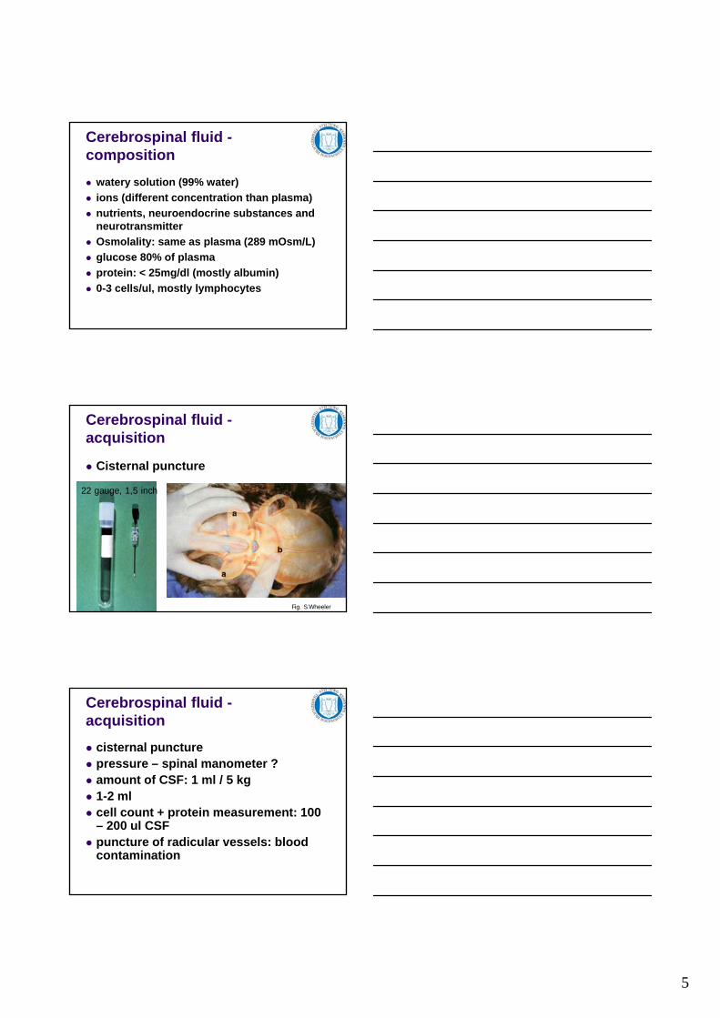
Cisternal puncture
22 gauge, 1,5 inch
Fig. S.Wheeler
Cerebrospinal fluid -acquisition
cisternal puncture pressure – spinal manometer ? amount of CSF: 1 ml / 5 kg 1-2 ml cell count + protein measurement: 100
– 200 ul CSF puncture of radicular vessels: blood
contamination
6
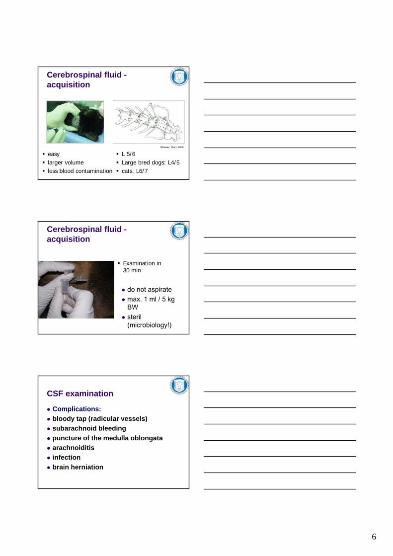
Cerebrospinal fluid -acquisition
easy larger volume less blood contamination
L 5/6 Large bred dogs: L4/5 cats: L6/7
Wheeler, Sharp 2000
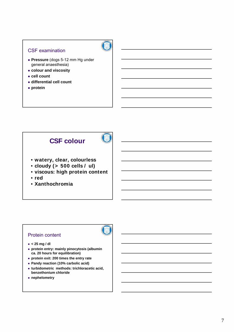
Cerebrospinal fluid -acquisition
do not aspirate
max. 1 ml / 5 kg BW
steril (microbiology!)
Examination in30 min
CSF examination
Complications:
bloody tap (radicular vessels)
subarachnoid bleeding
puncture of the medulla oblongata
arachnoiditis
infection
brain herniation
7
CSF examination
Pressure (dogs 5-12 mm Hg under general anaesthesia)
colour and viscosity
cell count
differential cell count
protein
CSF colour
• watery, clear, colourless• cloudy (> 500 cells / ul)• viscous: high protein content• red• Xanthochromia
Protein content
< 25 mg / dl
protein entry: mainly pinocytosis (albumin ca. 20 hours for equilibration)