96
Cerebrovascular Diseases
| Date post: | 25-Dec-2015 |
| Category: |
Documents |
| Upload: | christal-sims |
| View: | 218 times |
| Download: | 2 times |
Cerebrovascular Diseases

Vascular diseases of the nervous system are amongst the most frequent causes of admission to hospital
The annual incidence in the UK varies regionally between 150-200/100 000, with a prevalence of 600/100 000 of which one-third are severely disabled
Better control of hypertension, reduced incidence of heart disease and a greater awareness of all risk factors have combined to reduce mortality from stroke Despite this, stroke still ranks third behind heart disease and cancer as a cause of death in affluent societies

Risk Factors
Prevention of cerebrovascular disease is more likely to reduce death and disability than any medical or surgical advance in management.
Prevention depends upon the identification of risk factors and their correction

Hypertension :
Hypertension is a major factor in the development of thrombotic cerebral infarction and intracranial haemorrhage
There is no critical blood pressure level, the risk is related to the height of blood pressure and increases throughout the whole range from normal to hypertensive
A 6 mmHg fall in diastolic blood pressure is associated in relative terms with a 40% fall in the fatal and nonfatal stroke rate
Systolic hypertension (frequent in the elderly) is also a significant factor and not as harmless as previously thought

Cardiac disease :
Cardiac enlargement, failure and arrhythmias, as well as rheumatic heart disease, patent foramen ovale and, rarely, cardiac myxoma are all associated with an increased risk of stroke
Diabetes :
The risk of cerebral infarction is increased twofold in diabetes
More effective treatment of diabetes has not reduced the frequency of atherosclerotic sequelae

Heredity :
Close relatives are at only slightly greater risk than non-genetically related family members of a stroke patient
Diabetes and hypertension show familial propensity thus clouding the significance of pure hereidtary factors
Blood lipids, cholesterol smoking, diet/obesity, soft water :
These factors are much less significant than in the genesis of coronary artery disease

Race :
Alterations in life style, diet and environment probably explain the geographical variations more than racial tendencies
Haematocrit :
A high blood haemoglobin concentration (or haematocrit level) is associated with an increased incidence of cerebral infarction
Other haematological factors, such as decreased fibrinolysisi, are important also

Oral contraceptives :
The evidence of pill-related stroke is inconclusive
A recent prospective study has suggested an increased risk of subarachnoid haemorrhage rather than thromboembolic stroke

Cerebrovascular Disease - Mechanisms
‘Stroke’ is a generic term, lacking pathological meaning
Cerebrovascular diseases can be defined as those in which brain disease occurs secondary to a pathological disorder of blood vessels (usually arteries) or blood supply


Cerebrovascular Disease – Natural History
Approximately one-third of all ‘strokes’ are fatal
The age of the patient, the anatomical size of the lesion, the degree of deficit and the underlying cause all influence the outcome

Immediate outcome :
In cerebral haemorrhage, mortality approaches 50%
Cerebral infarction fares better, with an immediate mortality of less than 20%, fatal lesions being large with associated oedema and brain shift
Embolic infarction carries a better outcome than thrombotic infarction
Fatal cases of infarction die either at onset or else, more commonly, after the first week from cardiovascular or respiratory complications
The level of consciousness on admission to hospital gives a good indication to immediate outcome
The deeper the conscious level the graver the prognosis

Long-term outcome :
The prognosis following infarction due to thrombosis or embolisation from diseased neck vessels or heart is dependent on the progression of the underlying atherosclerotic disease
Recurrent cerebral infarction rates vary between 5%-15% per year
Symptoms of coronary artery disease and/or peripheral vascular disease may also ensue
Five year mortality is 44% for males and 36% for females
The long-term prognosis following survival from haemorrhage depends upon the cause and the treatment

Cerebrovascular Disease – Causes

Non-atheromatous diseases of the vessel wall :
Collagen disease e.g. rheumatoid arthritis
systemic lupus erythematosis (SLE)
Vasculitis e.g. Polyarteritis nodosa
temporal arteritis
Granulomatous vasculitis e.g. Wegener’s granulomatosis
Miscellaneous e.g. synphilitic vasculitis
fibromuscular dysplasia
sarcoidosis
trauma

Diseases of Blood :
E.g., Coagulopathies
Haemoglobinopathies
Venous Thrombosis :
Venous thrombosis may occur with infection and dehydration or in association with arterial occlusion when related to oestrogen excess
Decreased cerebral perfusion :
Infarction between arterial territories may results from impaired perfusion from
e.g. cardiac dysrhythmia GI blood loss

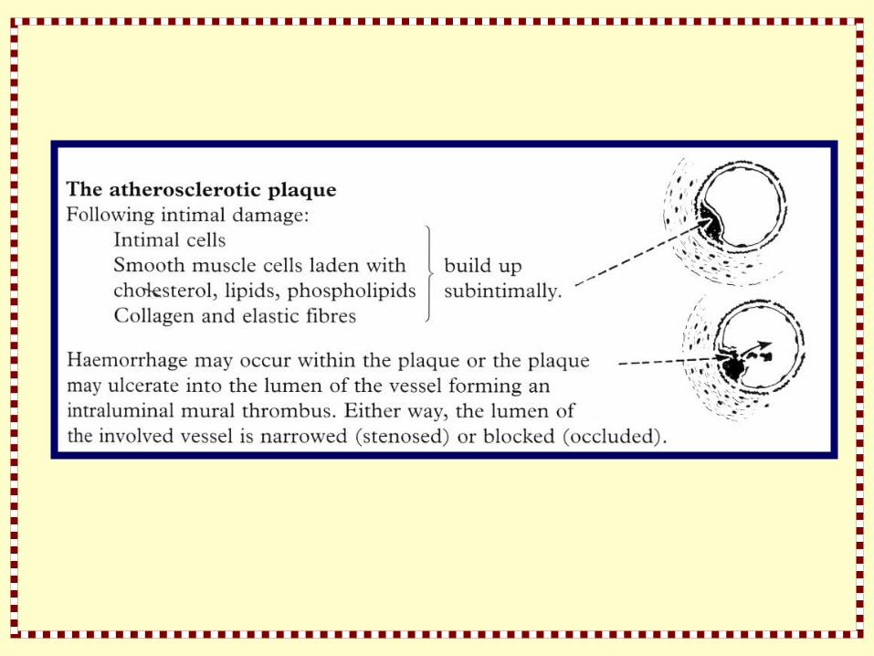
The plaque itself may give rise to emboli. Cholesterol is present partly in crystal form and fragments following plaque rupture may be sufficiently large to occlude the lumen of distal vessels
The cholesterol esters, lipids and phospholipids each play a role in the aggregation of such emboli
The carotid bifurcation in the neck is a frequent site at which the antheromatous plaque causes stenosis or occlusion

Platelet emboli arise from thrombus developed over the dam aged endothelium
The thrombus is produced partly by platelets coming into contact with exposed collagen fibres
Endothelial cells synthesise PROSTACYCLIN which is a potent vasodilator and inhibitor of platelet aggregation
THROMBOXANE A2, synthesised by platelets, has opposite effects
In thrombus formation these two PROSTAGLANDINS actively compete with each other

Transient Ischaemic Attacks (TIAs)
Transient ischaemic attacks are episodes of focal neurological symptoms due to inadequate blood supply to the brain
Attacks are sudden in onset, resolve within 24 hours or less and leave no residual deficit
These attacks are important as warning episodes or precursors of cerebral infarction
Before diagnosis TIAs, consider other causes of transient neurological dysfunction – migraine, partial seizures, hypoglycaemia, syncope and hyperventilation
The pathogenesis of transient ischaemic attacks :
A reduction of cerebral blood flow below 20-30 ml/100 g/min produces neurological symptoms
The development of infarction is a consequence of the degree of reduced flow and the duration of such a reduction
If flow is restored to an area of brain within the critical period, ischaemic symptoms will reverse themselves

TIAs may be due to

The synptomatology of TIAs :

The natural history of TIAs :
Following a TIA, between 5-10% of patients will develop infarction in each year of followup, irrespective of the territory involved
The risk of infarction is probably at its greatest in the first 3-6 months after the initial TIA
Not all patients who develop cerebral infarction have had a warning TIA

Clinical Syndromes – Large Vessel Occlusion


Clinical Syndromes – Large Vessel Occlusion

Clinical features :
The anterior cerebral artery may be occuluded by embolus or thrombus
The clinical picture depends on the site of occlusion (especially in relation to the anterior communicating artery) and anatomical variation, e.g. both anterior cerebral arteries may arise from one side by enlargement of the anterior communicating artery

Clinical Syndromes – Large Vessel Occlusion

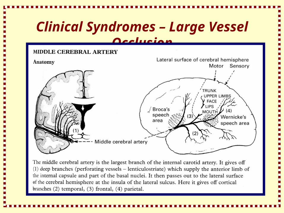
The middle cerebral artery is the largest branch of the internal carotid artery
It gives off
1 deep branches (perforating vessels – lenticulostriate) which supply the anterior limb of the internal capsule and part of the basal nuclei. It then passes out to the lateral surface of the cerebral hemisphere at the insula of the lateral sulcus. Here it gives off cortical branches
2. temporal
3. frontal
4. parietal

Clinical Features :
The middle cerebral artery may be occluded by embolus or thrombus
The clinical picture depends upon the site of occlusion and whether dominant or non-dominant hemisphere is affected

Occlusion at the insula :
- Contralateral hemiplegia (leg relatively spared)
- Contralateral hernianaesthesi and hemianopia
- Aphasia (dominant)
- Neglect of contralateral limbs
- Dressing difficulty(non-dominant)

When cortical branches are affected individually, the clinical picture is less severe, e.g. involvement of parietal branches alone may produce Wernicke’s dysphasia with no limb weakness or sensory loss
The deep branches (perforating vessels) of the middle cerebral artery may be a source of haemorrhage or small infarcts

Vertebral Artery Occlusion :

The vertebral artery arises from the subclavian artery on each side
Underdevelopment of one vessel occurs in 10%
The vertebral artery runs from its origin through the foramen of the transverse processes of the mid-cervical vertebrae
It then passes laterally through the transverse process of the axis, then upwards to the atlas accompanied by a venous plexus and across the suboccipital triangle to the vertebral canal
After piercing the dura and arachnoid matter, it enters the cranial cavity through the foramen magnum
At the lower border of the pons, it unites with its fellow to form the basilar artery
The vertebral artery and its branches supply the medulla and the inferior surface of the cerebellum before forming the basilar artery

Clinical Features :
Occlusion of the vertebral artery, when low in the neck, is compensated by anastomotic channels
When one vertebral artery is hypoplastic, occlusion of the other is equivalent to basilar artery occlusion
Only the posterior inferior cerebellar artery (PICA) depends solely on flow through the vertebral artery
Vertebral artery occlusion may therefore present as a PICA syndrome
The close relationship of the vertebral artery to the cervical spine is important

Rarely, damage at intervertebral foramina or the atlanto-axial joints following subluxation may result in intimal damage, thrombus formation and embolisation
Vertebral artery compression during neck extension may cause symptoms of intermittent vertebrobasilar insufficiency
Stenosis of the proximal left or right subclavian artery may result in retrograde flow down the vertebral artery on exercising the arm
The is commonly asymptomatic and demonstrated incidentally by Doppler techniques or angiography

Occasionally symptoms of vertebrobasilar insufficiency arise – subclavian steal syndrome
Surgical reconstruction or bypass of the subclavina artery may be indicated
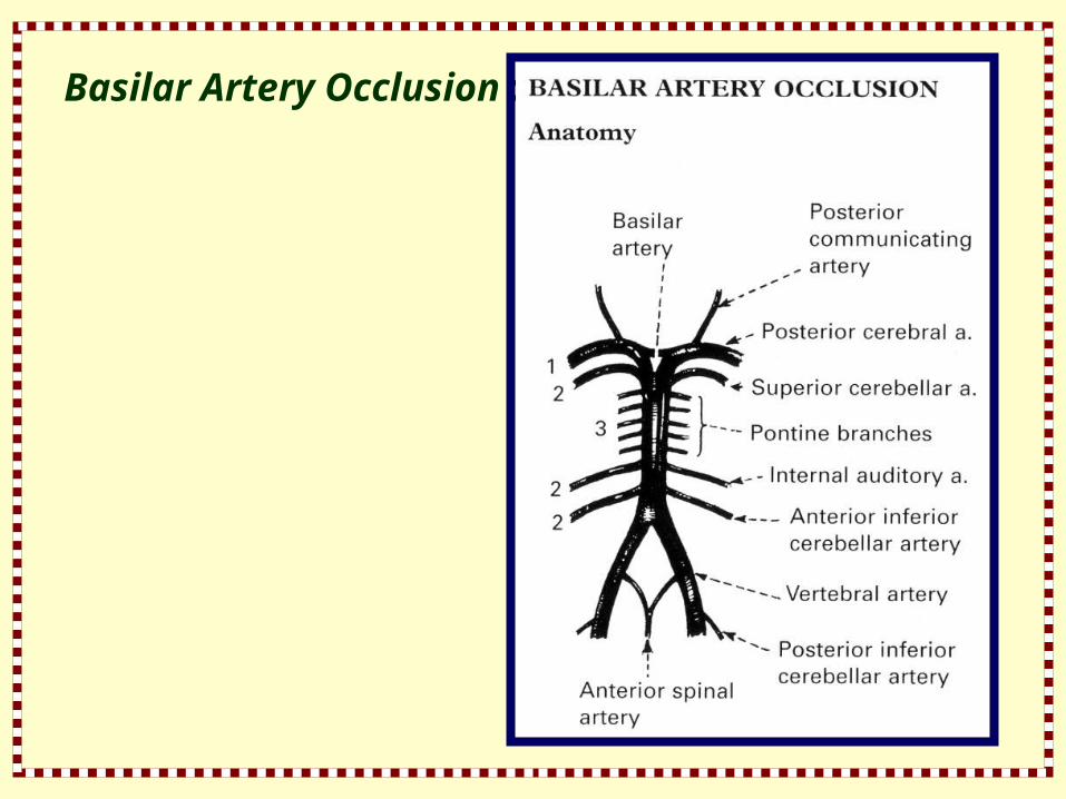
Basilar Artery Occlusion :

The basilar artery supplies the brain stem from medulla upwards and divides eventually into posterior cerebral arteries as well as posterior communicating arteries which run forward to join the anterior circulation
Branches can be classified into :
- Posterior cerebral arteires
- Long circumflex branches
- Paramedian branches

Clinical Features :
Prodromal symptoms are common and may take the form of diplopia, visual field loss, intermittent memory disturbance and a whole constellation of other brain stem symptoms
- Vertigo
- Ataxia
- Paresis
- Paraesthesia
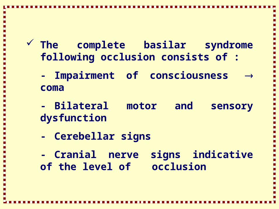
The complete basilar syndrome following occlusion consists of :
- Impairment of consciousness coma
- Bilateral motor and sensory dysfunction
- Cerebellar signs
- Cranial nerve signs indicative of the level of occlusion

The clinical picture is variable. Occasionally basilar thrombosis is an incidental finding at autopsy
‘Top of basilar’ occlusion : This results in lateral midbrain, thalamic, occipital and medial temporal lobe infarction.
Abnormal movements (hemiballismus) are associated with visual loss, pupillary abnormalities, gaze palsies, impaired conscious level and disturbances of behaviour
Paramedian perforating vessel occlusion gives rise to the ‘LOCKED IN’ SYNDROME and LACUNAR infarction

Posterior cerebral artery :

The posterior cerebral arteries are the terminal branches of the basilar artery
Small perforating branches supply midbrain structures, choroid plexus and posterior thalamus
Cortical branches supply the undersurface of the temporal lobe – temporal branch; and occipital and visual cortex – occipital and calcarine branches
Clinical features :
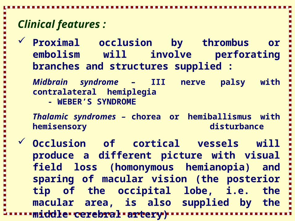
Proximal occlusion by thrombus or embolism will involve perforating branches and structures supplied :
Midbrain syndrome – III nerve palsy with contralateral hemiplegia - WEBER’S SYNDROME
Thalamic syndromes – chorea or hemiballismus with hemisensory disturbance
Occlusion of cortical vessels will produce a different picture with visual field loss (homonymous hemianopia) and sparing of macular vision (the posterior tip of the occipital lobe, i.e. the macular area, is also supplied by the middle cerebral artery)
Posterior cortical infarction in the dominant hemisphere may produce problems in naming colours and objects
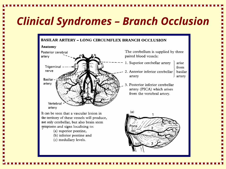
Clinical Syndromes – Branch Occlusion
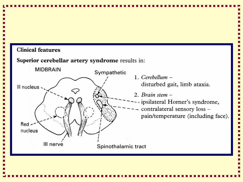

Clinical Features :


Clinical Syndromes – Lacunar Stroke (LACI)
Occlusion of deep penetrating arteries produces subcortical infarction characterised by preservation of cortical function – language, other cognitive and visual functions
Clinical syndromes are distinctive and normally result from long-standing hypertension
In 80%, infarcts occur in periventricular white matter and basal ganglia, the rest in cerebellum and brain stem
Areas of infarction are 0.5-1.5 cm in diameter and occluded vessels demonstrate lipohylinosis, microaneurysm and microatheromatous changes
Lacunar or subcortical infarction accounts for 17% of all thromboembolic strokes and knowledge of commoner syndromes is essential

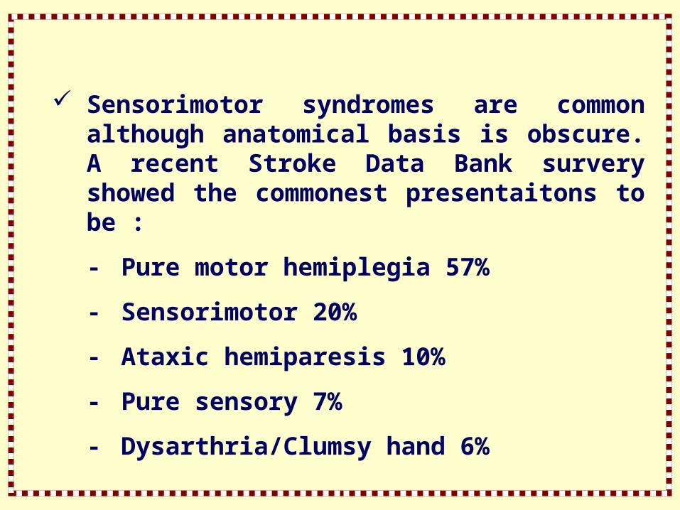
Sensorimotor syndromes are common although anatomical basis is obscure. A recent Stroke Data Bank survery showed the commonest presentaitons to be :
- Pure motor hemiplegia 57%
- Sensorimotor 20%
- Ataxic hemiparesis 10%
- Pure sensory 7%
- Dysarthria/Clumsy hand 6%
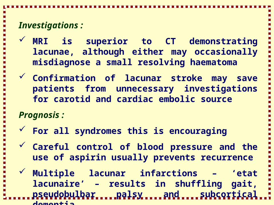
Investigations :
MRI is superior to CT demonstrating lacunae, although either may occasionally misdiagnose a small resolving haematoma
Confirmation of lacunar stroke may save patients from unnecessary investigations for carotid and cardiac embolic source
Prognosis :
For all syndromes this is encouraging
Careful control of blood pressure and the use of aspirin usually prevents recurrence
Multiple lacunar infarctions – ‘etat lacunaire’ – results in shuffling gait, pseudobulbar palsy and subcortical dementia

Classification of subtypes of cerebral infarction

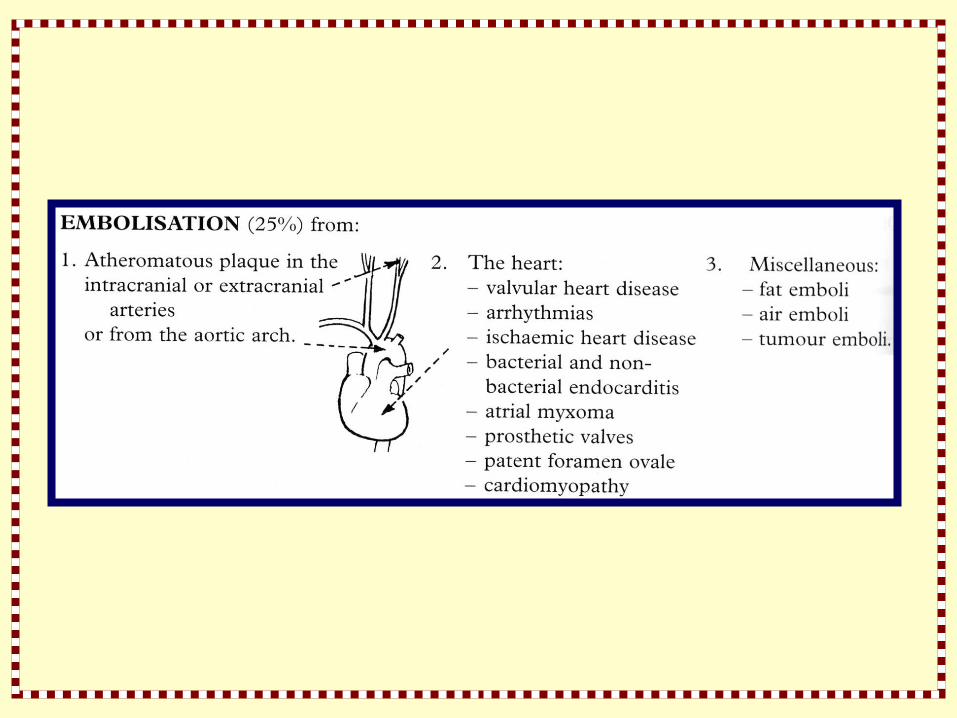
Embolisation
Emboli consist of friable atheromatous mateiral, platelet-fibrin clumps or well formed chrombus
The diagnosis of embolic infarction depends on :
The identification of an embolic source, e.g. cardiac disease
The clinical picture of sudden onset
Infarction in the territory of a major vessel or large branch
Clinical Picture :
Depends on the vessel involved
Emboli commonly produce transient ischaemic attacks (TIA) as well as infarction

Visual loss – transient, ie., amaurosis fugax or permanent
Hemisensory and hemimotor disturbance
Disturbance of higher function, e.g. dysphasia
Focal or generalised seizures – may persist for some time after the ischaemic episode
Depression of conscious level of major vessel occlusion occurs
Depression of conscious level if major vessel occlusion occurs
Emboli less frequently affect the posterior circulation
Symptoms are referable to the eye (retinal artery) and to the anterior and middle cerebral arteries, and take the form of :

Emboli from the internal carotid artery and aorta :
Emboli from these sources are commonest out with the heart
The majority of all cerebral emboli arise from ulcerative plaques in the carotid arteries
Embili arising from the aorta (atheromatous plaque or aortic aneurysm) often involve both hemispheres and systemic embolisation may coexist
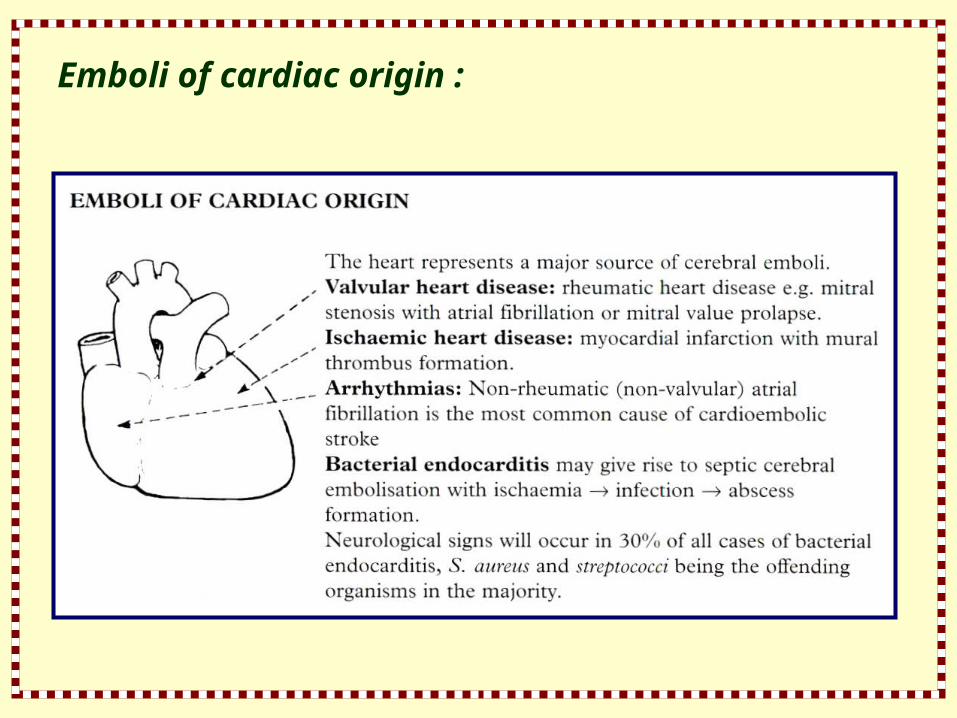
Emboli of cardiac origin :

Non-bacterial endocarditis :
Associated with malignant disease due to fibrin and platelet deposition on heart valves
Atrial myxoma :
Rare cause of recurrent cerebral embolisation Bihemisphere episodes with a perisistently elevated ESR should arouse suspicion which may be confirmed by cardiac ultrasound

Patent foramen ovale :
May result in paradoxical embolisation; suspect in patient with deep venous thrombosis who develops cerebral infarction
Emboli can also arise from intracardiac thrombus
New cardiac imaging techniques especially Transoesophageal Echocardiography (TOE) allow a more accurate detection of potential embolic source. Transcranial Doppler (TCD) may characterise emboli by analysing their signals and help quantify risk of recurrence

Emboli from other sources :
Fat emboli : following fracture, especially of long bones and pelvis, fat appears in the bloodstream and may pass into the cerebral circulation, usually 3-6 days after trauma. Emboli are usually multiple and signs are diffuse
Air emboli follow injury to neck/chest, or follow surgery. Rarely, air emboli complicate therapeutic abortion. Again the picture is diffuse neurologically. Onset is acute; if the patient survives the first 30 minutes, prognosis is excellent

Nitrogen embolisation or decompression sickness (the ‘bends’) produces a similar picture, but if the patient survives, neurological disability may be profound
Tumour emboli result in metastatic lesions; the onset is usually slow and progressive. Acute stroke-like presentation may occur, followed weeks or months later by the mass effects
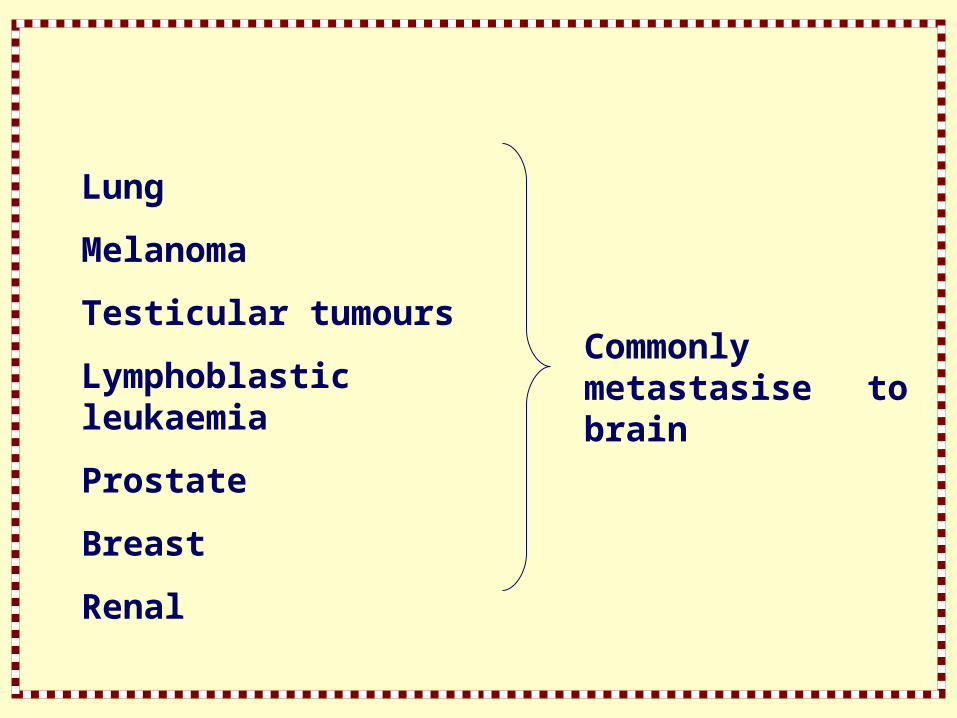
Lung
Melanoma
Testicular tumours
Lymphoblastic leukaemia
Prostate
Breast
Renal
Commonly metastasise to brain

Stenotic/Occulusive Disease - Investigations
CONFIRM THE DIAGNOSIS
Computerised tomography (CT Scan) :
All patients should have a CT scan, urgently if
- conscious level depressed
- Diagnosis uncertain
- On anticoagulants
- Before commencing/resuming antithrombotics
- If thrombolysis is considered

Infarction is evident as a low-density lesion which conforms to a vascular territory, i.e., usually wedge shaped
Subtle changes occur within 3 hours in some patients; most scans become abnormal within 48 hours
CT scan also identifies :
The site and size of the infarct, providing a prognostic guide
The presence of haemorrhagic infarction where bleeding occurs into the infarcted area
Intraacerebral haemorrhage or tumour

Magnetic resonance imaging (MRI) :
T2 prolongation (hyperintensity in relation to white and grey matter) occurs within hours of onset of ischaemic symptoms
Advanced techniques, diffusion weighted imaging (DWI) and perfusion imaging (PWI) show respectively early infarction (cytotoxic oedema) and ischaemic tissue at risk (the ischaemic penumbra)
These advanced techniques are valuable predictors of outcome and guide treatments directed as ‘ischaemic salvage’ e.g. thrombolysis

DEMONSTRATE THE SITE OF PRIMARY LESION
Non-invasive investigation
Ultrasound – Doppler/Duplex scanning : assesses extra – and intracranial vessels. A normal study precludes the need for angiography
Cardiac ultrasound (transthoracic or transoesophageal) : this often reveals a cardiac embolic source in young people with stroke, e.g. prolapsed mitral valve, patent foramen ovale
Magnetic resonance angiography (MRA) – ‘Time of flight’ or contrast enhanced techniques are used. Whilst of value in patients with heavily calcified carotid plaques, resistant to Doppler, it tends to overestimate the severity of stenosis. When assessing the carotid arteries it is best used in combination with Doppler. Its non-invasive nature makes it helpful in investigating the intracranial circulation
Computed tomographic angiography (CTA) – Dynamic helical CT, following bolus injection of non-ionic contrast, can be used to investigate both intracranial and extracranial vasculature. CTA compared with DSA correctly classifies the degree of carotid stenosis in 96% of cases but is insensitive to ulcerative plaque. Again it is best used in conjunction with Doppler
Digital intravenous subtraction angiography (DSA) :
The combination of the above techniques has decreased the need for invasive investigation but often cerebral angiography is still required to make a definitive diagnosis. The role and safety of angiography immediately following infarction is uncertain.
In the elderly or poor-risk patient, investigations to demonstrate the site of the primary lesion may be inappropriate


Suspected carotid disease :
Demonstrate both carotids, intracranial vessels, the aortic arch and origins of the vertebrals. Approximately two-thirds of patients with carotid territory attacks will have angiographic abnormality
Suspected vertebrobasilar disease :
Note the intracranial vessels and the course of the vertebral artery through the cervical foramina where osteophytic encroachment may occur
Note that proximal subclavian occlusion may result in retrograde flow down the vertebral arteries into the subclavian arteries, and cause TIAs aggravated by arm exercise – subclavian steal


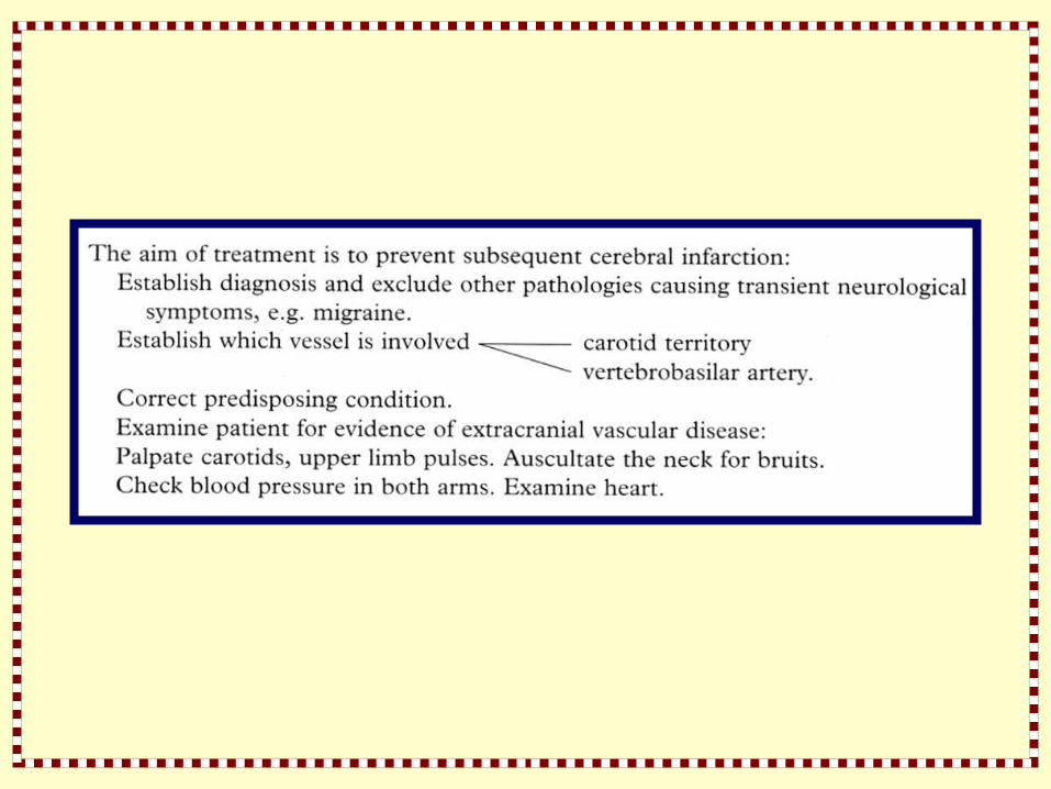
Cerebral Infarction - Management
The acute stroke :
Clinical history, examination and investigation will separate infarction and haemorrhage
Once the nature of the ‘stroke’ has been confidently defined, treatment should be instigated
Treatment aims :
- Prevent progression of present event
- Prevent immediate complication
- Prevent the development of subsequent events
- To rehabilitate the patient


Specific measures :
The following are generally ineffective, or are as yet inadequately evaluated
Anticoagulant therapy :
In patients with a known cardiac source of emboli, the risk of recurrent embolic infarction is high and anticoagulant therapy should be commenced once CT scan has ruled out haemorrhagic infarction
In chronic valvular disease, treatment is long term; following myocardial infarction (with mural thrombus) – 6 months
With mitral valve prolapse, antiplatelet drugs will suffice
In atrial fibrillation the overall annual risk of stroke is 5%
Several recent trials show highly significant benefit from long term oral anticoagulation with warfarin
Trials have shown no net benefit of heparin treatment in patients with acute infarction
Despite this, heparin is often used in the management of ‘stroke in evolution’.
The neurological deficit fluctuates but gradually worsens over some hours

The gradual progression is considered due to increasing thrombus formation with progressive ‘silting’ of collateral vessels
Studies of anticoagulant therapy produce conflicting results probably because of other potential mechanisms, e.g. collateral perfusion failure

Thrombolytic agents :
Recent experience with thrombolytic agents, especially recombinant tissue plasminogen activator (IVrTPA) suggests a sustained, significant neurological improvement when initiated within a few hours of infarction
Such agents are associated with rapid recanalisation of occluded vessels
Randomised clinical trials of rTPA and other thrombolytics are currently underway
Experience with streptokinase shows unacceptable risk of intracranial haemorrhage and studies have been suspended

Decreasing blood viscosity :
Improving hydration and venesection lower the haematocrit and reduce blood viscosity, thereby increasing cerebral blood flow (to a greater extent than the oxygen carrying capacity is reduced)
Studies of venesection alone have produced disappointing results
Plasma expanders, low molecular weight dextran and drugs that effect red blood cell deformity (pentoxifylline) lower blood viscosity but similarly appear to be of little value

Neuronal rescue :
Experimental work indicates a pathological role for intracellular calcium influx in neuronal injury
Excitatory amino acids – glutamate and glycine – promote calcium influx by acting on receptor – mediated membrane channels
The NMDA channel has at least 6 sites which may be pharmacologically blocked
Agents such as MK801, Mg2+, CGS-19755 and d-Methorphan have been evaluated in animal models
To date none have been effective in human clinical trials although Mg2+is still under evaluation
Voltage dependent calcium channel antagonists (Nimodipine, Diltiazem, Nifedipine and Verapamil) have been assessed, with, to date, disappointing results, in large multicentre studies of acute infarction

Treatment of oedema :
The degree of concomitant oedema relates to the magnitude of infarction
Oedema develops early and may cause ventricular displacement and transtentorial herniation with secondary brain stem damage
Controversy exists as to whether oedema is vasogenic or cytotoxic (as associated with metabolic encephalopathies), or a mixture of the two
Its effective treatment should lower morbidity and mortality but steroids and hyperosmolar agents (e.g. mannitol) have been used with little effect on outcome
The poor response probably reflects the ‘mixed’ nature of the oedema

Prevention of further stroke :
The recognistion of risk factors and their correction to minimise the risk of further events forms a necessary and important step in long-term treatment
- Control hypertension
- Emphasise the need to stop cigarette smoking
- Correct lipid abnormality
- Give platelet antiaggregation drugs (aspirin or in selected cases Dipyridamole or Clopidogrel) to reduce the rate of
reinfarction
- Remove or treat embolic source (long term anticoagulation in atrial fibrillation)
- Treat inflammatory or vascular inflammatory diseases
- Stop thrombogenic drugs, e.g. oral contraceptives

Surgical Treatment :
Carotid endarterectomy was introduced in 1954. Recent trials – European Carotid Surgery Trial (ECST) and North American Symptomatic carotid Endarterectomy Trial (NASCET) have defined its role in treatment
High grade (>70%) stenosis should be operated on by an experienced surgeon
Mild stenosis (<30%) should be treated with antiplatelet drugs
The place of surgery in moderate stenosis (30%-70%) remains unclear. The role of angioplasty with or without ‘stenting’ is currently being assessed
Trials show surgery for asymptomatic carotid disease produces negligible benefit
Most surgery is confined to the carotid territory, though osteophytic vertebral artery compression, subclavian steal syndrome and vertebral artery origin stenosis are all amenable to surgery
Superficial temporal to middle cerebral artery anastomosis (anteiror circulation)
Extracranial-intracranial (EC-IC) bypass aims at enhancing the collateral circulation in patients with carotid or middle cerebral artery occlusion to lessen the likelihood of further ipsilateral infarction
A randomised multicentre international study, however, demonstrated that ‘bypass was not superior to conservative treatment’
Despite many criticisms of the trial, this procedure has generally been abandoned
With the development of noninvasive techniques for assessing the intracranial collateral circulation, it is still possible that, with improved patient selection, this operation could gain favour in the future


