48
Cerebrovascular diseases
| Date post: | 24-Dec-2015 |
| Category: |
Documents |
| Upload: | vivian-lucas |
| View: | 231 times |
| Download: | 1 times |
Cerebrovascular diseases
Cerebrovascular diseases
• Vascular occlusive diseases (ischemic stroke)
• Intracerebral hemorrhage (hemorrhagic stroke)
Incidence of stroke
• 150-600 new cases per 100.000 population per year
• 2-3rd leading cause of death
• 1st leading cause disability
Ischemic stroke
• Atherosclerosis of great cerebral vessels 20-40%– Stenosis of vessels– Atherothromboembolism
• Cardiac embolism 15-30%
• Nonatherosclerotic vasculopaties and hematological abnormalities 10-20%
• Unknown 10-30%

Common sites of atherosclerotic disease.
Normal blood flow
• 55 ml/100g per min - average – 80-100 ml/100g per min for gray mater– 25-30 ml/100g per min for white matter
• <20 ml/100g - ischemic stroke
Acute ischemia
• Transient Ischemic attack – neurological deficit that resolves during 24 hours
• Reversible neurological deficit (minor stroke) – deficit that resolves completely during more then 24 hours
• Ischemic stroke – persistent neurological deficit
Clinical presentations of ischemic stroke
• Subacute begining (acute in cases of embosilsm)
• Consciousness is clear or short term lost of consiousness. Not often unconsciousness
• Focal neurological deficit – main in clinical picture
• Headaches, meningeal signs are not often
• History of TIAs, no history of hypertention
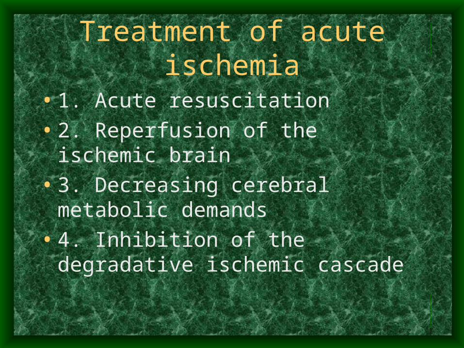
Treatment of acute ischemia
• 1. Acute resuscitation
• 2. Reperfusion of the ischemic brain
• 3. Decreasing cerebral metabolic demands
• 4. Inhibition of the degradative ischemic cascade
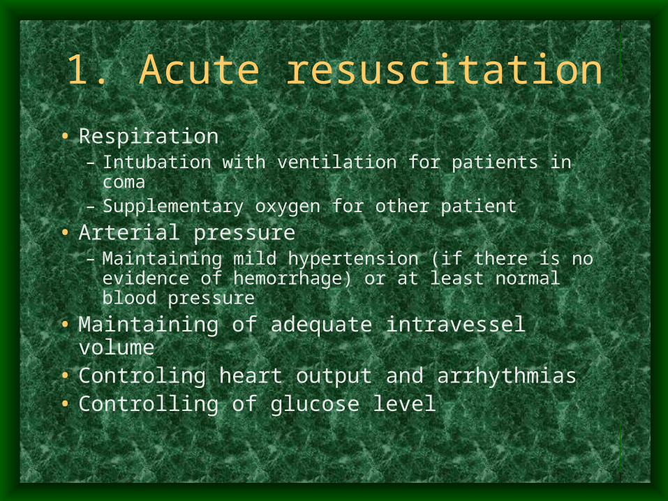
1. Acute resuscitation
• Respiration– Intubation with ventilation for patients in coma– Supplementary oxygen for other patient
• Arterial pressure– Maintaining mild hypertension (if there is no evidence
of hemorrhage) or at least normal blood pressure
• Maintaining of adequate intravessel volume• Controling heart output and arrhythmias• Controlling of glucose level
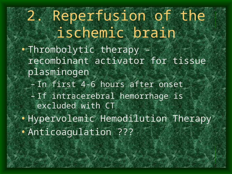
2. Reperfusion of the ischemic brain
• Thrombolytic therapy – recombinant activator for tissue plasminogen– In first 4-6 hours after onset– If intracerebral hemorrhage is excluded with
CT
• Hypervolemic Hemodilution Therapy
• Anticoagulation ???
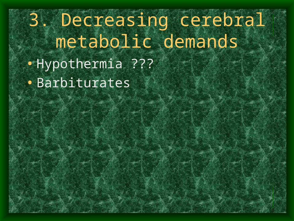
3. Decreasing cerebral metabolic demands
• Hypothermia ???
• Barbiturates
Surgical treatment for acute ischemia
• Possible only in cases of stenosis of great brain vessels (common carotid, internal carotid, middle cerebral arteries) – endarterectomia in first 2-3 hours.
Primary stroke prevention – controlling of risk factors
• Hypertension (increases risk of stroke in4-5 times)
• Smoking (1,5)
• Diabetes. (2,5-4)
• Lipids.
• Cardiac Disease.– Atrial fibrillation, (5)
– valvular heart disease, (4)
– myocardial infarction (5)
Secondary Stroke Prevention (After Transient Ischemic Attack or
Ischemic Stroke)• Aspirin 30-300 mg per day
• Or Ticlopidine
• Treatment or heart diseases
• Surgical
Surgical prevention of ischemia
• EXTRACRANIAL-TO-INTRACRANIAL CAROTID ARTERY BYPASS
• CAROTID ENDARTERECTOMY
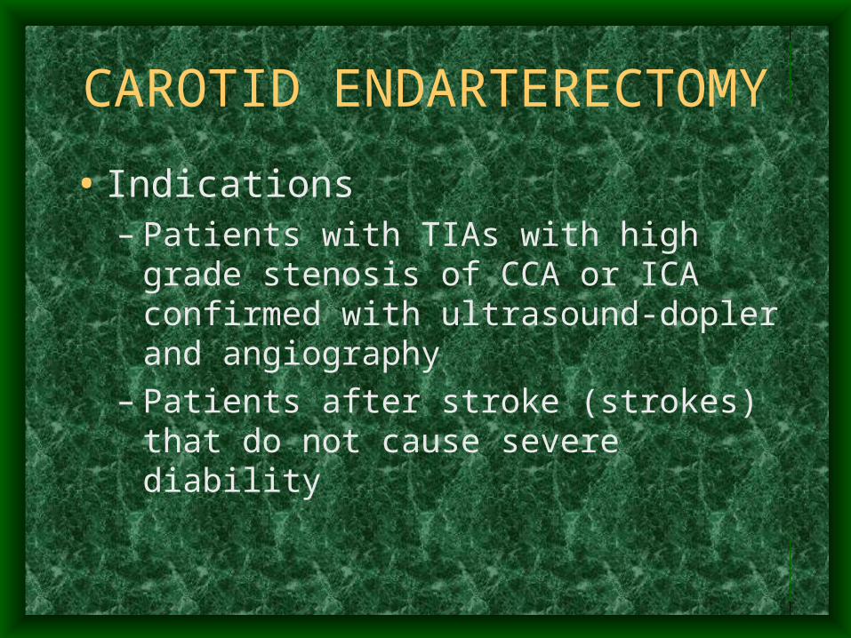
CAROTID ENDARTERECTOMY
• Indications– Patients with TIAs with high grade stenosis of
CCA or ICA confirmed with ultrasound-dopler and angiography
– Patients after stroke (strokes) that do not cause severe diability
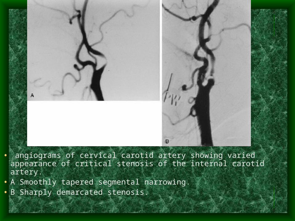
• angiograms of cervical carotid artery showing varied appearance of critical stenosis of the internal carotid artery.
• A Smoothly tapered segmental narrowing. • B Sharply demarcated stenosis.
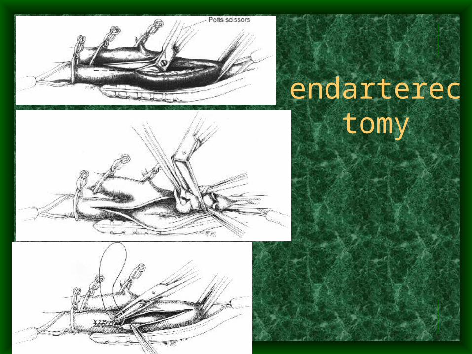
endarterectomy
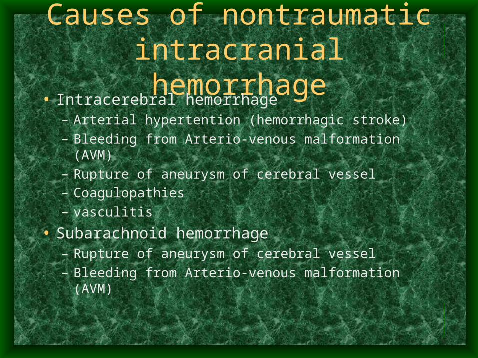
Causes of nontraumatic intracranial hemorrhage
• Intracerebral hemorrhage– Arterial hypertention (hemorrhagic stroke)
– Bleeding from Arterio-venous malformation (AVM)
– Rupture of aneurysm of cerebral vessel
– Coagulopathies
– vasculitis
• Subarachnoid hemorrhage– Rupture of aneurysm of cerebral vessel
– Bleeding from Arterio-venous malformation (AVM)
Clinical signs of hemorrhagic stroke due to hypertension
• Sudden and fast onset (seconds – minutes)
• Unconsciousness (semicoma-coma)
• Severe neurological deficit
• Vegetative symptoms: high arterial pressure; bradycardia, red face and cyanotic limbs, sweating.
• Severe headache in contact patients
Diagnostic procedures
• Computed tomography (CT)
• Angiography
• EchoEG
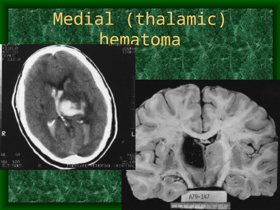
Medial (thalamic) hematoma
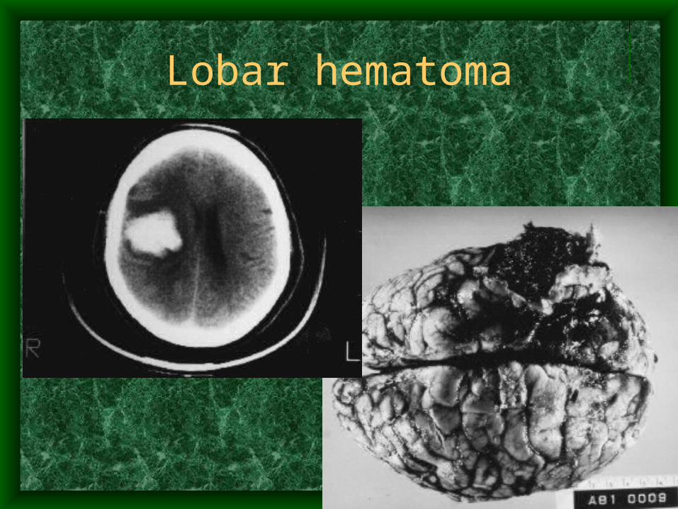
Lobar hematoma
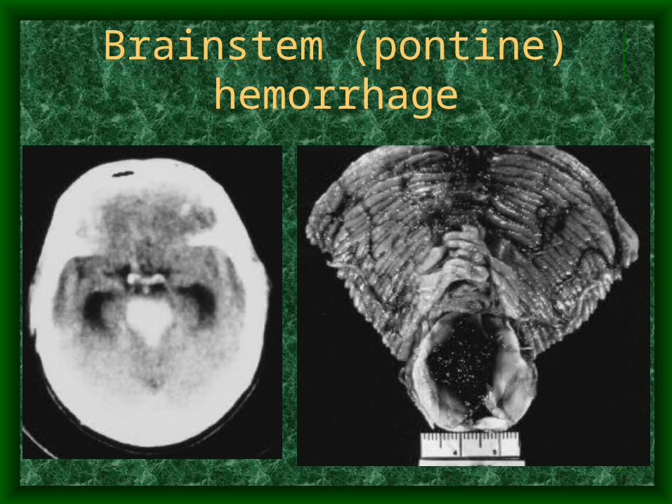
Brainstem (pontine) hemorrhage
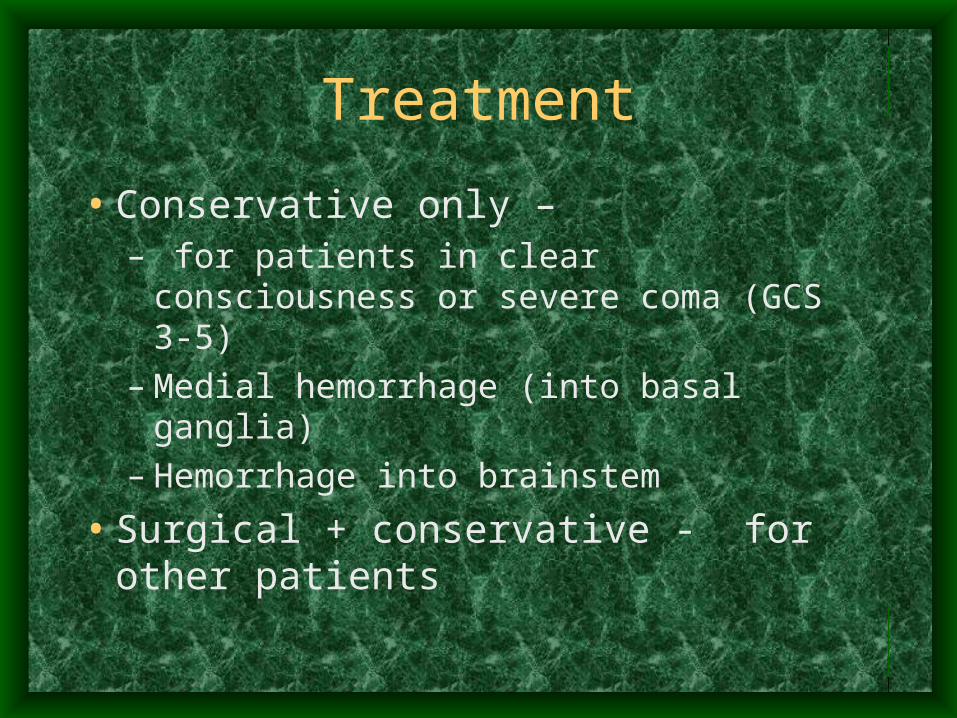
Treatment
• Conservative only –– for patients in clear consciousness or severe
coma (GCS 3-5)– Medial hemorrhage (into basal ganglia)– Hemorrhage into brainstem
• Surgical + conservative - for other patients
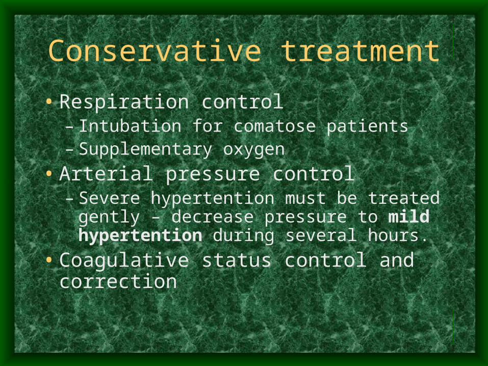
Conservative treatment
• Respiration control– Intubation for comatose patients– Supplementary oxygen
• Arterial pressure control– Severe hypertention must be treated gently –
decrease pressure to mild hypertention during several hours.
• Coagulative status control and correction
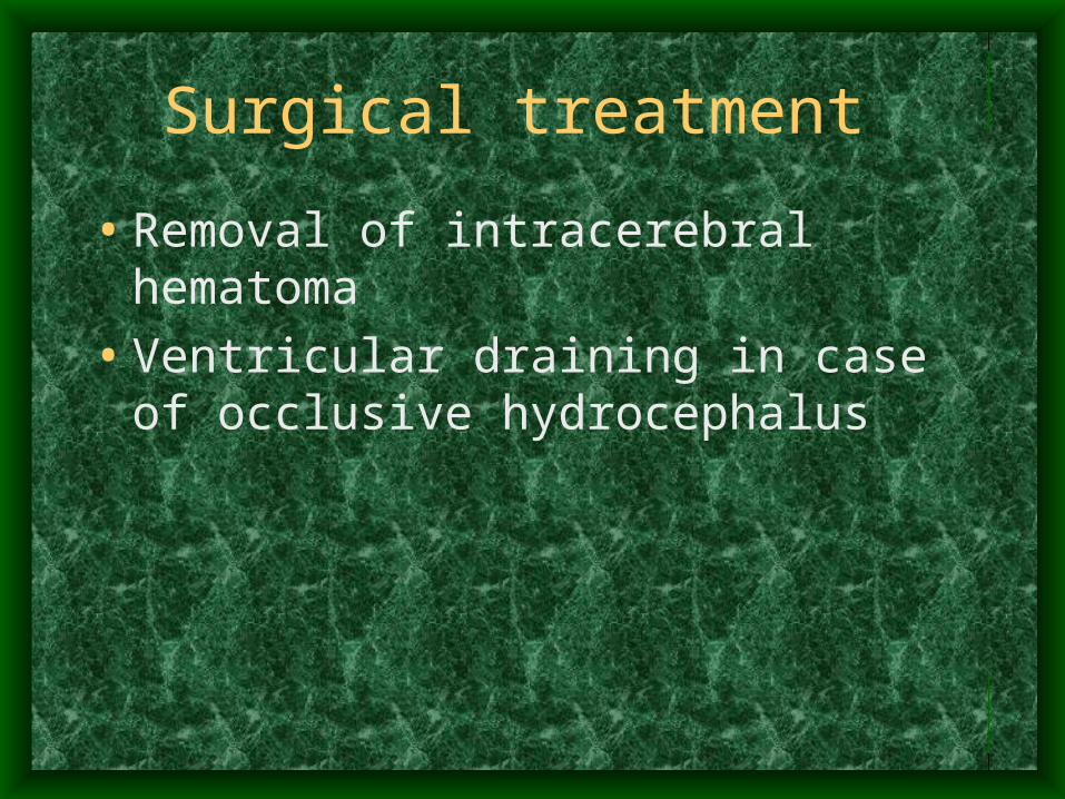
Surgical treatment
• Removal of intracerebral hematoma
• Ventricular draining in case of occlusive hydrocephalus
Clinical presentation of SAH
• Sudden onset• Severe headache• Meningeal signs• Minimal focal neurological deficit
• More rarely depressed level of consciousness and major neurological deficit
Diagnostic procedures for SAH
• CT • Lumbar puncture with CSF examination
– Blood in the CSF– High pressure of CSF– SAH and possible intracerebral hemorrhage
• Angiography – the main to reveal the cause of SAH – aneurisms and arterio-venous malformations
Aneurisms of cerebral arteries
• Localization– Anterior cerebral a. and anterior communicans .
- 45%– Internal carotid a. – 32%– Middle cerebral a. – 20%– Vertebrobasilar circulation – 4%
Aneurisms of cerebral arteries
• Saccular• Others (traumatic, atherosclerotic, mycotic,
neoplastic, inflamatory)
• Saccular aneurisms – ovoid-shaped outpouching of vessel wall, cased by congenital insufficiency of elastic component of vessel wall
SAH due to ruptured aneurism
• First rupture of aneurism – SAH only• Repeated rupture in 20-50% of cases, most
of them during 3-20 days after first • 50-85% mortality after repeated rupture, • Intracerebral hemorrhage are often at
repeated rupture• Often complicated with vasospasm and
consequent ischemical changes
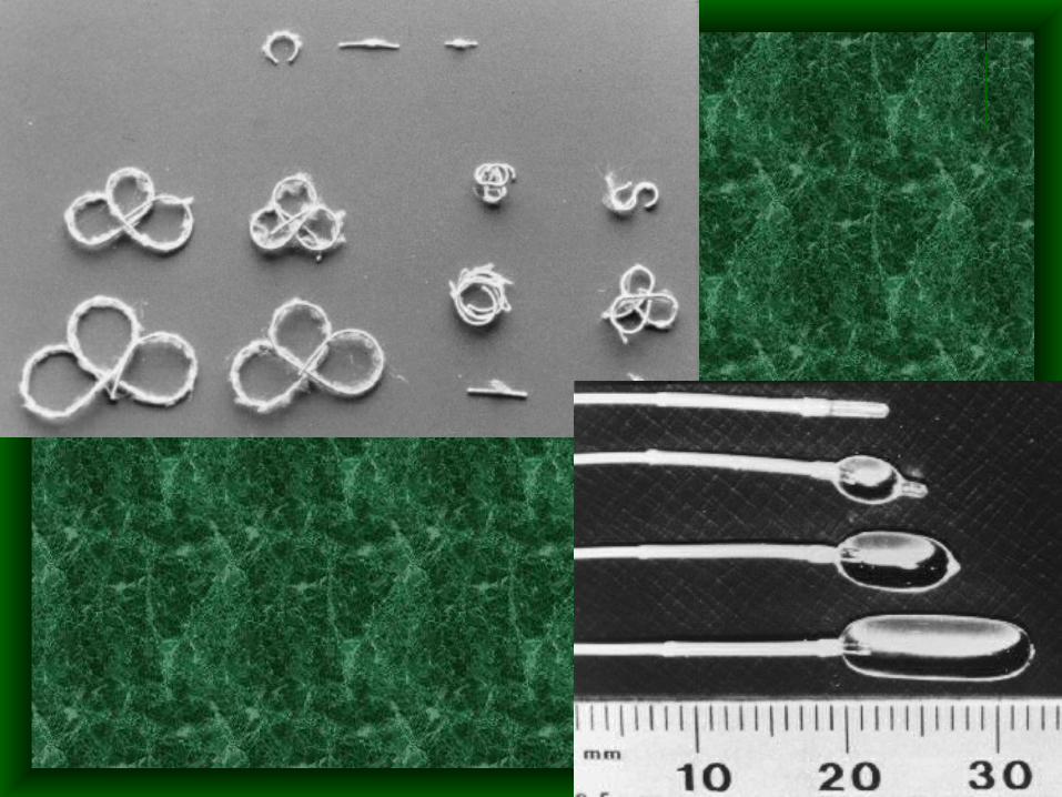
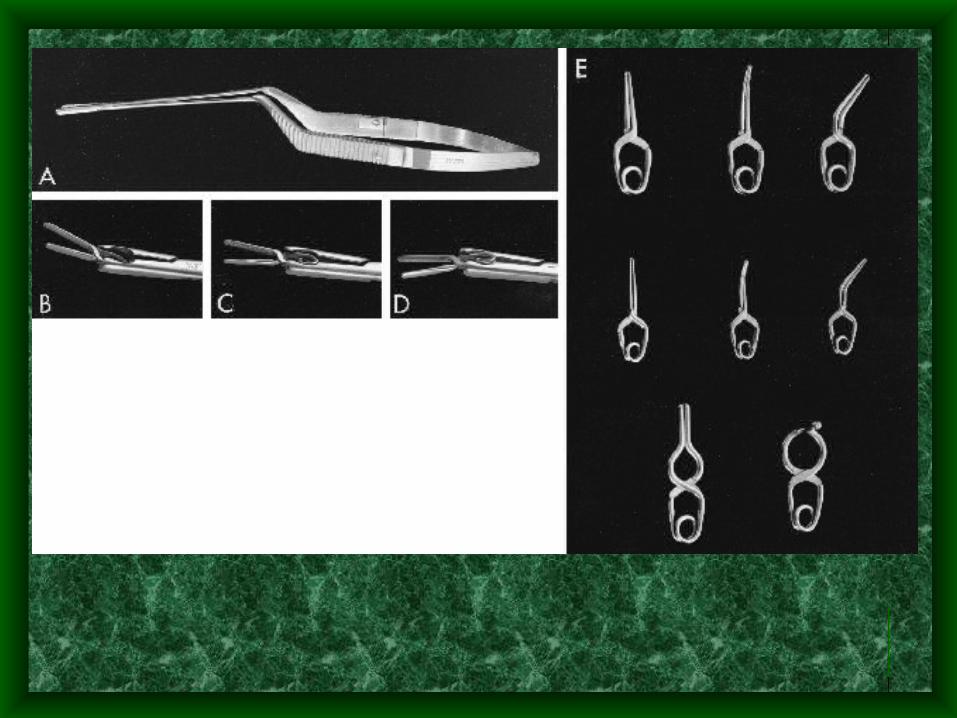
Surgical treatment of aneurism
• Any aneurism should be excluded from circulation as early as possible– Putting clips on the neck of aneurism– Endovascular embolisation of aneurism
• With coils
• With balloons
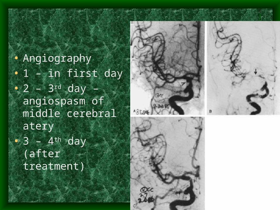
• Angiography
• 1 – in first day
• 2 – 3rd day – angiospasm of middle cerebral atery
• 3 – 4th day (after treatment)
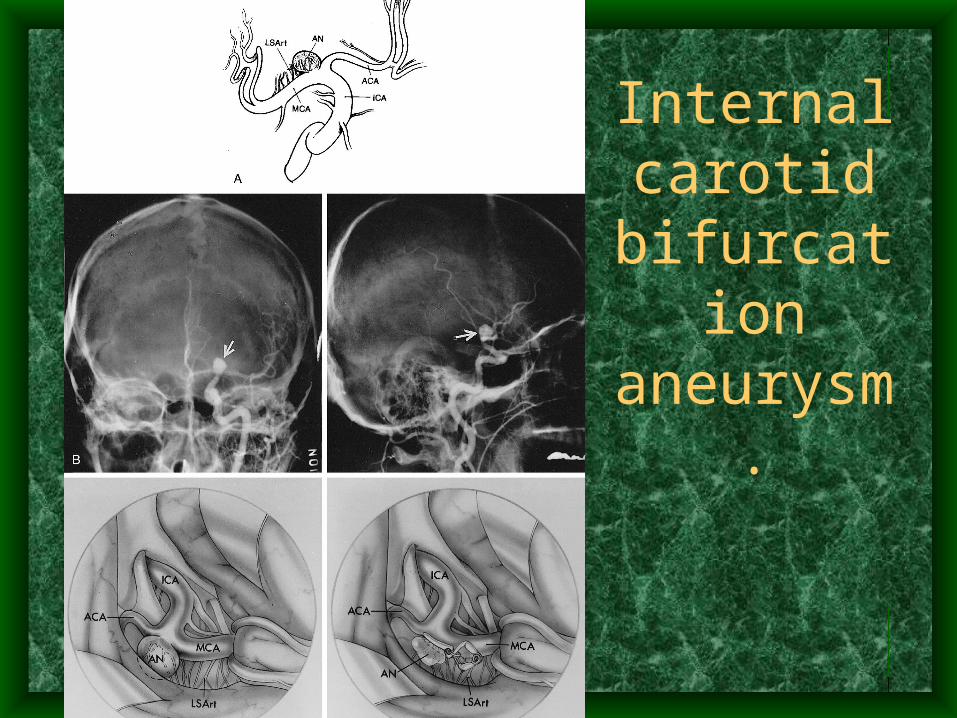
Internal carotid
bifurcation aneurysm.
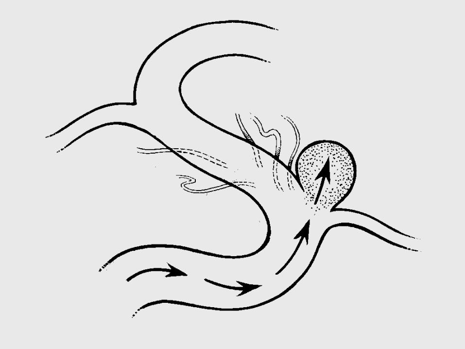
Arteriovenous malformations
• heterogeneous group of vascular developmental anomalies of the brain
• composed of a mass of abnormal arteries and veins of different sizes.
• Functionally, they represent direct artery-to-vein shunting with no intervening capillaries,
• angiographically are seen as early filling of veins.
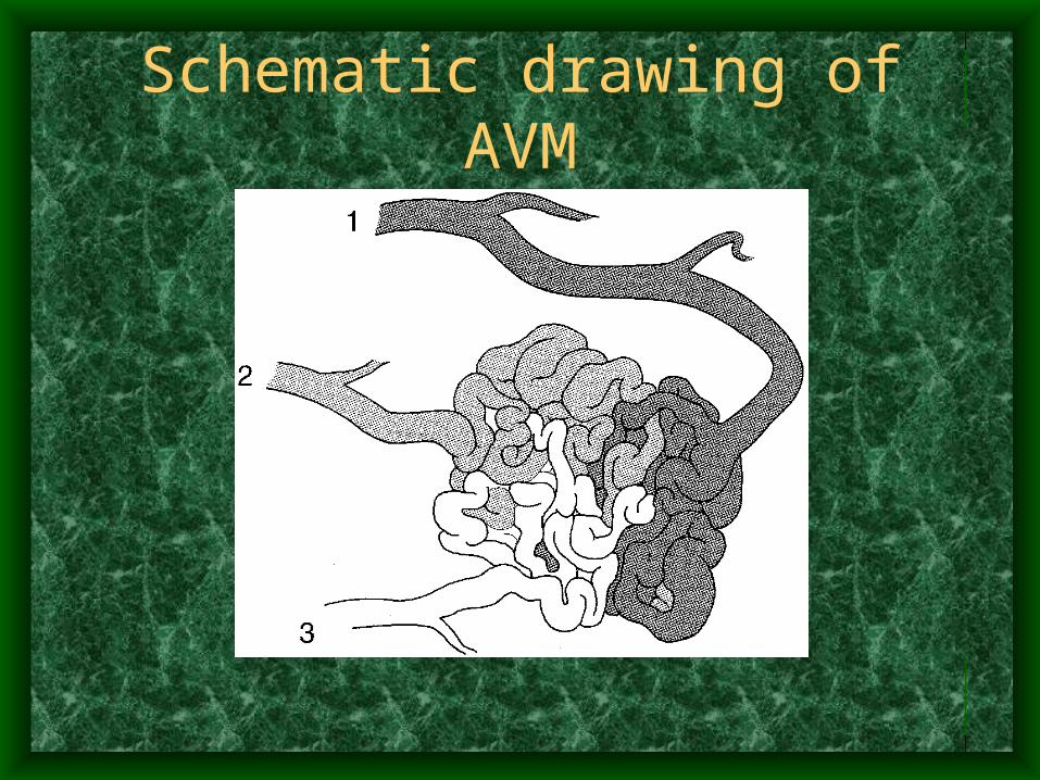
Schematic drawing of AVM
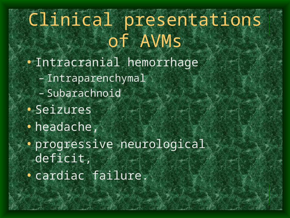
Clinical presentations of AVMs
• Intracranial hemorrhage– Intraparenchymal– Subarachnoid
• Seizures
• headache,
• progressive neurological deficit,
• cardiac failure.
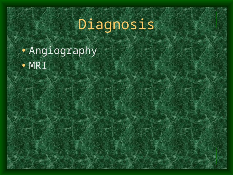
Diagnosis
• Angiography
• MRI
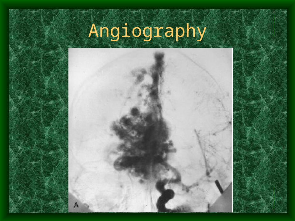
Angiography
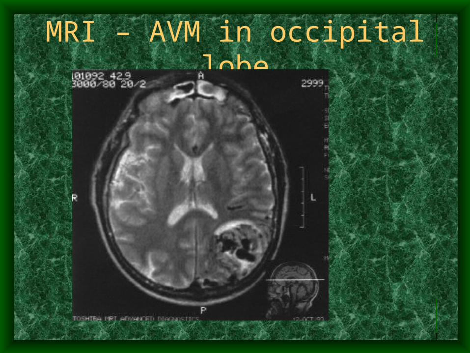
MRI – AVM in occipital lobe
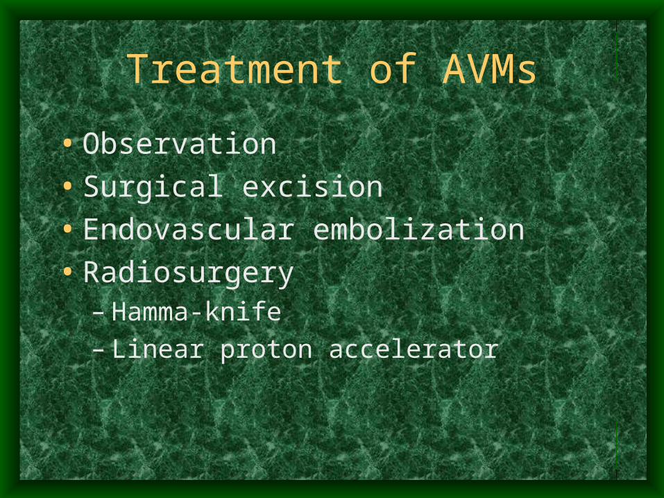
Treatment of AVMs
• Observation
• Surgical excision
• Endovascular embolization
• Radiosurgery– Hamma-knife– Linear proton accelerator
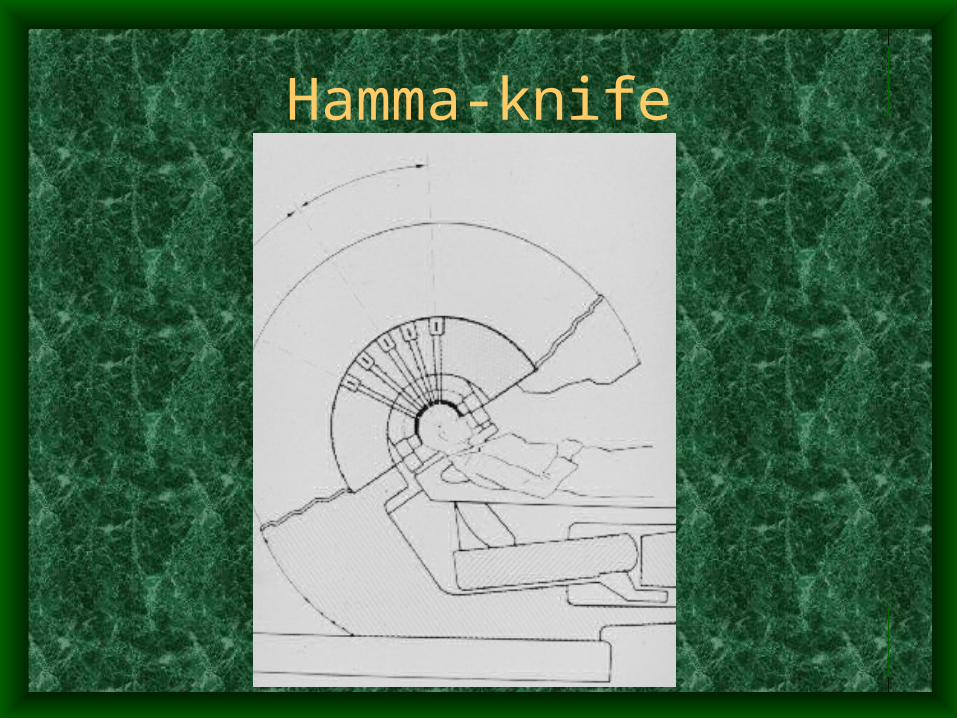
Hamma-knife


