Article ID: WMC004206 ISSN 2046-1690 Cervical Anatomy in Women at Risk of Preterm Labour Corresponding Author: Dr. Mohammad Othman, Consultant Obstetrician and Gynaecologist, School of Reeproductive and Developmental Medicine, University of Liverpool, 84 Bradfield Road, M32 9LE - United Kingdom Submitting Author: Dr. Mohammad Othman, Consultant Obstetrician and Gynaecologist, School of Reeproductive and Developmental Medicine, University of Liverpool, 84 Bradfield Road, M32 9LE - United Kingdom Article ID: WMC004206 Article Type: Clinical Trials Submitted on:17-Apr-2013, 10:16:36 AM GMT Published on: 18-Apr-2013, 08:32:04 AM GMT Article URL: http://www.webmedcentral.com/article_view/4206 Subject Categories:OBSTETRICS AND GYNAECOLOGY Keywords:Preterm birth, Power Doppler, 3D Ultrasound, 4D view, Cervical length, Cervical Volume How to cite the article:Othman M. Cervical Anatomy in Women at Risk of Preterm Labour . WebmedCentral OBSTETRICS AND GYNAECOLOGY 2013;4(4):WMC004206 Copyright: This is an open-access article distributed under the terms of the Creative Commons Attribution License(CC-BY), which permits unrestricted use, distribution, and reproduction in any medium, provided the original author and source are credited. Source(s) of Funding: None Competing Interests: None Additional Files: Figure 1 Figure 2 Table 1 Table 2 Table 3 Table 4 WebmedCentral > Clinical Trials Page 1 of 15
Transcript
Article ID: WMC004206 ISSN 2046-1690
Cervical Anatomy in Women at Risk of PretermLabourCorresponding Author:Dr. Mohammad Othman,Consultant Obstetrician and Gynaecologist, School of Reeproductive and Developmental Medicine, University ofLiverpool, 84 Bradfield Road, M32 9LE - United Kingdom
Submitting Author:Dr. Mohammad Othman,Consultant Obstetrician and Gynaecologist, School of Reeproductive and Developmental Medicine, University ofLiverpool, 84 Bradfield Road, M32 9LE - United Kingdom
Article ID: WMC004206
Article Type: Clinical Trials
Submitted on:17-Apr-2013, 10:16:36 AM GMT Published on: 18-Apr-2013, 08:32:04 AM GMT
Keywords:Preterm birth, Power Doppler, 3D Ultrasound, 4D view, Cervical length, Cervical Volume
How to cite the article:Othman M. Cervical Anatomy in Women at Risk of Preterm Labour . WebmedCentralOBSTETRICS AND GYNAECOLOGY 2013;4(4):WMC004206
Copyright: This is an open-access article distributed under the terms of the Creative Commons AttributionLicense(CC-BY), which permits unrestricted use, distribution, and reproduction in any medium, provided theoriginal author and source are credited.
WMC004206 Downloaded from http://www.webmedcentral.com on 18-Apr-2013, 08:32:07 AM
Cervical Anatomy in Women at Risk of PretermLabourAuthor(s): Othman M
Background
Preterm birth, defined as birth occurring after thegestational age of viability (23 weeks, 500 gramsweight) and before 37 completed weeks (259 days) ofpregnancy, is one of the most important problems inmedicine today. Preterm birth is the single largestcause of mortality and morbidity for newborns. Itaccounts for 5% to 11% of births in the world but isresponsible for 28% of all deaths within 28 days ofbirth and 50% of childhood neurological disabilities [1,2]. Other important adverse outcomes of preterm birthinclude respiratory distress syndrome, intraventricularhaemorrhage, leukomalacia, necrotizing enterocolitisand prolonged hospitalisation [2]. Survivors canexperience life-long complications including cerebralpalsy, blindness and deafness [1, 2].
Psychologically, giving birth to a preterm infant isconsidered to be a stressful event for parents. Manystudies have shown that mothers of these infantsexperience increased levels of stress in the neonatalperiod compared with mothers of term infants, andthey are more likely to suffer from depression andanxiety at the time of hospital discharge [3]. There isalso increased depressive symptoms among fathers ofpreterm infants during the neonatal intensive careunite stay [4]. It is assumed that increased parentingstress could interfere with the parent-child relationshipduring early childhood and consequently increase therisk for later behavioural problems [3, 5].
The direct and indirect costs of prematurity can beimmense [2]. The lifetime costs per preterm birth(baby's birth weight less than 2500 grams) have beenestimated at £511,614 [1, 6].
The incidence of preterm deliveries in developedcountries is 6% to 9%, currently it is 7% intheUKaffecting 21,000 babies each year inEngland.Preterm premature rupture of the membranes andspontaneous preterm labour accounts forapproximately 80% of preterm deliveries; theremaining 20% are planned deliveries for maternal orfetal reasons (for example, eclampsia) [7].
In the last 20 years it has become clear that infectionis an important cause of preterm labour and deliveryleading to more than 50% of the all preterm deliveries
world-wide [1, 8-14]. Infection has been recognised asan important and frequent mechanism of disease inpreterm birth with a firm link to prematurity. Theevidence that implicate infection as a cause of pretermlabour and birth includes:
Administration of microbial products to pregnant●
animals results in preterm birth.Systematic maternal infection, for example,❍
pyelonephritis, pneumonia or even Dental cariesare associated with preterm labour.Subclinical intrauterine infections usually trigger❍
preterm birth.Treatment of asymptomatic bacteriuria prevents❍
preterm labour.Clinical infection is increased in the infant and the❍
mother after preterm birth.10-15% of amniotic fluid cultures from■
preterm labour patients are positive formicroorganisms.Antibiotic treatment of intrauterine infections■
can prevent prematurity in experimentalmodels of chorioamnionitis [15].
Since infection is frequently difficult to confirm, weoften refer to women with positive amniotic culture,histological evidence of chorioamnionitis or elevatedcytokines in the amniotic fluid as having a subclinicalinfection. In this context, the organisms involved maynot be necessarily pathogenic; a change in vaginalflora may be enough to trigger the sequence of eventsleading to a preterm birth [1, 2, 8, 11, 14, 16-18].
The most common pathway for pathogens to causepreterm labour is the ascending route [2, 14]: severalmechanisms contribute to this pathway. Pathogensproduce proteolytic enzymes including different typesof mucinases, sialidases, peptidase and protease. Thepresence of bacterial sialidases facilitates theattachment of bacteria to cervical mucus and thebreakdown of mucin, while bacterial mucinases assistascent into uterine tissues [2, 12, 19]. Other enzymesmay act directly on cervical collagen leading topremature shortening and ripening cervix while alsoweakening the fetal membranes leading to pretermpremature rupture of the membranes [2, 12, 19].
Microorganisms stimulate maternal monocytes andmacrophages resulting in the production ofphospholipase A2. This is an enzyme that liberatesarachidonic acid from the phospholipids of themembranes leading to the synthesis of prostaglandins
WebmedCentral > Clinical Trials Page 2 of 15
WMC004206 Downloaded from http://www.webmedcentral.com on 18-Apr-2013, 08:32:07 AM
E2 and F2α by the placental membranes:prostaglandins are potent stimulator of uterinecontractions [14, 20-27]. Similarly, protease toxinsactivate the decidua and fetal membranes to produceCytokines such as Tumour Necrosis Factor (TNF),In te r leuk in ( IL1α , I L1β , I L6 , IL8 ) , andGranulocyte-Macrophage Colony Stimulating Factor(GM-CSF) [9, 12, 14, 20-22, 25-27]. The activation ofa local inflammatory reaction leads to prostaglandinsynthesis and release which subsequently stimulateuterine contractions [28-30]. Moreover, in infectedfoetuses, there is an increase in both fetalhypothalamic and placental product ion ofcorticotrophin releasing hormone leading to increasein fetal corticotrophin secretion, which in turnincreases fetal adrenal cortisol production leading toincreased production of prostaglandins [20, 25, 28].When the fetus is infected, there is a high increase inthe production of cytokines and marked decrease inthe delivery time with a high chance of direct fetaltissue damage (e.g. fetal brain or lung) [2, 14, 19, 20,24, 25, 31].
During pregnancy the primary function of the uterinecervix is to remain closed in order to retain the babywithin the uterus until fetal maturity and birth. Asecondary function of the cervix is to prevent infectionascending from the vagina into the uterus. Prior tonormal delivery at term, cervix shortens, softens andripens (becomes more distensible), to facilitatecervical dilatation by myometrial contractions duringlabour. The cervix consists mainly of connective tissue,principally, collagen fibres in a proteoglycan groundsubstance. The interaction between these twosubstances gives the cervix its unique characteristics,where the collagen fibres resist pulling forces and theground substance resists compressive forces [32].
Various methods have been used to try and detectcervical changes that predict preterm labour. Theseinclude manual vaginal examination, transabdominalultrasound, and transvaginal ultrasound [29, 33]. Ofthese modalities, measurements of cervical lengthusing transvaginal ultrasound scanning appear to havethe highest sensitivity, whereas transabdominalscanning was not predictive [29, 30]. There is however,no clear cut gestation at which the test should beperformed or what cervical length provides the bestcut-off for a diagnostic test [29, 33].
Rationale
We have analysed observational data on 106 womenwith a past history of preterm labour and birth [34] inorder to test the hypothesis that women with a past
history of preterm labour and birth have a moremarked inflammatory response in the cervical mucus.Strikingly, we found that in a subsequent pregnancy,women with a low macrophage count (<5% of cervicalepithelial cells expressing CD14 antigen) before 20weeks gestation were more likely to have recurrentpreterm birth compared with women who had normalcervical macrophage count (Odds Ratio 4.9, 95%CI1.5 to 18.7; P 0.0037). This prompted us to develop anew model for ascending infection, that ascendinginfection occurs in the presence of a defective cervicalbarrier.
Objective
To investigate to what extent a defective cervicalbarrier is a contributory factor to recurrent pretermlabour.
Hypothesis
Patients at high risk of preterm labour will have lowernumber of cervical leukocytes in general andmacrophages specifically. The lower the number ofmacrophages the less vascular the cervix would be,which would result in low readings on power Dopplerindices.
Methods and Materials
This is a hospital prospective observational cohort(non interventional study). Study was conducted inLiverpool Women's Hospital from October 2007 untilOctober 2009. Methods were explained in a previouspublication (Cervical Immunology in Women at Risk ofPreterm Labour).
3D ultrasound methods
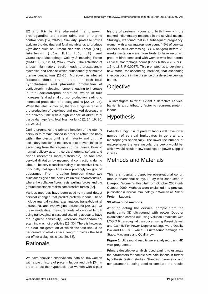
After collecting the cervical sample from theparticipants 3D ultrasound with power Dopplerexamination carried out using Voluson i machine withLOGIQ 9 transvaginal transducer, using Preset defaultand Gain 5. For Power Doppler settings were Qualitylow and PRF 0.6, while 3D ultrasound settings areStatic, Max angle and Quality low,
Figure 1. Ultrasound results were analysed using 4Dview programme.
Primary descriptive analysis used aiming to estimatethe parameters for sample size calculations in furtherhypothesis testing studies. Standard parametric andnonparametric testing used to compare the results
WebmedCentral > Clinical Trials Page 3 of 15
WMC004206 Downloaded from http://www.webmedcentral.com on 18-Apr-2013, 08:32:07 AM
between high risk group and controls. Conventionallevel of statistical significance (P<0.05) used.
Results
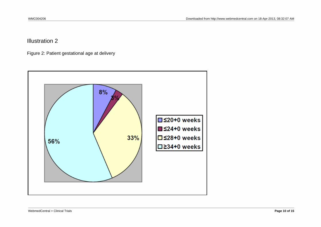
89 participants were recruited, 50 controls and 39patients. Demographically both groups were similar.The difference in the history of previous preterm birthwas statistically significant but this is expected sincepatients were defined as having history of previouspreterm birth while, this is an exclusion criteria in thecontrols, Table 1.
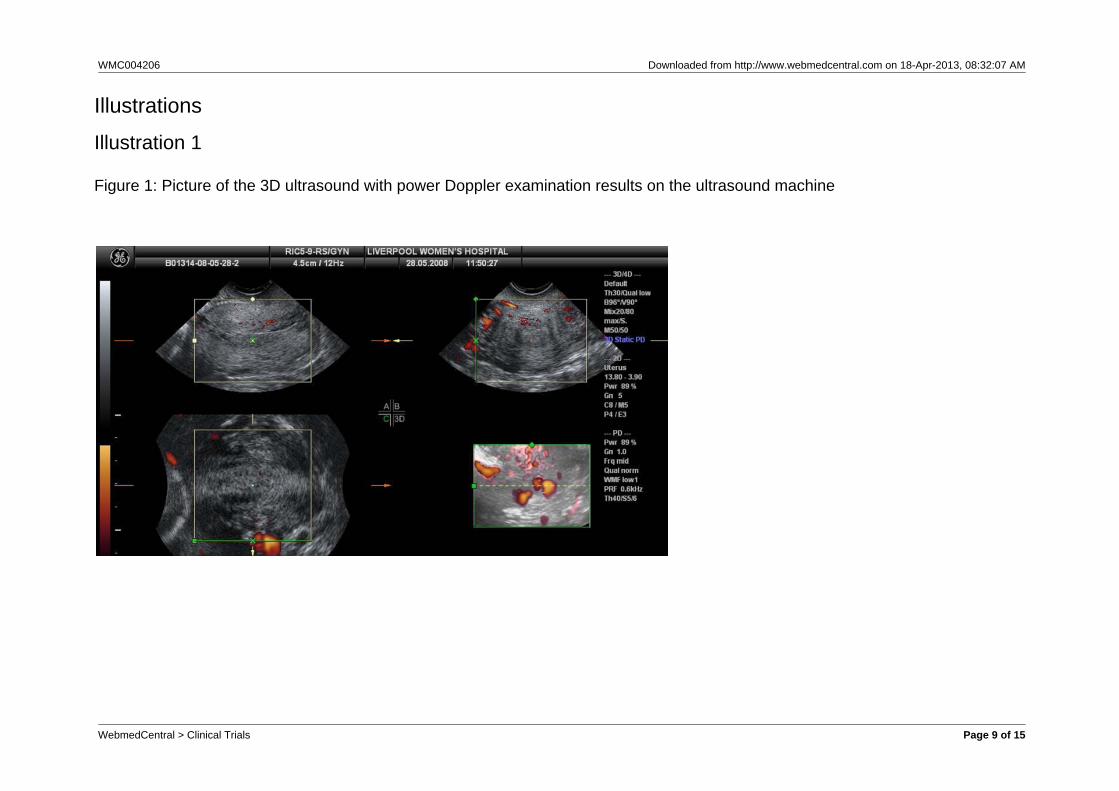
Of the 39 patients 22 delivered after 34+0 completedweeks and 17 delivered before 34+0 weeks. Of thosedelivered preterm, three delivered before 20+0completed weeks, one before 24+0 completed weeksand thirteen before 28+0 completed weeks, Figure 2.
Only one patient underwent caesarean section (forhistory of previous caesarean section). Another patientdeveloped pregnancy induced hypertension and onadmission she complained of abdominal pain. Shedelivered within 4 hours of admission at 26+3 weeksgestation.
Three babies delivered before 20+0 weeks died within30 minutes of delivery. One baby delivered before24+0 weeks was admitted to the NICU, butunfortunately developed infection and died at the ageof 2 weeks. The rest of the patient’s babies wereadmitted to the NICU and later discharged in goodcondition.
All controls delivered after 34+0 completed weeks ofthe pregnancy, Table 2. Six controls underwentcaesarean section. Three of those controls had ahistory of previous caesarean section. The other threehad caesarean sections for antepartum haemorrhage,one for abruptio placentae and two for placentapraevia. Two sets of twins delivered vaginally. One at36+4 weeks, the other at 37+2 weeks.
Of all babies delivered to controls, three babies wereadmitted to the NICU for meconium ingestion anddischarged later in good condition.
For the rest of the results, patients who delivered aftercompleted 34+0 weeks of gestation were excludedfrom the analysis.
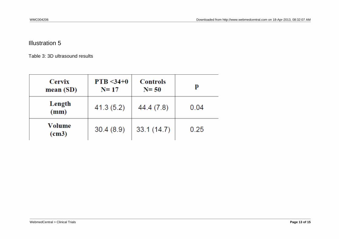
Ultrasound results showed a statistically significantassociation between decrease in cervical length andpreterm labour. On the other hand, a decrease incervical volume was not associated with pretermlabour, Table 3.
Settings for the ultrasound examination found to bedifferent from the agreed settings in two patients.
Added to that, art i facts affected two otherexaminations. Consequently, power Doppler datawere also affected. Those four patients were excludedfrom the analysis. For the controls, settings of theultrasound examination found to be different from theagreed settings in thirteen controls. In the other hand,artifacts did not affect any of the controls examinations.Those thirteen controls were excluded from theanalysis.
An increase in vascular index and vascular flow indexwere associated with preterm labour. The flow indexdid not show any significant difference between thetwo groups, Table 4.
Discussion
Twin pregnancies were included in this study becausethere were plans to perform a separate analysis fortwin pregnancies. This analysis would have been usedas a pilot for a future large study of twin pregnancies.However, recruiting twin pregnancies proved difficult. Itis recognised that the recruitment of twin pregnanciesin this study is inappropriate because twin pregnanciesare more prone to preterm labour than singletonpregnancy [36-38]. This wil l be taken intoconsideration in designing further studies.
The mucosal surface of the female genital tract servesas a potential site of entry for a variety of bacterial andviral pathogens [24, 26, 38-41]. Most infections areconfined to the lower genital tract. Consequently, ahigher level of immunological activity is associatedwith this region [24, 38, 40-42].
In the cervix, leukocytes are sparse prior to the onsetof labour [14, 40, 43]. There is increasing evidencethat the process of cervical dilatation resembles aninflammatory response [40, 43]. Leucocytes migrateinto cervical stroma and mucus during labour reachinga density 2–3-fold higher than that found in latepregnancy [7, 43, 44]. This stromal infiltrate iscomposed principally of neutrophils and macrophages[43]. In contrast, others reported an increase inmacrophage and neutrophil numbers in cervicalstroma during late pregnancy, but no further changesduring labour [40, 45]. This discrepancy may relate todifferences in cervical ripening at the time of thesample collection, but no clinical information on thestate of the cervix was provided in any studies. Thismeans that the identification of leukocyte phenotypesat the mucosal surface of the endocervix is integral tounderstanding the pathogenesis of genital infectionand the role of protective immunity.
Traditional methods to evaluate the cervix in
WebmedCentral > Clinical Trials Page 4 of 15
WMC004206 Downloaded from http://www.webmedcentral.com on 18-Apr-2013, 08:32:07 AM
pregnancy are limited and unsatisfactory. Attempts toscreen women efficiently with risk-scoring systems ordigital examination of the cervix, the standard method,suffers from large variation among examiners, andhave revealed low sensitivities and low predictivevalues [53-55]. Vaginal ultrasonography producesgood images and is well accepted by patients. Thereare no apparent risks associated with the examination[53-56]. In contrast, transvaginal ultrasonography is areproducible method of examination during pregnancy.The length of the cervix is directly correlated with theduration of pregnancy: the shorter the cervix, thegreater the likelihood of preterm delivery. This ismainly because uterine contractions, whetherperceived by the woman or not, shorten the cervix[53-60].
3D ultrasound was first developed atDukeUniversityin1987 by Olaf von Ramm and Stephen Smith [61]. Itwas available for clinical research in the beginningthen for clinical use in 1992. In 3D ultrasoundscanning, instead of sound waves being sent straightdown and reflected back, they are sent at differentangels. The returning echoes processed by computerprogram which reconstruct three dimensional volumeimage, but no movement is shown [61, 62]. 3Dultrasonography has the potential to provide moreaccurate volume measurements than conventional 2Dultrasound [56-58, 61, 63]. 3D imaging combined withpower Doppler became available for clinical use in1995 and provided the potential to quantify powerDoppler signals in a whole organ [56-59, 63, 64].
Results of this study showed a significant associationbetween cervical length and preterm labour, but notthe cervical volume. This could be due to the samplesize or due to the difficulty of estimating cervicalvolume. Since one of the methodological difficultieswhen estimating cervical volume and vascularity using3D ultrasound is defining landmarks when drawing thecontours of the cervix [53, 56-59, 64-66].Thedelineation between the cervix and the lower uterinesegment is particularly difficult, especially early inpregnancy and at mid-gestation when the loweruterine segment is thick and the cervix often curved. Itmay also be difficult to clearly separate the cervix fromthe surrounding vaginal tissue [53, 56-59, 64, 66].
Our hypothesis was that patients at high risk will havelower numbers of macrophages, and in turn lessvascular cervix which would be seen as low readingson the power Doppler indices, but against ourhypothesis, results of this study showed a significantinverse association between both vascular index andvascular flow index and preterm labour. On the otherhand, flow index also increased in preterm labour
patients than controls but this is not statisticallysignificant. This means there is an increase in thevascularisation of the cervix in preterm labour patients.This result could be due to the small sample size orthe difficulty in defining the landmarks when drawingthe contours of the cervix which in turn affects thecalculation of power Doppler indices. The last possibleexplanation is a premature remodeling of cervicalarchitecture and this may include an element ofpreconceptional hypervascularity since all thesewomen have previous history of preterm labour. Thecervix of a patient who has had a previous history ofpreterm labour may have permanent changes to hercervix [40, 67-69]. Part of the ripening process iscytokine stimulation of an influx of macrophages andneutrophils which in turn not only produce morecytokines but also induce angiogenic activity with theresult of newly formed microvessels which will allowmore influx of the macrophages and neutrophils [67,69]. Newly formed microvessels resolve soon afterdelivery but a small fraction remains as part of thenormal structure of the cervix. In the next pregnancythese microvesse ls are represented ashypervascularity of the cervix [40, 67-71]. Accordingly,a preconceptional 3D ultrasound with power Doppler isneeded to confirm or refute the presence of thisphenomenon.
Conclusion
There was a significant association between cervicallength and preterm labour, but not the cervical volume.
There is a real requirement for more research oncervical leukocyte population at reasonable time inpregnancy. Adding the 3D ultrasound element andpower Doppler at the same time will provide volubleinformation about the cervical morphology.
References
1. Kiss H, Petricevic L, Husslein P. Prospectiverandomised controlled trial of an infection screeningprogramme to reduce the rate of preterm delivery.BMJ. 2004;1136(10):1-5.2. McGregor J, French J. Pathogenesis to treatment:preventing preterm birth mediated by infection.Infectious Diseases in Obstetrics and Gynaecology.1997;5:106-14.3. Kaaresen P, Ronning J, Ulvund S, Dahl L. Arandomized, controlled trial of the effectiveness of anearly-intervention program in reducing parenting stressafter preterm birth. Pediatrics. 2006;118(1):9-19.
WebmedCentral > Clinical Trials Page 5 of 15
WMC004206 Downloaded from http://www.webmedcentral.com on 18-Apr-2013, 08:32:07 AM
4. Carter J, Mulder R, Bartram A, Darlow B. Infants ina neonatal intensive care unit: parental response.Archives of Disease in Childhood. 2005;90:F109-F13.5. Magill-Evans J, Harrison M. Parent-childinteractions and development of toddlers born preterm.Weste rn Journa l o f Nursery Research .1999;21:292-307.6. Petrou S, Sach T, Davidson L. The long term costsof preterm birth and low birth weight: results of asystematic review. Child: Care, Health andDevelopment. 2000;27(2):97-115.7. Yost N, Cox S. Infection and Preterm Labour.C l i n i ca l Obs te t r i c s and Gynaeco logy .2000;43(4):759-67.8. Chaim W, Mazor M, Leiberman J. The relationshipbetween bacterial vaginosis and preterm birth.Archives of Gynecology and Obstetrics. 1997;259:51-8.9. Cram L, Zapata M, Toy E, Baker B. Genitourinaryinfections and their association with preterm labor.American Family Physician. 2002;65(2):241-8.10. Crowther C, Thomas N, Middleton P, Chua M,Esposito M. Treating periodontal disease forpreventing preterm birth in pregnant women(Protocol). The Cochrane Database of SystematicReviews. 2005 issue 4.11. Hillier S, Nugent R, Eschenbach D, Krohn M,Gibbs R, Martin D, et al. Association between bacterialvaginosis and preterm delivery of a low birth weightinfants. The New England Journal of Medicine.1995;333(26):1737-42.12. Klein L, Gibbs R. Use of microbial cultures andantibiotics in the prevention of infection-associatedpreterm birth. American Journal of Obstetrics andGynecology. 2004;190:1493-502.13. Reid G, Bocking A. The potential for probiotics toprevent bacterial vaginosis and preterm labour.American Journal of Obstetrics and Gynaecology.2003;189(4):1202-8.14. Romero R, Espinoza J, Chaiworapongsa T,Kalache K. Infection and prematurity and the role ofpreventive strategies. Seminaries in Neonatology.2002;7:259-74.15. Romero R, Chaiworapongsa T, Kuivaniemi H,Tromp G. Bacterial vaginosis, the inflammatoryresponse and the risk of preterm birth: A role forgenetic epidemiology in the prevention of preterm birth.American Journal of Obstetrics and Gynecology.2004;190:1509-19.16. Leitich H, Brunbauer M, Bodner-Adler B, Kaider A,Egarter C, Husslein P. Antibiotic treatment of bacterialvaginosis in pregnancy: A meta-analysis. AmericanJournal of Obstetrics and Gynaecology. 2003;188(3):752-817. Reid G, Bruce A, Fraser N, Heinemann C, Owen J,
Henning B. Oral probiotics can resolve urogenitalinfections. FEMS Immunology and MedicalMicrobiology. 2001;30:49-52.18. Reid G, Jass J, Sebulsky M, McCormick J.Potential Uses of Probiotics in Clinical Practice.Clinical Microbiology Reviews. 2003;16(4):658-72.19. Kurki T. A survey of etiological mechanisms andtherapy of preterm labor. Acta Obstetricia etGynecologica Scandinavica. 1998;77:137-41.20. Bernal A, Watson S, Phaneuf S, Europe-Finner G.Biochemistry and physiology of preterm labour anddelivery. Baill iere's Clinical Obstetrics andGynaecology 1993:523-52.21. Goldenberg R, Hauth J, Andrews W. Intrauterineinfection and preterm delivery. The New EnglandJournal of Medicine. 2000;342(20):1500-7.22. Goldenberg R, Culhane J. Prepregnancy healthstatus and the risk of preterm delivery. Archives ofPediatric and Adolescent Medicine. 2005;159:89-90.23. Goncalves L, Chaiworapongsa T, Romero R.Intrauterine Infection and Prematurity. MentalRetardation and Developmental Disabilities.2002;8:3–13.24. Hay P. Bacterial vaginosis and pregnancy. Infection and Pregnancy 2001:158-71.25. Howe L, Wiggins R, Soothill P, Millar M, Horner P,Corfield A. Mucinase and sialidase activity of thevaginal micro flora: implications for the pathogenesisof preterm labour. International Journal of STD andAIDS. 1999;10:442-7.26. Locksmith G, Duff P. Infection, antibiotics, andpreterm delivery. Seminars in Perinatology.2001;25(5):295-309.27. Riggs M, Klebanoff M. Treatment of vaginalinfections to prevent preterm birth: a meta-analysis.C l i n i ca l Obs te t r i c s and Gynaeco logy .2004;47(4):796-807.28. Ganong W. Human reproduction. Review ofmedical physiology. 22nd ed: McGraw-Hill Companies2005:519-601.29. Norman J. Cervical function and prematurity. BestPractice & Research Clinical Obstetrics andGynaecology. 2007:1-16.30. Vonderpool B. Preterm labor: diagnosis andt rea tment . Amer ican Fami ly Phys ic ian .1998;57(10):2457-70.31. Lamont R. Infection in preterm labour. Infectionand Pregnancy 2001:305-17.32. Wilks W, Wiggins R, Whiley A, Hennessy E,Warwick S, Porter H, et al. Identification and H2O2Production of Vaginal Lactobacilli from PregnantWomen at High Risk of Preterm Birth and Relationwith Outcome. Journal of Clincal Microbiology.2004;42(2):713-7.
WebmedCentral > Clinical Trials Page 6 of 15
WMC004206 Downloaded from http://www.webmedcentral.com on 18-Apr-2013, 08:32:07 AM
33. Vause S, Johnston T. Management of pretermlabour. Archives of Disease in Childhood.2000;83:79-85.34. Whitworth M, Pafilis I, Vince G, Quenby S.Cervical leukocyte sub-populations in idiopathicpreterm labour. Journal of Reproductive Immunology.2007;75:48-55.35. Trotter J. WinMDI 2.9. 2.9 ed: Purdue University,USA 2000.36. Cunningham F, MacDonald P, Gant N, Leveno K,Gilstrap L, Hankins G, et al. Preterm birth. In: Lange A,ed. Williams Obstetrics. 20th ed: McGraw-HillCompanies 1997:797-826.37. Goldenberg R. The management of preterm labor.Obstetrics and Gynaecology. 2002;100(5):1020-1-37.38. Haram K, Mortensen J, Wollen A. Preterm delivery:an overview. Acta Obstetricia et GynecologicaScandinavica. 2003;82:687-704.39. Leitich H. Secondary predictors of preterm labour.BJOG. 2005;112(1):48–50.40. Liggins G. Cervical ripening as an inflammatoryreaction. The Cervix in Pregnancy and Labour. 3rd ed.Edinburgh: Churchill-Livingstone 1981:1-9.41. MacLean A. Asymptomatic bacteriuria duringpregnancy. Infection and Pregnancy 2001:85-95.42. Marelli G, Papaleo E, Ferrari A. Lactobacilli forprevention of urogenital infections: a review. EuropeanRevision of Medical Pharmacological Science.2004;8:87-95.43. Osman I, Young A, Ledingham M, Thomson A,Jordan F, Greer I, et al. Leukocyte density andpro-inflammatory cytokine expression in human fetalmembranes, decidua, cervix and myometrium beforeand during labour at term. Molecular HumanReproduction. 2003;9(1):41-5.44. Luo L, Ibaragi T, Maeda M, Nozawa M, KasaharaT, Sakai M, et al. Interleukin-8 levels and granulocytecounts in cervical mucus during pregnancy. AmericanJournal of Reproductive Immunology. 2000;43:78–84.45. Bokstrom H, Brannstrom M, Alexandersson M,Norstrom A. Leukocyte subpopulations in the humanuterine cervical stroma at early and term pregnancy.Human Reproduction. 1997;12(3):586-90.46. Gibbs R, Esehenbaeh D. Use of antibiotics toprevent preterm birth. American Journal of Obstetricsand Gynecology. 1997;177(2):375-80.47. Jacobsson B, Pernevi P, Chidekel L,Platz-Christensen J. Bacterial vaginosis in earlypregnancy may predispose for pretermbirth andpostpartum endometritis. Acta Obstetricia etGynecologica Scandinavica. 2002;81:1006–10.48. Lamont R. The prevention of preterm birth with theuse of antibiotics. European Journal of Pediatrics.1999;158(1):2-4.
49. Lamont R. Can antibiotics prevent pretermbirth—the pro and con debate. British Journal ofObstetrics and Gynaecology. 2005;112(1):67–73.50. Prakash M, Patterson S, Kapembwa M. Evaluationof the cervical cytobrush sampling technique for thepreparation of CD45q mononuclear cells from thehuman cervix. Journal of Immunological Methods.2001;258:37–46.51. Hume D. The mononuclear phagocyte system.Current Opinion in Immunology. 2006;18:49-53.52. Tacke F, Randolph G. Migratory fate anddifferentiation of blood monocyte subsets.Immunobiology. 2006;211:609-18.53. Hoesli I, Surbek D, Tercanli S, Holzgreve W. Threedimensional volume measurement of the cervix duringpregnancy compared to conventional 2D-sonography.International Journal of Gynecology & Obstetrics.1999;64:115-9.54. Iams J, Goldenberg R, Meis P, Mercer B, MoawadA, Das A, et al. The length of the cervix and the risk ofspontaneous premature delivery. The New EnglandJournal of Medicine. 1996;334(9):567-72.55. Iams J, Goldenberg R, Mercer B, Moawad A, MeisP, Das A, et al. The Preterm Prediction Study: Canlow-risk women destined for spontaneous pretermbirth be identified? American Journal of Obstetricsand Gynecology. 2001;184(4):652-5.56. Rovas L, Sladkevicius P, Strobel E, Valentin L.Reference data representative of normal findings atthree-dimensional power Doppler ultrasoundexamination of the cervix from 17 to 41 gestationalweeks. Ultrasound in Obstetrics and Gynecology.2006;28:761–7.57. Rovas L, Sladkevicius P, Strobel E, Valentin L.Reference data representative of normal findings attwo-dimensional and three-dimensional gray-scaleultrasound examination of the cervix from 17 to 41weeks’ gestation. Ultrasound in Obstetrics andGynecology. 2006;27:392–402.58. Rozenberg P, Rafii A, Senat M, Dujardin A, RaponJ, Ville Y. Predictive value of two dimensional andthree dimensional multiplaner ultrasound evaluation ofthe cervix in preterm labour. The Journal ofMaterna l -Feta l and Neonata l Medic ine .2003;13:237-41.59. Severi F, Bocchi C, Florio P, Picciolini E, D’anielloG, Petraglia F. Comparison of two-dimentsional andthree-dimensional ultrasound in the assessment of thecervix to predict preterm delivery. Ultrasound inMedicine and Biology. 2003;29(9):1261–5.60. Shennan A, Jones B. The cervix and prematurity:aetiology, prediction and prevention. Seminars in Fetal& Neonatal Medicine. 2004;9:471-9.61. Ramm O, Smith S, inventors; 4694434, assignee.
WebmedCentral > Clinical Trials Page 7 of 15
WMC004206 Downloaded from http://www.webmedcentral.com on 18-Apr-2013, 08:32:07 AM
Three dimentional imaging system USA. 1987.62. Michailidis G, Papageorgiou P, Economides D.Assessment of fetal anatomy in the first trimesterusing two and three dimentional ultrasound. BritishJournal of Radiology. 2002;75:215-9.63. Rovas L, Sladkevicius P, Strobel E, Valentin L.Intraobserver and interobserver reproducibility ofthree-dimensional gray-scale and power Dopplerultrasound examinations of the cervix in pregnantwomen. Ultrasound Obstet Gynecol. 2005;26:132–7.64. Rovas L, Sladkevicius P, Strobel E, SMET F,MOOR B, Valentin L. Three-dimensional ultrasoundassessment of the cervix for predicting time tospontaneous onset of labor and time to delivery inprolonged pregnancy. Ultrasound in Obstetrics andGynecology. 2006;28:306–11.65. Saarela M, Mogensen G, Fonden R, Matto J,Mattila-Sandholm T. Probiotic bacteria: safety,functional and technological properties. Journal ofBiotechnology. 2000;84:197–215.66. Strauss A, Heer I, Fuchshuber S, Janssen U,Hillemanns P, Hepp H. Sonographic CervicalVolumetry in Higher Order Multiple Gestation. FetalDiagnostic Therapy. 2001;16:346-53.67. Dace D, Apte R. Effect of Senescence onMacrophage Polarization and Angiogenesis.Rejuvination Research. 2008;11(1):177-86.68. Kelly R. Inflammatory mediators and cervicalripening. Journal of Reproductive Immunology.2002;57:217–24.69. Sunderk C, Steinbrink K, Goebeler TM, BhardwajR, Sorg C. Macrophages and angiogenesis. Journal ofLeukocyte Biology. 1994;55:410-22.70. Sennstrom M, Ekman G, Westergren-Thorsson G,Malmstrom A, Bystrom B, Endresen U, et al. Humancervical ripening, an inflammatory process mediatedby cytokines. Molecular Human Reproduction.2000;6(4):375-81.71. Stjernholm-Vladic Y, Stygar D, Mansson C,Masironi B, Akerberg S, Wang H, et al. Factorsinvolved in the inflammatory events of cervical ripeningin humans. Reproductive Biology and Endocrinology.2004;74(2):1-17.
WebmedCentral > Clinical Trials Page 8 of 15
WMC004206 Downloaded from http://www.webmedcentral.com on 18-Apr-2013, 08:32:07 AM
Illustrations
Illustration 1
Figure 1: Picture of the 3D ultrasound with power Doppler examination results on the ultrasound machine
WebmedCentral > Clinical Trials Page 9 of 15
WMC004206 Downloaded from http://www.webmedcentral.com on 18-Apr-2013, 08:32:07 AM
Illustration 2
Figure 2: Patient gestational age at delivery
WebmedCentral > Clinical Trials Page 10 of 15
WMC004206 Downloaded from http://www.webmedcentral.com on 18-Apr-2013, 08:32:07 AM
Illustration 3
Table 1: used CD antigens based on leukocyte types
WebmedCentral > Clinical Trials Page 11 of 15
WMC004206 Downloaded from http://www.webmedcentral.com on 18-Apr-2013, 08:32:07 AM
Illustration 4
Table 2: Distribution of participants in the study
WebmedCentral > Clinical Trials Page 12 of 15
WMC004206 Downloaded from http://www.webmedcentral.com on 18-Apr-2013, 08:32:07 AM
Illustration 5
Table 3: 3D ultrasound results
WebmedCentral > Clinical Trials Page 13 of 15
WMC004206 Downloaded from http://www.webmedcentral.com on 18-Apr-2013, 08:32:07 AM
Illustration 6
Table 4: 3D ultrasound with power Doppler results
WebmedCentral > Clinical Trials Page 14 of 15
WMC004206 Downloaded from http://www.webmedcentral.com on 18-Apr-2013, 08:32:07 AM
DisclaimerThis article has been downloaded from WebmedCentral. With our unique author driven post publication peerreview, contents posted on this web portal do not undergo any prepublication peer or editorial review. It iscompletely the responsibility of the authors to ensure not only scientific and ethical standards of the manuscriptbut also its grammatical accuracy. Authors must ensure that they obtain all the necessary permissions beforesubmitting any information that requires obtaining a consent or approval from a third party. Authors should alsoensure not to submit any information which they do not have the copyright of or of which they have transferredthe copyrights to a third party.
Contents on WebmedCentral are purely for biomedical researchers and scientists. They are not meant to cater tothe needs of an individual patient. The web portal or any content(s) therein is neither designed to support, norreplace, the relationship that exists between a patient/site visitor and his/her physician. Your use of theWebmedCentral site and its contents is entirely at your own risk. We do not take any responsibility for any harmthat you may suffer or inflict on a third person by following the contents of this website.