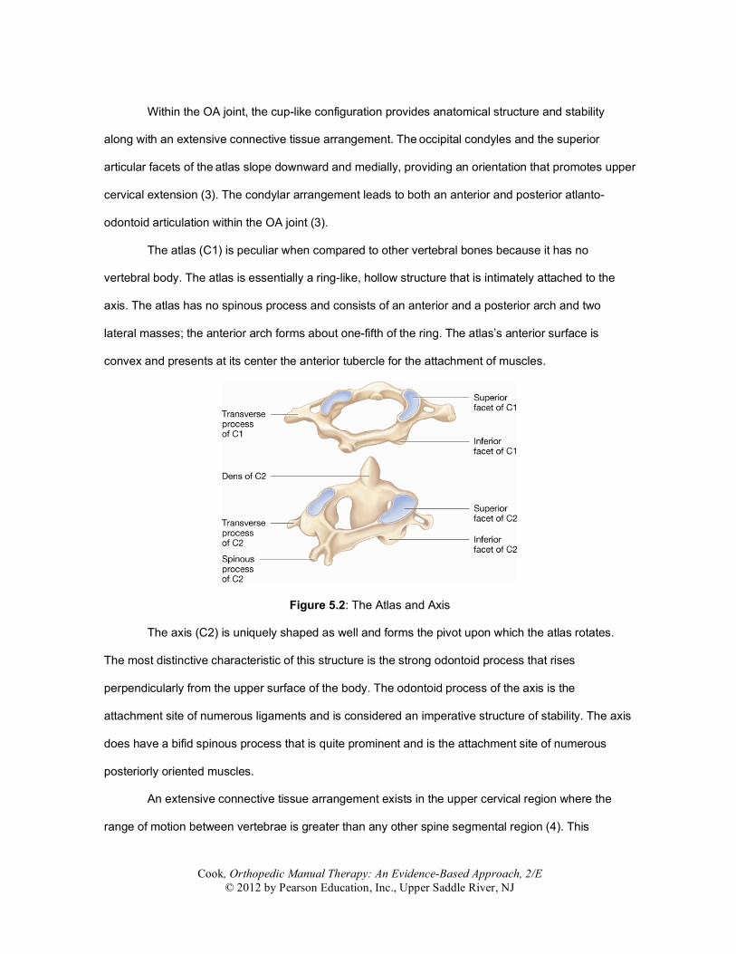
Intervertebral disc First disc at C2-3; intervertebral discs at all subsequent inferior levels Zygapophyseal (facet) joints
Synovial, planar, diarthrodial joints. All cervical joints are weight bearing in the upright postures. Facets are in a plane 45° between the frontal and horizontal planes. This allows significant movement in all 3 planes
Uncovertebral joints Also described as the joints of von Luschka; formed by the uncovertebral process—a raised lip on the superior-lateral surface of the vertebral bodies. Develop by age 6–10. Convert the planar superior vertebral plateau to a concave beveled surface. The concave superior surface of the vertebra receives the vertebral body above and articulates with it to form the uncovertebral joint. Debated as to what type of joint it is (i.e., synovial, type, etc.). Limit side bending and guide flexion and extension
Table 5.3: Ligaments and Connective Tissue of the Cervical Spine.
Name
Location
Function
Anterior longitudinal ligament
Attaches to the occiput, C1 and C2, the anterior occipitoatloid membrane, and the anterior aspects of the discs and vertebrae of the cervical spine
Crucial contributor to stability. Helps retain form of intervertebral disc
Articular capsular ligaments Surround the condyles of the occipital bone and connect them with the articular processes of the atlas
Assist in stabilizing joint capsules
Apical Extends from tip of the dens to anterior edge of foramen magnum
Controversial contributor to stability. Limits traction-based movements
Alar Two ligaments that arise one on either side of the upper part of the odontoid process and, passing obliquely upward and lateral, are inserted into the rough depressions on the medial sides of the condyles of the occipital bone
Crucial contributor to stability. During side bending of the head, the alar ligament on the side that is side flexed toward is relaxed, while the alar ligament on the side opposite is tightened. During rotation, the alar tightens on the side opposite of the direction of rotation. Subsequently, the left alar ligament tightens and restricts rotation of the head and C1 to the right
Transverse Attaches to the occiput, C1 and C2 (10), and the anterior occipitoatloid membrane
Most important ligament of the C0-C1-C2 complex. Prevents the atlas from translating anterior on the axis during flexion
Ligamentum flavum Overlies the space between the laminae of adjacent vertebrae and the neural arches
Helps restrain hyperflexion
Posterior longitudinal ligament
Attaches to the occiput, C1 and C2, the anterior occipitoatloid membrane, and the posterior aspects of the discs and vertebrae of the cervical spine
Stabilizes flexion and reinforces posterior aspect of annulus
The muscles of the upper cervical spine are compartmentalized into three layers—the
superficial, the middle, and the deep layer (7), each layer contributing different elements of mobility
and stability to the upper cervical spine. Panjabi (8) described a form of dynamic stability response,
which is provided by muscular control during movements in mid or early ranges. This concept is
distinguished from passive stability, in which stabilization is obtained through passive structures such
as ligaments, discs, bones, and joint capsules.
Summary
• The upper cervical spine demonstrates unique bony and ligamentous stabilization systems. • The complex arrangement of ligamentous structure in the upper cervical spine is responsible
for movement control and stability. • It is theorized that different muscles of the upper cervical spine are responsible for
stabilization and movement initiation.
LOWER CERVICAL SPINE ANATOMY
The lower cervical segments include the segmental levels of C2-3 to C7-T1 and are noted
more for their similarities than differences. All the segments exhibit intervertebral discs, uncinate
processes, and spinous processes (5). Passive spine integrity is imposed by a combined stabilization
effort of the zygapophyseal joints, uncinate processes, intervertebral discs, and other passive
structures. Although all vertebrae demonstrate significant similarities, Lysell (9) considered the C2
vertebral body unique as a transitional vertebra that divides the functions of the cervical spine.
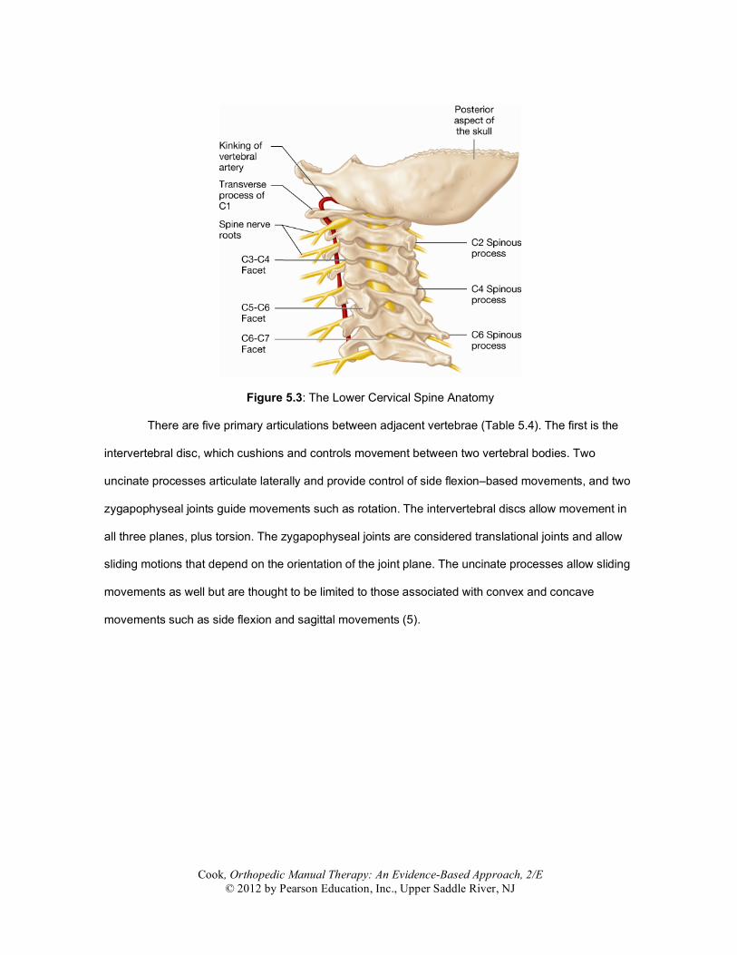
Figure 5.6: The Kinking of the Vertebral Artery during Rotation
The spinal column is covered by a variety of structures. The lateral aspect is covered
posteriorly by the lateral aspects of a superior and inferior lamina. The ligamentum flavum provides
the ventral cover and is attached to two-thirds of the undersurface of the superior lamina. Inferiorly,
the ligamentum flavum is attached to the superior edge of the lower lamina (25). Posteriorly, from
cephaled to caudal, the neural foramen’s parameter is bound by 1 to 2 mm of the superior
(descending) and inferior (ascending) facets. The superior and inferior boundaries of the neural
foramen are formed by the superior and inferior vertebral pedicles (25). Of all the vertebral spinal
foramina, the largest diameter is at C2-C3, which progressively decreases in size to the C6-C7 level.
In a nondamaged segment, the nerve root occupies between 25 and 33% of the foramina space (8).
Summary
• The lower cervical anatomy is dissimilar to the upper cervical anatomy. • The cervical intervertebral discs exhibit dissimilar characteristics to those of the lumbar spine. • The uncovertebral joints develop in the second decade of life and become more prominent
with degenerative changes in the intervertebral disc. • The intervertebral foramen and spinal column are predisposed to anatomical changes and
may be the origins of cervical radiculopathy and myelopathy.
Fundamentally, there are two disparate regions to the cervical spine—the upper cervical
spine and the lower cervical spine. Within this textbook, there are two points of consideration
associated with biomechanics of the cervical spine—range of motion and coupling behavior. (Table
5.5). Range of motion is presented in two forms—gross cervical movement and individualized
segmental movements. Using the 3-Space Isotrak measurement system, Trott et al. (26) outlined
normative, average gross range-of-motion values for the age groups 20–59. They found the average
flexion values were 45.1 to 57.5 degrees, average extension 60 to 76.1 degrees, average left rotation
63.4 to 71.7 degrees, average right rotation 70.4 to 78 degrees, average left lateral flexion, 32.4 to
45.5 degrees, and average right lateral flexion 35.4 to 47.6 degrees. The lower values represent the
age group of 50–59, identifying a notable decline of range of motion with age.
Table 5.5: General Biomechanics and Movements of the Cervical Spine. Topic
General Biomechanics and Movement
Limitation of movement
The facet orientation of the cervical spine tends to limit extension and guides side bending and rotation. Flexion and extension are pure movements because they do not include secondary motions such as side bending or rotation. Rotation occurs as a unit between O-C1-C2. The head and the atlas move together on the axis. Alar ligaments resist movement of the head, not movement of the atlas, as they connect C2 to the head and not to C1.
Several methods of measurement for cervical range of motion are presented within the
literature (27,28), the more sophisticated and expensive the method, the more reliable and valid the
reported range-of-motion scores. Traditionally, most manual therapy clinicians measure three cardinal
planes of motion (sagittal, coronal, and frontal) with a standard goniometer—a method that has
exhibited fair reliability when specific guidelines are followed (Table 5.6). Nilsson et al. (29) suggest
that goniometric measurements exhibit good reliability when combined movements are measured (for
instance, total range of motion of flexion and extension) versus selecting an arbitrary neutral or zero
point as a starting reference. Others whom have used the zero point method (29,30) have reported
interclass coefficient values of 0.6 to 0.8, values that are moderately acceptable. For research
purposes, a device that provides more accuracy in range of motion is beneficial, but for clinical
purposes, the goniometric measure appears to be efficient and reliable.
Table 5.6: Range-of-Motion Values for the Cervical Spine (in degrees).
Upper Cervical Spine
Range of Motion
The primary planar motion at C0-1 is flexion/extension, characterized by total segmental
range of motion values of 25 degrees (5). Unilateral lateral flexion is limited to 5 degrees, as is
unilateral rotation (5) (Table 5.7).
Table 5.7: Joint Specific Biomechanics and Movement of the Occipital-Atlanto Joint. Region
Biomechanics and Movement
OA flexion and extension
This joint generally follows the convex-concave rule, with the convex portion describing the OA joint motion with the glide opposite the direction of movement of the bone. For example, during flexion, the occipital condyles roll anterior (forward) and glide posterior (backward). The condyles roll backward during extension and slide in the direction opposite of the roll
OA rotation
There is no significant rotation at the OA joint
OA joint lateral flexion
A small amount of side-to-side rolling of the occipital condyles occurs over the superior articular facets of the atlas. At the extremes of lateral flexion there is a slight unilateral joint approximation on the side of the lateral flexion and a slight joint separation on the side opposite the lateral flexion. Thus, the head slides upon the atlas away from the direction of side flexion. Lateral glide occurs with the atlas shifting to the side of tilt (i.e., the TP of C1 translates into the concavity)
Joint Flexion Extension Rotation Side bending OA 5 10 0 5 AA 5 10 40–45 0 C2-7 35 70 45 35 Total 45–50 85 90 40
The axial-atlanto (AA) joint allows for 50% of all cervical rotation motion (5). The occipital
atlantal joint (OA) is responsible for 50% of flexion and extension of the complete cervical spine (1).
Unilateral side flexion accounts for 5 total degrees whereas flexion/extension measures 20 degrees
during combined values (5). It has been stated that the essential movement of the upper cervical
spine occurs between the occiput and C2 and is regulated by the atlas (2). In all motion initiations,
whether proximal or distal to the upper cervical spine, C1 is mobile and the movement of the occiput
and C2 predicates on the initiation of the movement (2).
Table 5.8: Joint Specific Biomechanics and Movement of the Atlanto-Axial Joint.
Region
Biomechanics and Movement
AA Flexion and extension
Has ~15 degrees of flexion and extension, with the atlas pivoting forward during flexion and backward during extension
AA Rotation
The inferior facet of the atlas glides across the superior facet of the axis. These surfaces are slightly convex when the articular cartilage is considered. This rotation is coupled with a significant amount of extension (and sometimes flexion) depending on the alignment of forces through the axis
AA Joint lateral flexion
There is essentially no lateral flexion at the AA joint. If lateral flexion is seen on a radiograph, it may be indicative of a fracture
Lower Cervical Spine
Range of Motion
White and Panjabi (5) reported the mid-lower cervical range-of-motion values with a wide
degree of variability (Table 5.9). C2-3, C3-4 C6-7 and C7-T1 display the lowest segmental combined
flexion/extension ranges, while C4-5 and C5-6 exhibit the highest values. Unilateral side flexion
progressively declines from cephaled to caudal, dropping from a peak of 10–11 degrees at C2-3, C3-
4, and C4-5 to a low of 4 degrees at C7-T1. Unilateral rotation is greatest at C3-4 to C6-7, with nearly
comparative values throughout. One exception is the lowest recorded value for unilateral rotation at
Table 5.9: Joint Specific Biomechanics and Movements of the C2-C7 Region of the Cervical Spine. Region
Biomechanics and Movement
C2-C7 Flexion and extension
Up to 20–25% of total cervical flexion takes place at the OA and AA joints, leaving the remaining 75–80% at C2-C7. Extension is limited by the inferior articular facet of the superior vertebra as this vertebra slides inferior and posterior relative to the superior facet of the inferior vertebra. Flexion is the reverse of the above process and is limited by ligamentous and capsular tension. On average there is approximately 20° of sagittal plane motion between each of the segments from C2-3 and C6-7. The largest displacement tends to occur at C5-6.
C2-C7 Rotation
Rotation is guided primarily by the spatial orientation of the facet joints. The inferior facets slide posteriorly and somewhat inferiorly on the same side as rotation and anteriorly and somewhat superiorly on the side opposite the rotation. Rotation is greatest in the more cranial segments.
C2-C7 Lateral flexion
Lateral flexion occurs primarily in the C2-7 segments. There is ~5° lateral flexion at the OA joint and none at the AA joint.
Coupling Biomechanics
Cook et al. (31) reported on the coupling patterns of the cervical spine in a detailed summary
of three-dimensional (3-D) investigations. Five studies qualified as 3-D analyses of coupling motion
with side-bend initiation, and Table 1 outlines those findings. Every study identified the simultaneous
occurrence of the coupled movements of flexion and rotation at all levels tested. Generally, there was
remarkable agreement among all studies at the majority of segmental levels. All studies that tested
CO-1, C2-3, C3-4, C4-5, C5-6, C6-7, and C7-1 found consistent side-bend and axial rotation to the
same side. Two studies reported that side-bend and axial rotation occurred in opposition at C1-2, and
two others found coupling movement to the same side.
Five studies measured rotation,; two of which measured all levels. Similar to side-bend
initiation, axial rotation initiation demonstrated strong agreement among researchers. All studies that
and C1-2 exhibited side bend to the opposite direction as the initiated movement of axial rotation. The
spinal levels C2-3, C3-4, C4-5, C5-6, C6-7 and C7-1 exhibit side bend to the same direction as the
initiated movement of axial rotation. Only two studies reported the movement values of C2-3 and C3-4
and found rotation and side flexion occurred to the same side.
It is worth noting that Fryette’s laws of physiological motion (32) are not included in this
chapter’s discussion of coupling. In 1954, Fryette’s findings were published and were largely based
upon the findings of Lovett (33). Fryette’s perception of coupling of the cervical region was that, “side
bending is accompanied by rotation of the bodies of the vertebrae to the concavity of the lateral curve,
as in the lumbar (spine)” (32). The findings of Fryette are not included because they did not contain a
systematic, investigatory method of evaluation and were 2-D at best.
Lastly, the ability to perform a specific movement by use of apposition during coupling has
recently been questioned (34). The concept of “locking” the cervical spine to allow movement to one
area was recently tested and movement occurred in other aspects of the spine. Although this
mechanism (locking) may be useful for a more focused, target-specific technique, apposition (which is
based on the concept of coupling) does not appear to wholly stabilize segments outside the targeted
segment.
Summary
• With reference to biomechanical movement, the upper cervical spine is mostly responsible for
physiological rotation, and flexion and extension movements. • Coupling patterns of the upper cervical spine at C1-2 are somewhat unpredictable during side
flexion initiation but are predictable during rotation initiation. • C1 is mobile during all forms of movement whether caudally or cephalically initiated. • The lower cervical spine demonstrates equivocal percentages of biomechanical movements
in all ranges of physiological motion. • The lower cervical spine demonstrates a consistent and predictable coupling pattern
regardless of the initiation of motion; side flexion and rotation occur to the same side.
Online References
1. Malanga GA: The diagnosis and treatment of cervical radiculopathy. Med Sci Sports Exerc.1997;29(7 Suppl):S236–245.
2. Penning L. Normal movements of the cervical spine. Am J Roentgenology. 1978;130:317-326. 3. Leone A, Cerase A, Colosito C, Lauro L, Puca A, Marano P. Occipital Condylar Fractures: A
Review. Radiology. 2000;216:635–644 4. Dean NA, Mitchell BS. Anatomic relation between the nuchal ligament (ligamentum nuchae)
and the spinal dura mater in the craniocervical region. Clin Anat. 2002;15(3):182–185. 5. White A, Panjabi M. Clinical biomechanics of the spine. Philadelphia; J.B. Lippincott Co: 1990. 6. Dvorak J, Schneider E, Saldinger P, Rahn B. Biomechanics of the craniocervical region: the
Alar and transverse ligaments. J Orthop Res. 1988;6(3):452–461. 7. Lewis H. Gray’s anatomy. 20th ed. Philadelphia; Lea & Friberger: 2001. 8. Panjabi M. The stabilizing system of the spine. Part I. Function, dysfunction, adaptation, and
enhancement. J Spinal Disord. 1992;5:383–389. 9. Lysell E. Motion in the cervical spine. An experimental study on autopsy specimens. Acta
Orthop Scand. 1969;Suppl 123:1+. 10. Bogduk N. Functional and applied anatomy of the cervical spine. In: Grant R. Physical therapy
of the cervical and thoracic spine. 3rd ed. New York; Churchill Livingstone: 2002. 11. Mercer S, Bogduk N. The ligaments and annulus fibrosus of human adult cervical intervertebral
discs. Spine. 1999;24(7):619–626. 12. Mercer SR, Jull GA. Morphology of the cervical intervertebral disc: implications for McKenzie's
model of the disc derangement syndrome. Man Ther. 2000;1(2):76–81. 13. Bogduk N, Mercer S. Biomechanics of the cervical spine. 1: Normal kinematics. Clin Biomech.
2000;15:633–648. 14. Russell SM, Benjamin V. The anterior surgical approach to the cervical spine for intervertebral
disc disease. Neurosurgery. 2004;54(5):1144–1149. 15. McAfee PC, Cunningham B, Dmitriev A, et al. Cervical disc replacement-porous coated motion
prosthesis: a comparative biomechanical analysis showing the key role of the posterior longitudinal ligament. Spine. 2003;28(20):S176–185.
16. Williams P, Warwick R, Dyson M, Bannister L. Gray’s anatomy. 37th ed. Edinburgh; Churchill Livingstone: 1989.
17. Pal GP, Routal RV, Saggu SK. The orientation of the articular facets of the zygapophyseal joints at the cervical and upper thoracic region. J Anat. 2001;198(Pt 4):431–441.
18. Penning L, Tondury G (Abstract). Enststehung, Bau and Funktion der meniskoiden Strukturen in den Halswirbelgelenken. Z Orthop. 1964;1:14.
19. Dvorak J. Epidemiology, physical examination, and neurodiagnostics. Spine. 1998;23:2663–2672.
20. Inami S, Kaneoka K, Hayashi K, Ochiai N. Types of synovial fold in the cervical facet joint. J Orthop Sci. 2000;5(5):475–480.
21. Sherk H. Disorders of the cervical spine. Clin Orthop. 1999;(359):2–3. 22. Chen TY, Crawford NR, Sonntag VK, Dickman CA. Biomechanical effects of progressive
anterior cervical decompression. Spine. 2001;1;26(1):6–13 23. Bergmark A. Stability of the lumbar spine. A study in mechanical engineering. Acta Orthop
Scand Suppl. 1989;230:1–54. 24. Falla D. Unraveling the complexity of muscle impairment in chronic neck pain. Man Ther.
2004;9(3):125–133. 25. Russell S, Vallo B. Posterior surgical approach to the cervical neural foramen for intervertebral
disc disease. Neurosurgery. 2004;54(3):662–665. 26. Trott PH, Pearcy MJ, Ruston SA, Fulton I, Brien C. Three-dimensional analysis of active
cervical motion: the effect of age and gender. Clin Biomech. 1996;11(4):201–206. 27. Haynes MJ, Edmondston S. Accuracy and reliability of a new, protractor-based neck
goniometer. J Manipulative Physiol Ther. 2002;25(9):579–586. 28. Antonaci F, Ghirmai S, Bono G, Nappi G. Current methods for cervical spine movement
evaluation: a review. Clin Exp Rheumatol. 2000;18(2 Suppl 19):S45–52 29. Nilsson N, Hartvigsen J, Christensen HW. Normal ranges of passive cervical motion for women
and men 20–60 years old. J Manipulative Physiol Ther. 1996;19(5):306–309.
30. Youdas JW, Carey JR, Garrett TR. Reliability of measurements of cervical spine range of motion—comparison of three methods. Phys Ther. 1991;71(2):98–104.
31. Cook C, Hegedus E, Showalter C, Sizer PS Jr. Coupling behavior of the cervical spine: a systematic review of the literature. J Manipulative Physiol Ther. 2006;29(7):570–5.
32. Fryette H. The principles of osteopathic technique. Carmel, CA; Academy of Applied Osteopathy: 1954.
33. Cook C. Lumbar coupling biomechanics—A literature review. J Man Manipulative Ther. 2003;11(3):137–145.
34. Cattrysse E, Baeyens JP, Clarys JP, Van Roy P. Manual fixation versus locking during upper cervical segmental mobilization. Part 2: an in vitro three-dimensional arthrokinematic analysis of manual axial rotation and lateral bending mobilization of the atlanto-axial joint. Man Ther. 2007;12(4):353–62.


![Chapter 2ems.jbpub.com/chapleau/firstresponder/docs/PPT_Lectures/Chapter_0… · Title: Microsoft PowerPoint - Chapter_002 [Compatibility Mode] Author: Jennifer.Meltz Created Date:](https://static.documents.pub/doc/80x56/5ad016617f8b9aca598d40d3/chapter-2emsjbpubcomchapleaufirstresponderdocspptlectureschapter0title.jpg)


