REVIEW Cervical spine clearance: a review Paula J. Richards a,b a X-ray Department, University Hospital of North Staffordshire NHS Trust (UHNS), Princes Road, Hartshill, Stoke on Trent ST4 7LN, UK b Keele University, Staffordshire, UK Accepted 22 July 2004 Injury, Int. J. Care Injured (2005) 36, 248—269 www.elsevier.com/locate/injury KEYWORDS Clinical cervical spine clearance; Radiological cervical spine clearance; Cervical radiology; Spine trauma; Unconscious multiple trauma; Spine protocols Summary Ethical concerns have hindered any randomised control blinded studies on the imaging required to assess the cervical spine in an unconscious trauma patient. The issue has been contentious for many years and has resulted in burgeoning but inconclusive guidance. MRI and multislice CT technology have made rapid advances, but the literature is slower to catch up. Never the less there appears to be an emerging consensus for the multiply injured patient. The rapid primary clinical survey should be followed by lateral cervical spine, chest and pelvic radiographs. If a patient is unconscious then CT of the brain and at least down to C3 (and in the USA down to D1) has now become routine. The cranio-cervical scans should be a maximum of 2 mm thickness, and probably less, as undisplaced type II peg fractures, can be invisible even on 1 mm slices with reconstructions. If the lateral cervical radiograph and the CT scan are negative, then MRI is the investigation of choice to exclude instability. Patients with focal neurological signs, evidence of cord or disc injury, and patients whose surgery require pre-operative cord assessment should be imaged by MRI. It is also the investigation of choice for evaluating the complications and late sequela of trauma. If the patient is to have an MRI scan, the MR unit must be able to at least do a sagittal STIR sequence of the entire vertebral column to exclude non-contiguous injuries, which, since the advent of MRI, are now known to be relatively common. Any areas of oedema or collapse then require detailed CT evaluation. It is important that cases are handled by a suitably skilled multidisciplinary team, and avoid repeat imaging due to technical inadequacies. The aim of this review is to re-examine the role of cervical spine imaging in the context of new guidelines and technical advances in imaging techniques. # 2004 Published by Elsevier Ltd. E-mail address: [email protected]0020–1383/$ — see front matter # 2004 Published by Elsevier Ltd. doi:10.1016/j.injury.2004.07.027
Transcript
Injury, Int. J. Care Injured (2005) 36, 248—269
www.elsevier.com/locate/injury
REVIEW
Cervical spine clearance: a review
Paula J. Richardsa,b
aX-ray Department, University Hospital of North Staffordshire NHS Trust (UHNS), Princes Road, Hartshill,Stoke on Trent ST4 7LN, UKbKeele University, Staffordshire, UK
Summary Ethical concerns have hindered any randomised control blinded studieson the imaging required to assess the cervical spine in an unconscious trauma patient.The issue has been contentious for many years and has resulted in burgeoning butinconclusive guidance. MRI and multislice CT technology have made rapid advances,but the literature is slower to catch up. Never the less there appears to be an emergingconsensus for themultiply injured patient. The rapid primary clinical survey should befollowed by lateral cervical spine, chest and pelvic radiographs. If a patient isunconscious then CT of the brain and at least down to C3 (and in the USA down toD1) has now become routine. The cranio-cervical scans should be a maximum of 2 mmthickness, and probably less, as undisplaced type II peg fractures, can be invisibleeven on 1 mm slices with reconstructions. If the lateral cervical radiograph and the CTscan are negative, then MRI is the investigation of choice to exclude instability.Patients with focal neurological signs, evidence of cord or disc injury, and patientswhose surgery require pre-operative cord assessment should be imaged by MRI. It isalso the investigation of choice for evaluating the complications and late sequela oftrauma. If the patient is to have an MRI scan, the MR unit must be able to at least do asagittal STIR sequence of the entire vertebral column to exclude non-contiguousinjuries, which, since the advent of MRI, are now known to be relatively common. Anyareas of oedema or collapse then require detailed CTevaluation. It is important thatcases are handled by a suitably skilled multidisciplinary team, and avoid repeatimaging due to technical inadequacies. The aim of this review is to re-examine therole of cervical spine imaging in the context of new guidelines and technical advancesin imaging techniques.# 2004 Published by Elsevier Ltd.
Historically imaging the cervical spine in blunttrauma has been controversial. The debate has beendominated by the problem of ruling out a spinalinjury in the unconscious trauma patient. Therehave been several reports of spinal instabilitydespite normal radiographs, but maintaining immo-bilisation on the intensive care unit ‘just in case’ hasbeen associated with significant morbidity. Newimaging techniques have become available, butdid not solve the problem, adding their own ‘bag-gage’, such as cost, availability, logistic difficulties,radiation dosage, lack of specificity and evidenceof effectiveness or safety. The plethora of gui-dance84,125,154,75,83,126 reflected the inability tocompromise between timely yet complete examina-
tions, on a background distinctly lacking in highquality research, resulting in widely varying prac-tices.76,118,82,112 A consensus is now emerging fromthe uncertainty, with a practical set of options toguide clinical practice.26,47
Background
The incidence of major trauma in the UK is relativelylow and trauma system development has been slow,with no legal mandate to enforce change.200 Prior to1988, there was a wide variation in practice and themortality for blunt trauma was significantly higherthan in a comparable group in the United States.220
Since their introduction into the UK in 1988, therehas been a huge demand for Advanced Trauma Life
250 P.J. Richards
Support (ATLS) courses, together with an expecta-tion that this would lead to a higher and moreuniform standard of care.6 There has also been anincreased understanding of the ‘‘Golden Hour’’ andpre-hospital trauma care,131 although occasionallyapplying a hard collar is deleterious as in someankylosing spondylitis injuries.152 Although theTrauma Audit and Research Network has indeedshown significant improvements in mortalitybetween 1989 and 1997, there is still a markedvariation in outcome between hospitals, even afteradjusting for case-mix differences.112 The majorityof patients are first seen in district general hospi-tals, with no national, integrated system of care forthe severely, multiply injured patients, in spite of 8different reports in the last 14 years from the RoyalCollege of Surgeons of England and The BritishOrthopaedic Association since 1988, culminatingin their joint report of 2000 Better Care of theSeverely Injured.1 However, government legislationon seat belts and alcohol limits is believed to havereduced the deaths from road trauma and seriousinjury in the UK in the last decade. The UK incidenceof severe trauma, with an injury severity score (ISS)over 15, is estimated to be 4 per million per week or1 emergency case per 1000, so that the averageacute hospital may not even see one severelyinjured patient each week (Better Care for theSeverely Injured, 2000).1 This level of experiencemay be insufficient to maintain skills. The injuryseverity score (ISS)12 is for assessing the multiplyinjured, modified from the abbreviated injury scalewhich has been validated to correlate with mortal-ity, severe disability and length of hospital stay.32,187
An ISS over 25 is associated with an increased risk ofpermanent impairment, and people do not usuallysurvive with an ISS of more than 50. An ISS of 20 ormore is fatal in 50% of those aged 65 or over, while1% of all multiple injury patients die with an ISS of 14or less.32
The prevalence of trauma is greater in America,particularly of penetrating injury (Better Care forthe Severely Injured, 2000).1 In the USA, trauma hashad a higher profile since Vietnam, with federalfunding, shock trauma ATLS and regional traumacentres. Level I centres have all the acuteservices available on site, and have handled mostmultiply injured patients in America for manyyears. The incidence of cervical spine injury inLevel I trauma centres is 2—4.2% of blunt traumavictims.55,37,72
Recent trends in practice
The ATLS courses spread through the UK, generallyimproving care,6 and raising awareness of occult
spinal injury, but also raised concern, fuelled byincreasing litigation. One result has been thatunconscious patients with normal plain films mayremain in a rigid collar for days or weeks on theintensive care unit. CT scanning, with multislicetechnology, is now widely available 24 h per dayand is being used more liberally. MRI scanning isstill of limited availability, especially out of hours.Meanwhile, clinicians have become confused in thetransition from somewhat slipshod practice to nearparanoia.
In the USA, medico-legal concerns emerged ear-lier where the new technology was embraced morevigorously. Over-investigation of conscious patientsled to spiralling costs but little benefit to the patient‘‘much ado about nothing’’,207 where plain radio-graphy was overused by up to a third.134 The NorthAmerican Spine Society and the Orthopaedic TraumaAssociation were unable to reach a consensus onhow to exclude spinal injury in both conscious andunconscious patients.82 In unconscious patients,ATLS teaching no longer went far enough. Whilethe course emphasised spinal precaution, providedinstruction on clinical examination and on the inter-pretation of plain films, it was misleading in theunconscious stating, ‘‘Patients who are comatose,have an altered level of consciousness or are tooyoung to describe their symptoms may be clearedafter normal three-view cervical spine series and anappropriate clinical evaluation by an orthopaedicsurgeon or neurosurgeon’’, which is not true. ManyNorth American Level I trauma centres turnedinstead to ‘‘evidence-based’’ recommendationsfrom the Eastern Association for the Surgery ofTrauma (EAST),125 even though there were no RCTs.They relied on plain radiographs of the cervicalspine and targeted CT scans of abnormal and poorlyvisualised areas to clear the cervical spine inpatients with an altered conscious level. Theydemanded a rigorous technical approach to imagingand reporting, but pragmatically overlooked theissue of ligamentous instability when there was nodemonstrable fracture or soft tissue swelling.Before revising their guidelines, EAST carried outa postal survey of practice in 31 Level I traumacentres in North America to check compliance withtheir previous recommendations. It became appar-ent that several centres were using MRI scanningand, more controversially, some used flexion—extension fluoroscopy to clear the cervical spinein unconscious patients. When the revised guide-lines were published in 2000, flexion—extensionimaging was overtly recommended for patientswho were predicted to remain unconscious for morethan 24 h.126 This promised rapid spinal clearancehowever, the safety of moving the neck in these
Cervical spine clearance: a review 251
patients, even under expert supervision, has beenseriously questioned. In the revised guidelines, theapparent ‘‘safety’’ was based on just three papers,each reporting a very small number of patients withnormal plain views and/or CT scans followed by nodemonstrable instability on flexion—extension ima-ging. In the UK, radiological clearance was oftentechnically inadequate and spinal precautions weresometimes withdrawn on the basis of a single lateralplain radiograph or on clinical assessment aloneafter consciousness was eventually regained.76,118
In 2002 the British Trauma Society responded withthree, practical management options, of increasingcomplexity, for institutions to discuss and choose apolicy suitable for their case mix and thereforeexpectation of injury (BTS Injury, 2003).26
Anatomical distribution of injury
The NEXUS study confirmed the typical distributionof fractures (C2 23.9%, C6 20.25%, C7 19.08%, C514.98%) and dislocations or subluxations (C5/625.11%, C6/7 23.77%, C4/5 16.96%).72 However, halfof the cranio-cervical injuries may not be suspectedclinically,30,139 so identification requires a highindex of suspicion and little reliance on an apparentlack of symptoms or signs. In 202 consecutive uncon-scious patients, 28 (13.9%) had C1 or C2 fracturesand 11 of these had normal cervical radiographs, asdid all except 1 of the 9 (4.4%) cases with anoccipital condyle fracture.117 Others have doubtedsuch a high incidence of occipital condyle frac-tures,66 but it is likely that the true incidencedepends not only on the case mix, but also on theexpectation of the clinician, which will determinehow carefully they are looked for. In order to make areliable diagnosis of an occipital condyle fracture, itis necessary to perform high resolution, thin section(1 or 2 mm) CT scan (or multislice CT scan) of thebase of the skull, with both orthogonal reconstruc-tions. The diagnosis and classification of occipitalcondyle fractures guides treatment for instabil-ity213,7 and may explain persistent symptoms.
In the elderly, domestic falls are the commonestcause of cervical injury, where two-thirds ofcervical fractures in this age group involve theoccipital condyles or the upper three vertebrae.Such injuries are commonly associated with spon-dylosis or osteoporosis,124 which complicate theinterpretation plain films and contribute to delayeddiagnosis in 15—40%. In the over 60s devastatingcord injury may occur without fractures or dis-locations, spinal cord injury without obvious radi-ological abnormality, or SCIWORA is relativelycommon.150,107,48
The relevance of mechanism of injury
It is intuitive that the mechanism of injury influ-ences the risk of cervical spine trauma, but there isinsufficient documented evidence to rigidlystratify risk accordingly. While the mechanismmay raise the level of suspicion, it rarely allowsspinal injury to be excluded.19,21,79 This means thatone cannot predict spinal injury on the basis of otherinjuries or vice versa. Each patient needs full eva-luation of the whole spine. It must be rememberedthat cervical spine trauma is associated with upperrib fractures, pneumothoraces and damage to thegreat vessels and/or trachea, which need activeexclusion in cases with a spinal injury.38 A mechan-ism involving high-energy transfer merely reinforcesthe need to investigate thoroughly. Several authorshave determined mechanisms of injury and clinicalparameters which allow patients to be divided intohigh or low risk, with imaging reserved for theformer (Table 1). If the circumstances of the injuryare unclear, it is wise to err on the side of cautionand investigate carefully, particularly in blunttrauma.
High velocity bullets that miss but pass close tothe spinal column may cause spinal injury as a resultof the associated shock wave.16,132 On the otherhand, gunshot wounds to the head rarely have anyassociated spinal injury98 and it is not necessary totake spinal precautions if there is no evidence ofbullets passing close to the spinal column or of aseparate blunt mechanism of injury.
The incidence of spinal injuries depends on thegroups studied: They were seen in 3.4% of motorvehicle occupants, 2.8% of pedestrians, 1.9% ofmotorbike riders and 0.9% of falls of all attendancesto a major urban trauma centre.55 Post-mortemstudies of fatal motor vehicle collisions (MVCs), onthe other hand show up to 24% have cervical spineand up to 40% have head injuries of varying sever-ity.49,5,30,31 Frontal airbags cause all types of cervi-cal injury if the occupants are unrestrained or ifchildren in rear facing car seats are too close to theactivated air bag.105 Rear passenger’s fair worst inMVCs because they are most often unrestrained,with three point seat belts offering most pro-tection.43,192 High-speed MVCs and falls from aheight are associated with a high risk of spinalinjury.95,10,170 Patients with clinically significanthead injuries are at increased risk of cervical spineinjury.184,167,215,174,88,123,142 Up to a third of Level Itrauma cases requiring head CT in Chicago for headtrauma or retrograde amnesia, had fractures of C1or C2.104 The incidence of cervical spine injuries isinversely related to the GCS (10.2% of those withGCS 8, 6.8% of those with GCS 9—12 and 1.4% of
252 P.J. Richards
Table 1 Summary of criteria for cervical spine injury.47
Vandemark: criteria forhigh-risk patients
High velocity blunt trauma; multiple fractures;evidence of direct cervical injury(cervical pain, spasm, obvious deformity); altered mental status(loss of consciousness, alcohol and/or drug abuse);drowning of diving accident Fall of >10 ft;significant head or facial injury; thoracic or lumbar fracture;rigid vertebral disease (AS, DISH);paresthesias or burning in extremities
206
University of Washington criteriaMechanism parameters High-speed (>35 mph) MVA; crash with death at scene;
fall from height >10 ftClinical parameters Closed head injury; neurologic symptoms or signs
referred to the cervical spine;pelvic or multiple extremity fractures
Steill: Canadian rules, no radiography 195
Absent high-risk factors Age >65 years; dangerous mechanism (see Vandemark ofUniversity of Washington criteria);paresthesias in extremities
Low-risk factors which allow safeassessment of range of motion
Simple rear end MVC; sitting position in ED;ambulatory at any time;delayed onset of neck pain;absent midline cervical tenderness;able to actively rotate neck 458 left and right
NEXUS criteria (low risk) Absence of midline cervical tenderness;absence of focal neurologic deficits;absence of intoxication;absence of painful distracting injuries;normal alertness
72
Hanson validated high risk cervical spine80
Mechanism ClinicalSpeed >35 mph;
fall >10 ft;death at scene
Cervical spine pain, spasm, deformity or neurologysignificant closed head injury;pelvic or multiple extremity #
those with GCS 13—15),55 however there is no directassociation between the severity of head injury andthe incidence and nature of cervical spine193 oroccipital condyle injury.144
The role of clinical assessment in cervicalspine clearance
In the field, opportunities for reliable clinical evalua-tion are limited and it is generally advisable to immo-bilise the spine in significant blunt trauma cases untilthe patient is in a more conducive environment inhospital. In the alert patient, there is agreement onhow to clear the cervical spine if the conscious levelhas not been altered by head injury, drugs or alco-hol33,147,121 and there is nodistractingpain fromotherinjuries. Then a history and clinical examination canrule out significant injury.62,10,147,170,136,108,13,130,180,88,116,176,61,207,221,73 This was validated, in a prospec-tive, multi-centre, observational study in North
America: the National Emergency X-radiography Uti-lisation Study (NEXUS). It looked only at low prob-ability injuries, to try and identify those in whomradiography can be safely omitted.89 Of the 34,069patients from 21 centres, 818 (2.4%) had a radio-graphic cervical spine injury. Two hundred and fortyof the 818 patients (29.3%) met the 5 criteria ofinsignificant injury: no midline cervical tenderness,no focal neurological deficit, normal alertness, nointoxication or painful distracting injury,72 i.e. nodepressed consciousness sometimes called the ‘‘FiveNos’’. This practice is more akin to the British prac-tice. The Canadian cervical spine rule195 looked atstable patients with a normal GCS of 15, excludedthose with high risk factors, and set out low riskcriteria including delayed onset of neck pain whichthen allowed active rotation of the neck of up to 458bilaterally. Controversially if they could move theneck, even if it was painful they reported that noradiography was indicated.
Cervical spine clearance: a review 253
In an alert patient without neurological features,clinical examination should be repeated if the radio-graphs are normal, this time including active move-ments. If pain or tenderness is still a problem,flexion—extension radiographs should be consid-ered, but may cause false negatives in neck musclespasm.137
If the patient has an altered level of conscious-ness or has received sedative drugs, includingopioids, the clinical examination may be unreliable.Similarly, distracting pain from a separate (non-spinal) injury may cause the patient to disregardsymptoms from an unstable neck injury.4 Local pain,tenderness and neurological symptoms or signs(such as segmental weakness, numbness or para-esthesia) must be assumed to indicate a potentiallyunstable injury. In all these circumstances, it isessential to image the spine beforemoving the neck.However, clinical examination (short of moving theneck) remains an important part of the assessmentand should not be omitted simply because radio-graphs are indicated.
Most units receiving the multiply injured rapidlyperform a CXR, pelvis and lateral cervical spineradiographs after the primary survey. At a clinicallyappropriate time the whole neural axis must becleared.
Plain radiographs
Despite the availability of newer technologies,there is still an important role for plain films andall staff need a basic understanding of the princi-ples. They are ubiquitous, cheaper than CT and theradiation dose is much less for the full spine, only0.069 mSv in our A & E. The role of plain films islikely to diminish in the unconscious as multisliceMS-CT technology spreads. Previous guidelinesrecommend that swimmer’s views are replaced bytrauma obliques, which are of lower radiation dosesand show the posterior elements more exten-sively,202,94 allowing fracture and facet joint dislo-cation diagnosis. Our own experience of apparentlystable radiographic uni-facetal dislocations is thatthere is often extensive fracturing at the level and/or adjacent vertebra on CT, the pattern of which isassociated with instability. Thus, even in the con-scious limited MS-CT is now indicated more often.Although all the multislice sagittal and coronalreconstructions give more information than plainfilms111, the single radiograph is the baseline forfollow up, and remains invaluable in MS-CT unitshenceforth radiography will probably be limited to alateral with an AP.47 However, in technically difficultpatients, repeated plain films followed by extensiveCT scanning is hardly saving on radiation.
Adequacy of the films
There is often a difference in quality between por-table films and those taken on a fixed departmentalmachine, although new portable digital units are agreat improvement. Good radiographic technique isessential if subtle signs are to be revealed. To beadequate, the films should show the full extent ofthe cervical spine, from the occiput to the upperborder of T1, and should not be rotated. The pene-tration should be sufficient to show bone architec-ture without losing soft tissue detail.
Systematic radiological evaluation
The films must be evaluated by a competent practi-tioner who maintains sufficient activity to maintainskills. Clinicians may find it useful to have a simplesystem for analysing plain films, Courtesy of Dr.Peter Oakley the radiological ABC.
A(ii): Adequacy–—extent (occiput to T1 upperborder, penetration, rotation/projection).
A(iii): Alignment–—anterior aspect of vertebralbodies, posterior aspect of vertebral bodies, poster-ior pillar line, spino-laminar line; cranio-cervicaland other lines and relationships.
A technically adequate lateral cervical spine radio-graph will include down to the upper first dorsalvertebra, with clear bony detail. The pre-vertebralsoft tissue should be assessed, even though swellingis an unreliable sign of injury,133 as it is not alwayspresent and may require a bright light to be seen. Ifpresent it is a specific sign for ligamentous disrup-tion. This is worth assessing early on in the radi-ological evaluation, as it is often otherwiseoverlooked and gives vital clues to the site ofinjuries. Adenoidal tissue in the retropharyngealspace can appear bulky, particularly in childrenwho are crying or swallowing. Below the orophar-ynx, the pre-vertebral soft tissue stripe is usuallyless than 4 mmwide down to the level of the fourthcervical vertebra, where it widens to approxi-mately the width of a vertebral body due to theoesophagus.87 Using absolute measurements asan indicator of abnormality is unreliable, and
254 P.J. Richards
localised bulging due to haematoma is generallymore revealing.
The classical way to assess the radiology is inrelation to the smooth bony alignment Fig. 1a and b,but it is important to understand what other struc-tures make up each line of alignment, as assessed onthe lateral radiograph, displayed in Fig. 1b. The firstline to be assessed on the lateral radiograph extendsfrom the anterior margin of the atlas down the
Figure 1 (a) The lateral cervical radiograph; (b) the an
anterior margins of the other cervical vertebrae,which corresponds to the anterior longitudinal liga-ment. Analysis of intervertebral body movementoverall is required, and usually there is a smooth‘‘C’’, and no ‘‘foraminae’’ are seen and the facetsare not superimposed on the vertebrae. Minordegrees of tilt cause either a smooth, regularchange of successive levels of vertebrae or a pro-gressive one (Fig. 2). A disproportionate amount of
atomy compromising the radiological lines seen (a).
Cervical spine clearance: a review 255
Figure 2 Normal cervical intervertebral body move-ment: The effect of flexion/extension on the radiographs.
rotation or forward subluxation at one level ispathological. Usually there is less than 3 mm offorward displacement of successive vertebrae, butmore than 3.5 mm or an 118 angle between adjacentendplates suggests instability, be it degenerant ortraumatic. The anterior ligaments contribute moreto stability in extension and the posterior ones inflexion.214 Pathologically widened and narroweddiscs may be traumatic in origin.
The posterior margins of the vertebral bodiesform the second line and includes the posterior
Figure 3 Lateral cranio
aspect of the odontoid process. At the cranio-cer-vical junction, the second line forms an angle of150—1808, Wackenheim Clivus baseline (Fig. 3),which extends up to the back of the clivus to theodontoid peg. The vertical distance from the basion(the anterior margin of the foramen magnum) to thepeg, the basion-dental interval, should be less than12.5 mm. The anterior margin of the foramen mag-num sits just above the tip of the odontoid process.In normal cranio-cervical alignment, the tip of theodontoid lies below Chamberlain’s line. The latter,extends from the posterior pole of the hard palate tothe opisthion, the posterior margin of the foramenmagnum. The atlanto-dental distance, the spacebetween the posterior margin of the anterior archof the axis and the anterior margin of the odontoidprocess, is less than or equal to 3 mm in adults. Itmay be between 4 and 5 mm in a child due toincomplete ossification. When the transverse liga-ment is intact, the distance should remain unalteredon flexion/extension views. On the AP the jointspaces between the lateral masses of C1 and C2are bilateral and symmetrical, as is the distancefrom C1 to the peg (Fig. 4). There is much normalvariation in the size of C1 and C2 lateral masses. Intrauma this must be investigated with high resolu-tion CT to exclude fractures. The commonest causeof offset of the lateral masses of C1 and C2 ispositional, as part of the normal tilting process,and should move in the same directions, if notinvestigation is indicated, similarly asymmetry atthe peg and C1. The centre of rotation of C2 isnormally within the odontoid peg172 on CT.
The next line, is the posterior pillar line, which ismade up of the backs of the lateral masses, whichhave the facet joints obliquely orientated between
adjacent levels.189 If the facet joints fail to lie likeroof tiles, with the superior one overlapping the onebelow then disruption should be excluded.
Next is the spinolaminar line, which passes downin a smooth arc from the posterior margin of theforamen magnum along the anterior aspect of thespinous processes, where the laminae fuse. It isalong this line, that the distance between the spinusprocesses is assessed most accurately. It is unreli-able to assess the line joining the tips of the spinusprocesses, as the radiographic appearance of thespinous processes are highly variable as the tips arebifid. The disruption of the spinolaminar line at aspecific level, implies complex spinus process frac-tures, which extend to the laminae and thereforeare likely to effect the spinal canal. They are oftenassociated with posterior longitudinal ligamentinjury and therefore have the potential for instabil-ity and neurological deficit.128
Uni-facetal dislocation can cause anterior displa-cement of one vertebra, by up to 25% of the width ofthe vertebral body, whereas between 40 and 50% orworse overlap suggest bilateral facet dislocation.Uni-facetal rotatory subluxation closes the spacebetween the spinolaminar line and the cortex ofthe facet, causing a ‘butterfly’ or ‘bow tie’ appear-ance of the facet, which are locked. In addition thedegree of rotation may mean that pedicles andforaminae, not normally seen on the lateral,become visible. Conventionally uni-facetal disloca-tions are considered stable, whilst bilateral ones areunstable.14 The use of CT at our institution com-monly finds other injuries both bony and ligamen-tous, not visible on the radiographs, which oftenmake uni-facetal injuries unstable.
The hyperflexion teardrop fracture is the hall-mark for severe soft tissue injury resulting in
ligamentous disruption, and disc rupture with rela-tively little bony injury. The anterior triangle or‘‘flake’’ of bone as it is miss named, may be theonly clue to a complete three column soft tissueinjury causing an unstable spine. The ruptureddisc is usually narrow, but may be pathologicallywidened.
Disruption of the ‘ring’, or junction of the bodyand lateral structures of C2, as seen on a lateralradiograph, indicates a low (Type III) odontoid frac-ture.81
A radiologically ‘fat C2’, where there is increaseddistance between the anterior and posterior mar-gins of C2 on a lateral, is due to an oblique fractureof the body of C2,155 which may not be visible onplain films and requires a CT.
On the AP the lateral extent of the vertebrallateral masses make a ‘‘wavery’’ contour. The unci-nate processes, lateral vertebral body margins andtips of the spinous processes make straight lines,with malalignment indicating malrotation. Facetjoints are not seen on an AP.
Radiological analysis of the cranio-cervical junction
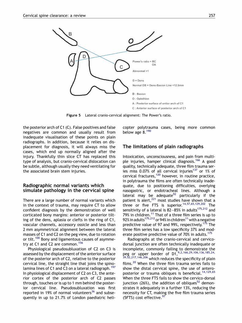
Powers et al. described the plain radiographic cri-teria for the diagnosis of atlanto-occipital disloca-tion on a lateral radiograph (Fig. 5).159 The ratio ofBC/OA > 1.0 is consistent with atlantoocipital dis-location and less than 1.0 are normal. This methodrelies on being able to identify clearly the anteriormargin of the foramen magnum (basion: B) which isat the distal end of the clivus, the posterior marginof the foramen magnum (Opisthion: O), the poster-ior margin of the anterior (A) and anterior margin of
Cervical spine clearance: a review 257
Figure 5 Lateral cranio-cervical alignment: The Power’s ratio.
the posterior arch of C1 (C). False positives and falsenegatives are common and usually result frominadequate visualisation of these points on plainradiographs. In addition, because it relies on dis-placement for diagnosis, it will always miss thecases, which end up normally aligned after theinjury. Thankfully thin slice CT has replaced thistype of analysis, but cranio-cervical dislocation canbe subtle, although usually they need ventilating forthe associated brain stem injuries.
Radiographic normal variants whichsimulate pathology in the cervical spine
There are a large number of normal variants whichin the context of trauma, may require CT to allowconfident diagnosis by the demonstration of wellcorticated bony margins: anterior or posterior tilt-ing of the dens, aplasia or clefts in the ring of C1,vascular channels, accessory ossicle and less than2 mm asymmetrical alignment between the lateralmasses of C1 and C2 on the peg view, due to rotationor tilt.100 Bony and ligamentous causes of asymme-try at C1 and C2 are common.156
Physiological pseudosubluxation of C2 on C3 isassessed by the displacement of the anterior surfaceof the posterior arch of C2, relative to the posteriorcervical line, the straight line that joins the spino-lamina lines of C1 and C3 on a lateral radiograph.197
In physiological displacement of C2 on C3, the ante-rior cortex of the posterior arch of C2 passesthrough, touches or is up to 1 mm behind the poster-ior cervical line. Pseudosubluxation was firstreported in 19% of normal volunteers36 and subse-quently in up to 21.7% of London paediatric heli-
copter polytrauma cases, being more commonbelow age 8.190
The limitations of plain radiographs
Intoxication, unconsciousness, and pain from multi-ple injuries, hamper clinical diagnosis.166 A goodquality, technically adequate, three film trauma ser-ies miss 0.07% of all cervical injuries137 or 1% ofcervical fractures,120 however, in routine practice,in polytrauma the films are often technically inade-quate, due to positioning difficulties, overlyingnasogastric, or endotracheal lines. Although alateral may be adequate92 particularly if thepatient is alert,221 most studies have shown that athree or five FTS is superior.14,57,65,129,202 Thesensitivity of a lateral is 82—85% in adults175,212 and79% in children.11 That of a three film series is up to92% inadults175,212 or 94% inchildren11withanegativepredictive value of 97 and 99%, respectively.175 Thethree film series has a low specificity 37% and mod-erate positive predictive value of 70% in adults.175
Radiographs at the cranio-cervical and cervico-dorsal junction are often technically inadequate orincomplete, commonly failing to demonstrate thepeg or upper border of D1,9,2,166,29,106,136,185,24,39,52,217,146,206 which reduces the specificity of plainfilms.20 When the three film trauma series fails toshow the distal cervical spine, the use of antero-posterior or trauma obliques is beneficial.14,129,65
When the three FTS fails to show the cervico-dorsaljunction (26%), the addition of obliques52 demon-strates it adequately in a further 13%, reducing thenecessity for CT, making the five film trauma series(5FTS) cost effective.97
258 P.J. Richards
Missed injuries on plain radiographs
The precise incidence of missed fractures varieswith the patient population studied, and in manypapers it is not clear exactly which radiographs wereperformed routinely or who was evaluating them.Causes of missed injuries and delayed diagnosisinclude failure of the patient to seek medical atten-tion, failure to take the relevant radiographs, andfailing to identify fractures which were visible onthe films165,166,52,27,138,140,206 and film misinterpre-tation.53 Delayed diagnosis of cervical spine frac-tures may occur in up to 23% of patients, especiallywhen complicated by other factors such as intoxica-tion or, altered level of consciousness.166
A retrospective study in Phoenix, with a four filmcervical trauma series (AP, peg, lateral and swim-mer’s views) supplemented by directed CT in 1331multiple injuries patients, mean ISS of 30 and GCS of11, found cervical fractures or dislocations in 61(4.6%).69 There were nine fatal atlanto-axial dislo-cations and, of the 50 survivors, there were neuro-logic deficits in 15 with complete cord lesions in 8.69
Five patients had delayed diagnosis of their cervicalinjuries (2—21 days), due to inadequate or incom-plete plain radiography.69
In a retrospective study in Philadelphia, of 372spinal injury cases to the regional spinal centre,3.2% of spinal injuries were missed on radiographs,and 25% of these were associated with a progressiveneurological defect attributed to incorrect initialimmobilisation.203
In the National Spinal Injuries Centre, Stoke Man-deville, UK, of 353 consecutive admissions withspinal cord injury, 11 cervical injuries were missedat presentation (3.1%) and 10 of these (2 paraplegic,8 tetraplegic) were considered to have deterioratedas a result of the initial management.165
Noncontiguous vertebral injuries, which arereported between 4.5 and 15.2% cases are oftenmissed at presentation.34,78,76,85,203 The NEXUS,90
study which avoids radiographing those with clini-cally insignificant injury, found that plain radio-graphs failed to detect 10.5% (60/570) of cervicalinjuries, but in the majority, 89.5% the cervicalspine was not the primarily injured area.137 Thus,the whole vertebral column should be assessed in amultiply injured patient in a timely fashion, depend-ing on the nature of the other injuries.
The evolving role of CT scanning
Patients with clinically significant head injuries areat increased risk of cervical spine injury,184,167,174,215,88,123 inversely related to the GCS.55 In a
prospective study of blunt trauma patients requiringintubation and ventilation, spiral CT of the cervico-dorsal junction detected fractures in 10%, whichwere occult on lateral and both oblique plain radio-graphs.96 Thirty four percent of ICU blunt traumaadmissions (21/58), who could not be evaluatedclinically, had cervical fractures on CT.18 In uncon-scious patients at the time of the initial brain CT,cranio-cervical junction CT should be performedwith sagittal and coronal reconstructions as a mini-mum, assuming that the AP and lateral whole cer-vical spine are adequate and normal (NICE, 2003;RCR 2003).143,177 The technique must be meticulouswith 1—3 mm axial slices.117,22,23,125 Multislice CTallows axial reconstructions at 1 mm or submilli-metre widths, which allows one to diagnose smallcortical breaks invisible even on 2 mm slices.178 Itmust be remembered that CTwill miss up to 10% offractures especially if in the plane of the axial CTslice, if both reconstructions are omitted196 typi-cally at the peg. Axial fractures are missed, whenslices are over 3 mm thick, usually at the dens, orbetween C6 and D1.218,178
Good quality, thin-section spiral CT is the optimalmeans of imaging fractures, particularly where plainradiography is poor, at the cranio-cervical and cer-vico-dorsal junction.199 In addition, for high riskcases of cervical spine fractures, the specificity ofradiography is relatively low.20 Although plain filmsshow dislocations more reliably,218 sagittal recon-structions from spiral CT give similar information.Spiral CTof thewhole cervical spine is used routinelyin most high risk or polytrauma cases in NorthAmerica,146,21,45,79,113,161 where MS-CT has beenfreely available for 3 or 4 years, and has just beenrecommended by the American College of Radiol-ogy,47 and AP radiography can probably be safelyomitted.179 The diagnostic performance of conven-tional CT for injury is good,18 sensitivity 95% (95% CI,90—100%) specificity 93% (95% CI, 91—95%) accuracy93% (95% CI, 91—95%), but false negatives areusually ligamentous, and false positives are com-mon, also often ligamentous.79
The cost effectiveness of CT
In North America, in high risk trauma patients,whole cervical spine spiral CT at the time of theinitial body or head CT is quick, and cost effec-tive.145,104,18,21 In children, CT of the cervical spinewhen the head injury was scanned resulted in fewercervical spine radiographs.101 CT only minimallyincreases the total imaging time by around20 min,45,46 but is much more expensive than plainfilms alone,21 particularly in the USA. CT Cost effec-tiveness depends on the probability of injury and the
Cervical spine clearance: a review 259
consequences of misdiagnosis in the group under-going the scan. In America, in patients with severehead injury, the probability of detecting concurrentcervical spine injury is 11.2%,21 where the cost caneasily be justified. If the probability of an injury isless than 4%, CTscanning is not cost effective in theAmerican health system, even though it has beenconsidered to contribute to preventing paralysis.21
It has been suggested that if plain radiographicanalysis of the cervico-dorsal junction is inade-quate, localised CT is also cost effective, becausethe patients are often young,199,198 with large finan-cial implications over many years for any missedinjuries.
In Britain however, CTof the whole cervical spineis rarely performed,76,118 reflecting fewer multisliceCTs, the low incidence of major trauma in DGHs,with only six North American style Level I TraumaCentres (Better Care for the Severely Injured,2000).1 No cost effectiveness analysis is availablein the UK, where the NHS health economics meanthat preventing disability is likely to be cost effec-tive, even for low probability cervical injury cases,as CT is relatively cheap, and the huge long termrehabilitation costs are usually state funded.
CT even with volume scanning or isotopic imagesand reconstructions in two planes, can only diagnosesignificant disc or ligament injury if there is mala-lignment. More experience with multislice CT maychange this view, but there are no comparative orrandomised controlled studies as yet. However, itmust be remembered that intervertebral malalign-ment of or over 4 mmmay be inadvertently reducedby long spinal board extraction and head blocks. Theadvantages of MS-CT are clear, but the radiationdose savings though small may be lost with increas-ing coverage (Table 2). In the UK the simple increasein CT usage has increased the effective dose ofradiation from 20 to 40% in the 1990s, which pre-dates multislice CT,141,93 and every doctor mustweigh the benefits and risks of such exposure.148,40
Table 2 Comparison of CT radiation doses at UHNS, a) con
Active F/E is a safe, good test in conscious, co-operative patients to screen for ligamentousinstability.157,115,158,8 Instability is confirmed ifthere is more than 3.5 mm of intervertebral bodymotion or more than 118 of relative angulation.Normally there is either a smooth, fixed step invertebral body alignment or a progressive change(Fig. 2). Cervical instability occurred in 8% of alert,trauma patients in a Missouri Level I Trauma Centre,nearly half of whom had a normal three film ser-ies.115 The addition of F/E views to a three filmseries increases sensitivity (99%), specificity (93%)with a high positive (89%) and negative (99%) pre-dictive value, with false negatives largely due tospasm.115 F/E radiography is unable to excludeinstability, even if the other radiographs are normaluntil the spasm has resolved.137,138
Passive F/E views or fluoroscopy in unconscious orsedated patients are technically inadequate in up toa third,8 and they may cause devastating neurolo-gical deficit, and remains controversial.115,50 It isavoided in the overwhelming majority of UK cen-tres.77,118 Fortunately the incidence of isolatedligamentous injury is rare,37 in a retrospectivereview of 14,577 blunt trauma victims in a tertiaryreferral centre in Baltimore, 614 (4.2%) had cervicalspine injuries, of which only 87 (0.6%) had isolatedligamentous injuries. There were 2605 patients inthe series with a GCS less than 15 and only 14 (0.5%)had isolated ligamentous injuries. Interestingly, 13were identified on the initial lateral radiograph andthe other was diagnosed on CT. In these cases ofisolated ligamentous injury, flexion—extensionviews were not needed to reveal instability. Thepre-vertebral soft tissue swelling on plain films or CTscans implies ligamentous disruption, but may beabsent in rapid helicopter transfers. In a series of14,755 trauma cases in Los Angeles, 292 patients
ventional spiral b) multislice.
Entrance surfacedoes (mGy)
Effectivedose (mSV)
m, pitch 1—3 mm,mm, 2 mm
Not applicable 0.8
m, 3 mm slices Not applicable 1.9cm, 3mm slices Not applicable 2.7.88 coll. 0.75 mm,radiated slice width
Not applicable 0.6
.88 coll. 0.75 mm,radiated slice width
Not applicable 1.5
.88 coll. 0.75 mm,radiated slice width
Not applicable 2.1
260 P.J. Richards
had cervical spinal injuries. Of these, 250 (85.6%)had fractures, 10% had subluxations (presumablywith ligamentous disruption) and 3.8% (11 patients)had isolated cord injury without fracture or obviousligamentous damage.55 There are few advocates,but with less than 20 cases of instability out ofless than 2000 pooled cases of unconsciouspatients,51,186,41,50,28,8 the numbers are too smallfor it to be recommend. The ACR47 no longer recom-mend F/E views in those with altered consciousness,and they should be reserved for follow up assess-ment of ligamentous instability in conscious patientswhose muscle spasm has resolved. Somatosensoryevoked potentials (SSEP) need further evaluation asa monitoring method182 for F/E in the unconscious,as the delayed or decreased amplitude SSEPs due tocompression or ischaemia of the dorsal column,which means the damage to the cord has alreadyoccurred, so fails to prevent damage at all. Thus, F/E should only be done after informed consent of therelatives of an unconscious patient, when part of arandomised and controlled study, assuming ethicalapproval is obtained. In the current legal climate, itis doubtful if any ethics committee would allow sucha study. In addition as so few UK institutions use it, ifa disaster occurred, the patient may be able to suefor malpractice, as it probably does not pass theBowlam test25 of reasonable practice. In today’sworld it cannot be recommended.
The place of MRI
MRI is unequivocally the modality of choice forevaluation of patients with neurological signs orsymptoms:
1. T
o assess soft tissue injury of the cord, disc andligaments. MRI gives excellent soft tissue and corddetail, showing cord compression from haema-toma and disc prolapses, often allowing the causeof focal neurology to be analysed.110,109,135,15,71,60,63,168,56,17,102,205,98 To assess soft tissue injurywithout MRI the extent of disc and ligamentousinjury are underestimated.64,98,194
Previously asymptomatic necks with spondylo-sis causing spinal stenosis may cause direct impin-gement on the cord at the time of injury. Cordinjury is more likely with spondylosis (P = 0.5) andcanal stenosis (P = 0.001),98 where acute centralcord injury is particularly associated with a poorprognosis in the over 1960s.58,48 Although cordinjuries are associated with acute cervical frac-tures (P = 0.001),98 there may be no relationshipbetween the extent of bone and soft tissueinjury.64 In as many as 3.5% of spinal injuriesisolated cervical cord injury may occur usually
due to established spondylosis, without fracturesor subluxations.55 In children the relatively largesize of the head and inherent skeletal mobility,leaves the cord particularly vulnerable to damageseen on MRI with normal radiography, called sig-nificant cord injury without obvious radiologicalabnormality or SCIWORA.151,150,107 Interestingly inthe 34,069 patient NEXUS study, with over 3000children, there were no cases of SCIWORA.208,86
Evenminor hyperextension in spondylotic cervicalspines may cause cord injury. This is becauseosteophytes narrow the spinal canal and bucklingof the ligamentum flavum occurs on hyperxten-sion,163 without there necessarily being frac-tures55 or obvious cord abnormality on theinitial MRI.71
Hyperextension injuries may be unstablebecause of ligamentous or acute disc ruptures.53
Even if no specific intervention is indicated onthe basis of the scans, the prognosis is oftenclarified,109,219,183,127,114 as acute cord haema-toma at presentation is predictive of a completelesion and has a poor outcome,63,219,64,164 whilstextradural haematoma evacuation149 or discresection is associated with a lower morbidity.135
MRI may be beneficial in hyperextension injuries,due to direct craniofacial trauma or whiplash,where the plain radiography abnormalities maybe subtle,53,182 but this is controversial.173
Pathological studies have shown that cervicalspine ligaments can be disrupted with and with-out vertebral fractures and rarely in isolation.49
MRI showed disc injury in blunt trauma patientspresenting to a neurosurgical unit with cervicalinjury in 23% overall and in 36% of cases withcomplete and 54% of incomplete cord lesions.169
MRI showed that 47% of unstable cervical spineinjuries (9/19) had herniated intervertebraldiscs.160 Ligamentous, disc and soft tissue injuryis often extensive, and account for 89% of post-traumatic cervical spine injuries in post-mortemseries.194 Benzel et al. used an ultra low fieldmagnet to evaluate patients whose physicalexamination or plain radiography was equivocal.They found that 15.5% had both disc and ligamen-tous disruption, whilst 20% had isolated ligamen-tous abnormality.17 Anterior longitudinal ligament(ALL)disruption, diagnosedonMRI,was associatedwithpre-vertebral soft tissue swelling inmost (13/14) cases hospitalised following cervical injury.191
Hence, in the absence of soft tissue swelling onplain radiographs, asmay occur in rapid helicoptertransfers may be a false negative for ALL disrup-tion and occult fractures.
The spinal ligaments can be assessed onMRI,60,106,201,102,98,68,194 which is sensitive and
Cervical spine clearance: a review 261
has a high negative predictive value, but as yet areported suboptimal specificity and positive pre-dictive value.211,201,216,102,68 Few studies havesurgical follow up, but where available MRI diag-noses all the unstable ligamentous injuries, withsome false positives and no false nega-tives,211,216,4 but these papers predate Saiffud-din et al. He showed that disruption of the blackstripe of ligaments is not a reliable sign of rup-ture, when taken in isolation.181 On MRI thediscontinuity of interspinous ligaments must bevisualised and not simple haemorrhage alone arerequired to diagnose rupture.216,181 It is likelythat subsequent papers will show a higher spe-cificity and positive predictive value, and is ourexperience of MRI.
2. M
RI may show vertebral artery trauma, associatedwith facet or foramina transversaria fractures,whose effect otherwise may be incorrectly attrib-uted to cerebral or cord injury.119,54,70,153 Interrup-tion to flow is surprisingly uncommon, in practice.
3. M
RI is a good method, to diagnose traumaticmeningoceles or CSF escaping from the neuralforamen, after nerve root avulsions35 or briachialplexus injury.147,209,67
4. T
o diagnose noncontiguous vertebral fractures.Plain films in tertiary spinal units find 15.2%,85
but this is an underestimate as MRI has shownnearly double at (29%), on whole spine T2 fatsuppressed MRI.74 This implies that current ima-ging strategies do not fully evaluate noncontig-uous injuries which are often unsuspected162 anddiagnosed late.34 If MRI is indicated for focalneurology in a conscious patient, rapidMRI assess-ment of the whole spine is prudent,162 eventhough the injuries are less significant clinically.
5. M
RI allows accurate pre-operative cord assess-ment, surgical planning in unstable cervical spineinjuries and prevents iatrogenic worsening of theneurological defect,160,59,171,122,169,204 and isnow mandatory.
6. M
RI can evaluate complications and late symp-toms after trauma such as cord atrophy (62%),myelomalacia (54%), minicystic degeneration(9%) or post-traumatic syrinx formation(22%).219,210 Spinal injury patients are probablymost cost effectively followed up with MRI.188
MRI diffusion imaging may allow more confidentdifferentiation between traumatic and meta-static vertebral collapse,91 but in practical termsthis is rarely an issue in this group.
Unconscious patient and MRI
When ventilated, multiple injury patients withobvious cervical spine injuries on plain radiographs
and focal neurology are excluded, MRI finds a highincidence (25.6%) of significant ligamentous, disc orbony injury, and can be used to direct areas for CT,where up to 10.7% have previously unsuspectedfractures.44 When a good quality helical or multi-slice cervical CT is normal, MRI may find abnormal-ities and ligamentous injury in 10%.103 Accuratediagnosis of cervical bony or ligamentous injuriesallows appropriate management of the unconsciouspatient by nursing staff, obviates the need for logrolling and allows the early removal of collars, thusreduces the likelihood of pressure sores, deep veinthromboses and chest infections. With hard collarsyet more complications become common after 72 h,including pressure sores, rash and difficult intrave-nous access.3 In addition cervical immobilisationnecessitates more attempts at intubation with morerisks,99 and delays tracheostomies.3
MRI, which uses magnetisation and no radiation,sounds like a good screening tool for bone, ligamentand disc injury. However, MRI is unsuitable forunstable polytrauma, because of the difficulties inmonitoring ventilated patients, in spite of theexpensive specialised equipment. In addition, thescanner is often remote from the emergency depart-ment, necessitating further hazardous transfers andconsequent delay. In a small study, Vaccaro et al.203
found that routine MRI screening of both consciousand unconscious cases, was cost effective in Amer-ica only where there was a neurological deficit. Inthis group MRI changed the management of 25%, or4/55 cases however, more than half of the patients,77 were excluded, making accurate analysis of thebenefits of MRI impossible from their data. From theAmerican College of Radiology (ACR) for uncon-scious patients with a normal CT and radiographs,MRI is now the investigation of choice for instabil-ity,47 and on direct questioning at the InternationalSkeletal Society in 2003, Dr. Daffner felt that lessthan 10% of unconscious polytrauma cases actuallyrequired MRI, and our experience is less than that.
The unconscious patient clinicalperspective
Careful progressive evaluation of the cervical spineis needed, rather than a rush to clear it. All manage-ment needs clinical prioritisation by a multi-disci-plinary team. Unstable patients need immediate lifesaving clinical intervention followed by appropriatetimely spine imaging. There is little controversyabout CTing the base of the skull to C3 at the sametime as the brain CT, in unconscious patients. Thispractice is not widespread in the UK, but is to berecommended as long as the technique is good and
262 P.J. Richards
reconstructions are available immediately, and thereport is issued immediately. It is not appropriate todelay assessment of the reconstructions.
The current position
There is now a consensus forming on how to clear thecervical spine, and as randomised, controlled trialswill probably never be allowed ethically, then thepragmatic approach will prevail. The British TraumaSociety, acknowledging the uncertain evidence, in2002 emphasised the clinical evaluation in consciouscases. We also recommended three managementoptions in the unconscious patient (Fig. 6) where,in the first two, if radiographs and targeted CTscans
Figure 6 BTS 2002 cervical s
are normal,26 gentle in-line handling is permitted onthe intensive care unit while the patient remainsunconscious, or is deeply sedated allowing the hardcollar to be taken off. The collar is replaced whenthe sedation is reduced and the patient is re-eval-uated clinically when awake. This option is pre-ferred if the patient is unlikely to remainunconscious for more than 24 h. The second optionadded MRI, accepting the logistic problems asso-ciated with transporting a ventilated traumapatient. The relatively high rate of false positiverate predated the paper by Suffiadin et al. whoshowed that in non-trauma ligaments may not nor-mally be visible and that to diagnose rupture bothends of the torn ligament must be seen, not simply
pine clearance algorithm.
Cervical spine clearance: a review 263
the presence of haematoma/oedema.181 In ourinstitution I have also found these criteria of benefitas evidenced by the cases which subsequently wentto surgery. The final option requires a thin-slice CTofthe whole cervical spine together with a singlelateral plain radiograph. A major advantage of thisoption is that it can be carried out convenientlybefore admission to the intensive care unit, at thesame time as the head CT. This avoids the logisticdifficulties associated with MRI and the potentialrisk of moving the neck to perform flexion—exten-sion views. The last option, of dynamic fluoroscopyto clear the spine, cannot be supported.
The British Trauma Society recognised that theideal protocol for a particular institution variesaccording to its status (University, DGH), locationand case mix. It is wise for multidisciplinary agree-ment on one protocol in which the merits and risksare understood, accepted and audited.
More recently, in June 2003, NICE’s Guidance onHead Injury (National Institute for Clinical Excel-lence, 2003)143 reinforced the Royal College of Radi-ologists guidance to CT the brain down to C3 withorthogonal reconstructions set out in the 5th editionof ‘‘Making the best use of a department of radi-ology’’.177 In 2003, the American College of Radi-ology reviewed practice and new guidance led byRichard H. Daffner of Allegheny General Hospital,Pittsburgh recommend for unconscious patients: anAP and lateral radiograph of the cervical spine,spiral CT of the entire cervical spine at the sametime as the brain CT and use MRI for suspectedligamentous instability, if the radiographs and/orthe CT were negative. He also warns caution inrelation to the massive doses in relation to multi-slice CT even for the multiply injured patient, withCT now making up 40% of the total annual dose tothe public from medical procedures.42 The MS-CTwith reconstructions remains incomplete until areport is issued, at which time the clinicians canact for the patients’ best interest. In practice Dr.Daffner told the International Skeletal Society,August 2003 that this had resulted in an MRI in about10% of cases.
Conclusions
Much has improved in recent years, both in decision-making and in the technology itself.42,142 Consciouspatients are no longer subjected to unnecessaryinvestigation, just because spinal immobilisationhas been applied by pre-hospital personnel in thefield. Within the hospital, improved resolution andsensitivity of CT and MRI scanning have facilitatedthe definitive care of specific injuries.
This paper has inevitably focused on cervicalspinal clearance in the unconscious patient. It isimportant to understand the balance between miss-ing injuries, delaying diagnosis or risking secondaryspinal cord damage and performing unnecessary,potentially harmful spinal precautions at unjustifiedcost. At the same time, the potential benefits andlimitations of new imaging techniques were dis-cussed. I strongly recommend that all unconsciouspatients undergoing brain CT should continue 2 mmslices reformatted at 1 mm, to incorporate the bodyof the axis (C2) with reconstructions in the other twoplanes. This document is intended to summarise thebalancing demands of the clinical and radiologicalevaluation, to move the debate forward allowingmultidisciplinary teams to evaluate and agree thebest policy for their unit.
Acknowledgments
To Dr. Peter Oakley, Consultant in anaesthesia andtrauma, a great colleague, mentor and opinion. ToMrs. Cynthia Jackson for preparation of the manu-script and friendship.
References
1. A Joint Report from The Royal College of Surgeons ofEngland and the British Orthopaedic Association. BetterCare for the Severely Injured, July 2000.
2. Acheson MB, Livingston RR, Richardson ML, Stimac GK. High-resolution CT scanning in the evaluation of cervical spinefractures: comparison with plain film examinations. AJR1987;148:1179—85.
3. Ajani AE, Copper DJ, Scheinkestel CD, et al. Optimal assess-ment of cervical spine trauma in critically ill patients: aprospective evaluation. Anaesth Intensive Care 1998;26:487—91.
4. Albrect RM, Kingsley D, Schermer CR, et al. Evaluation ofcervical spine in intensive care patients following blunttrauma. World J Surg 2001;25:1089—996.
5. Alker Jr GJ, Young S, Leslie EV, et al. Postmortem radiologyof head and neck injuries in fatal traffic accidents. Spine1975;114:611—3.
6. Anderson ID, Anderson IWR, Clifford P, Gentleman D, LawLH, Ryan J, Stoneham J. Advanced trauma life support in theUK: 8 years on. Brit J Hosp Med 1997;57(6):272—3.
7. Anderson PA, Pasquale X, Montesano PX. Morphology andtreatment of occipital condyle fractures. Spine 1988;13(7):731—6.
8. Anglen J, Metzler M, Bunn P, Griffiths H. Flexion extensionviews are not cost-effective in a cervical spine clearanceprotocol for obtunded trauma patients. J Trauma Infect CritCare 2002;52:54—9.
9. Annis JAD, Finlay BL, Allen MJ, Barnes MR. A review ofcervical—spine radiographs in casualty patients. The BritJ Radiol 1987;60:1059—61.
264 P.J. Richards
10. Bachulis BL, Long WB, Hynes GD, Johnson MC. Clinicalindications for cervical spine radiographs in the traumatizedpatient. Am J Surg 1987;153:473—8.
11. Baker C, Kadish H, Schunk JE. Evaluation of paediatriccervical spine injuries. Am J Emerg Med 1999;17:230—4.
12. Baker SP, O’Neill B, Haddon W, Long WB. The injury severityscore: a method for describing patients with multiple inju-ries and evaluating emergency care. J Trauma 1974;14:187—96.
13. Bayless P, Ray VG. Incidence of cervical spine injuries inassociation with blunt head trauma. Am J Emerg Med1989;7:139—42.
14. Beatson TR. Fractures and dislocations of the cervical spine.J Bone Joint Surg 1963;45B1:21—35.
17. Benzel EC, Hart BL, Ball PA, Baldwin NG, Orrison WW,Espinosa MC. Magnetic resonance imaging for the evaluationof patients with occult cervical spine injury. J Neurosurg1996;85:824—9.
18. Berne JD, Velmahos GC, El-Tawil Q, Demetriades D, AsensioJA, Murray JA, Cornwell EE, Belzberg H, Berne TV. Value ofcomplete cervical helical computed tomographic scanningin identifying cervical spine injury in the unevaluable blunttrauma patient with multiple injuries: a prospective study.The J Trauma Injury Infect Crit Care 1999;47(5):896—902.
19. Blackmore C, Emerson SS, Mann FA, Koepsell TD. Cervicalspine imaging in patients with trauma: determination offracture risk to optimize use. Radiology 1999;211:759—65.
20. Blackmore CC, Deyo RA. Specificity of cervical radiography:importance of clinical scenario. Emerg Radiol 1997;4:283—6.
21. Blackmore CC, Ramsey SD, Mann FA, Deyo RA. Cervical spinescreening with CT in trauma patients: a cost-effectivenessanalysis. Radiology 1999;212:117—25.
22. Blacksin MF, Lee HJ. Frequency significance of fractures ofthe upper cervical spine detected by CT in patients withsevere neck trauma. AJR 1995;165:1201—4.
23. Bloom AI, Neeman Z, Slasky BS, Floman Y, Milgrom M,Rivkind A, Bar-Ziv J. Fracture of the occipital condylesand associated craniocervical ligament injury: incidence,CT imaging and implications. Clin Radiol 1997;52:198—202.
24. Borock EC, Gabram SGA, Jacobs LM, Murphy MA. A prospec-tive analysis of a two-year experience using computedtomography as an adjunct for cervical spine clearance. JTrauma 1991;31(7):1001—5.
25. Bowlam. The test of liability, Bolam V Friern Hospital Man-agement Committee Queen’s Bench Division of the HighCourt of Justice. All Engl Law Rep 1957;2:118—28.
26. British Trauma Society (BTS 2002). Guidelines for initialmanagement and assessment of spinal injury. Injury2003;34:405—25.
27. Brohi K,Wilson-Macdonald J. Evaluation of unstable cervicalspine injury: a 6-year experience. The J Trauma Injury InfectCrit Care 2000;49:76—80.
28. Brooks RA, Willett KM. Evaluation of the Oxfo protocol fortotal spinal clearance in the unconscious trauma patient. JTrauma Injury Infect Crit Care 2001;50(5):862—7.
29. Bryan AS. A review of cervical spine x-rays from a casualtydepartment. J Roy Coll Surg Edin 1988;33:143—5.
30. Bucholz RW, Burkhead WZ, Graham W, Petty C. Occultcervical spine injuries in fatal traffic accidents. J Trauma1979;19(10):768—71.
32. Bull JP. The injury severity score of road traffic casualties inrelation to mortality, time of death, hospital treatmenttime and disability. Accid Anal Prev 1975;7:249—55.
33. Cadoux CG, White JD, Hedberg MC. High-yield roentgeno-graphic criteria for cervical spine injuries. Ann Emerg Med1987;16(7):738—42.
34. Calenoff I, Chessare JW, Rogers LF, Toerge J, Rosen JS.Multiple level spinal injuries: importance of early recogni-tion. AJR 1978;130:665—9.
35. Carvalho GA, Nikkhah G, Matthies C, Penkert G, Samii M.Diagnosis of root avulsions in traumatic brachial plexusinjuries: value of computerized tomography myelographyand magnetic resonance imaging. J Neurosurg 1997;86:69—76.
36. Cattell HS, Filtzer DL. Pseudosubluxation and other normalvariations in the cervical spine in children. The J Bone JointSurg 1965;47(7):1295—309.
37. Chiu WC, Haan JM, Cushing BM, Kramer ME, Scalea TM.Ligamentous injuries of the cervical spine in unreliableblunt trauma patients: incidence, evaluation, and outcome.The J Trauma Injury Infect Crit Care 2001;50(3):457—64.
38. Cogan PM. Is there an association between fractures of thecervical spine and first and second rib fractures? Can AssocRadiol J 1999;50(1):41—3.
40. Council Directive 97/43, Ionising Radiation (Medical Expo-sure) Regulations, Euratrom of 30.6.97 Annex p1-12, legis-lated 2000.
41. Cox MW, McCarthy M, Lemmon G, Wenker J. Cervical spineinstability: clearance using dynamic fluoroscopy. Curr Surg2001;58(1):96—100.
42. Crawley MT, Rogers AT. A comparison of CT practice in 1989and 1991. Brit J Radiol 1994;67:872—6.
43. Cuerden R. The risk of car occupant spinal injury bycrash type and severity. Accident Research Group. 2002www.trl.co.uk.
44. D’Alise MD, Benzel EC, Hart BL. Magnetic resonance imagingevaluation of the cervical spine in the comatose or obtundedtrauma patient. J Neurosurg 1999;91:54—9.
46. Daffner RH. Helical CT of the cervical spine for traumapatients. AJR 2001;177:677—9.
47. Daffner RH, DalinkaMK, Alazaki N, Desmet AA, El-Khoury GY,Kneeland JB, Manaster BJ, Pavlov H, Rubin DA, SteinbachLS, Sundaram M, Weissman BN, Haralson RH, McCabe JB.Suspected cervical spine trauma. American College of Radi-ology ACR Appropriateness Criteria 2003. http://www.acr.org.
48. Dai L. Acute central cervical cord injury: the effect of ageupon prognosis. Injury Int J Care Injured 2001;32:195—9.
49. Davies D, Bohlman H, Walker E, Fisher R, Robinson R. Thepathological findings in fatal craniospinal injuries. J Neuro-surg 1971;34:603—13.
50. Davis JW, Kaups KL, Cunningham MA, Parks SN, Nowak TP,Bilello JF, Williams JL. Routine evaluation of the cervicalspine in head injured patients with dynamic fluoroscopy: areappraisal. J Trauma 2001;50:1044—7.
51. Davis JW, Parks SN, Detlefs CL, Williams GG, Williams JL,Smith RW. Clearing the cervical spine in obtundedpatients: the use of dynamic fluoroscopy. J Trauma1995;39(3):435—8.
55. Demetriades D, Charalambides K, Chahwan S, Hanpeter D,Alo K, Velmahos G, Murray J, Asensio J. Nonskeletalcervical spine injuries: epidemiology and diagnosticpitfalls. The J Trauma Injury Infect Crit Care 2000;48(4):724—7.
56. Doran SE, Papadopoulos SM, Ducker TB, Lillehei KO. Mag-netic resonance imaging documentation of coexistent trau-matic locked facets of the cervical spine and discherniation. J Neurosurg 1993;79:341—5.
57. Doris PE, Wilson RA. The next logical step in the emergencyradiographic evaluation of cervical spine trauma: the five-view trauma series. J Emerg Med 1985;3:371—85.
58. Ehara S, Shimamura T. Cervical spine injury in the elderly:imaging features. Skeletal Radiol 2001;30:1—7.
59. Eismont FJ, Arena MJ, Green BA. Extrusion of an interver-tebral disc associated with traumatic subluxation or dislo-cation of cervical facets. J Bone Joint Surg 1991;73A:1555—60.
61. Ersoy G, Karcioglu O, Enginbas Y, Eray O, Ayrik C. Arecervical spine x-raysmandatory in all blunt trauma patients?Eur J Emerg Med 1995;2:191—5.
62. Fischer RP. Cervical radiographic evaluation of alertpatients following blunt trauma. Ann Emerg Med1984;13(10):905—7.
63. Flanders AE, Schaefer DM, Doan HT, Mishkin MM, GonzalezCF, Northrup BE. Acute cervical spine trauma: correlation ofMR imaging with degree of neurologic deficit. Radiology1990;177:25—33.
64. Flanders AE, Spettell CM, Tartaglino LM, Friedman DP, Her-bison GJ. Forecasting motor recovery after cervical spinalcord injury: value of MR imaging. Radiology 1996;201:649—55.
65. Freemyer B, Knopp R, Piche J, Wales L, Williams J. Compar-ison of five-view and three-view cervical spine series in theevaluation of patients with cervical trauma. Ann Emerg Med1989;18(8):818—21.
66. Frye G, Wolfe T, Knopp R, Lesperance R, Williams J. Intra-cranial haemorrhage as a predictor of occult cervical—spinefracture. Ann Emerg Med 1994;23(4):797—801.
67. Gasparotti R, Ferraresi S, Pinelli L, Crispino M, Pavia M,Bonetti M, Garozzo D, Manara O, Chiesa A. Three-dimen-sional MR myelography of traumatic injuries of the brachialplexus. AJNR 1997;18:1733—42.
68. Geck NJ, Yoo S, Wang JC. Assessment of cervical ligamen-tous injury in trauma patients using MRI. J Spinal Disorders2001;14(4):371—7.
69. Gerrelts BD, Petersen EU, Mabry J, Petersen SR. Delayeddiagnosis of cervical spine injuries. The J Trauma1991;31(12):1622—6.
70. Giacobetti FB, Vaccaro AR, Bos-Giacobetti MA, et al. Ver-tebral artery occlusion associated with cervical spinetrauma. Spine 1997;22(2):188—92.
71. Goldberg AL, Rothfus WE, Deeb ZL, Daffner RH, Lupetin AR,Wilberg JE, Prostko ER. The impact of magnetic resonanceon the diagnostic evaluation of acute cervicothoracic spinaltrauma. Skeletal Radiol 1988;17:89—95.
72. Goldberg W, Mueller C, Panacek E, Tigges S, Hoffman JR,Mover WR. NEXUS Group. Distribution and patterns of blunttraumatic cervical spine injury. Ann Emerg Med 2001;38(1):17—21.
73. Gonzalez RP, Fried PO, Bukhalo M, Holevar MR, FalimirskiME. Role of clinical examination in screening for bluntcervical spine injury. J Am Coll Surg 1999;189(2):152—7.
74. Green RAR. Saifuddin A. MRI assessment of the whole spinein acute spinal injury patients. Skeletal Radiol 2000;29:486.
75. Grossman MD, Reilly PM, Gillett T, Gillett D. National surveyof the incidence of cervical spine injury and approach tocervical spine clearance in U.S. trauma centers. The JTrauma Injury Infect Crit Care 1999;47(4):684—90.
76. Gupta A, Masriws EI. Multilevel spinal injuries, incidence,distribution and neurological patterns. JBJS Brit 1989;71:692—5.
77. Gupta KJ, Clancy M. Discontinuation of cervical spine immo-bilisation in unconscious patients with trauma in intensivecare units telephone survey of practice in south and westregion. BMJ 1997;314:1652—5.
78. Hadden WA, Gillepiew J. Multiple level injuries of thecervical spine. Injury 1985;16:628—33.
79. Hanson JA, Blackmore CC, Mann FA, Wilson AJ. Cervicalspine injury: accuracy of helical CT used as a screeningtechnique. Emerg Radiol 2000;7(1):31—5.
80. Hanson JA, Blackmore CC, Mann FA, Wilson AJ. Cervicalspine injury: a clinical decision rule to identify high-riskpatients for helical CT screening. AJR 2000;174:713—8.
81. Harris JH, Burke JT, Ray RD, Nichols-Hostetter S, Lester RG.Low (Type III) odontoid fracture: a new radiographic sign.Radiology 1984;153:353—6.
82. Harris JH, Yeakley JW. Hyperextension–—dislocation of thecervical spine. J Bone Joint Surg 1992;74:567—70.
83. Harris MB, Kronlage SC, Carboni PA, Robert KQ, Menmuir B,Ricciardi JE, Chutkan NB. Evaluation of the cervical spine inthe polytrauma patient. Spine 2000;25(22):2884—92.
84. Harris MB, Waguespack AM, Kronlage S. ‘Clearing’ cervicalspine injuries in polytrauma patients: is it really safe toremove the collar? Orthopedics 1997;20:903—7.
86. Hendy GW, Wolfson AB, Mower WR, Hoffman JR. Spinal cordinjury without radiographic abnormality: results of thenational emergency X-radiography utilization study in bluntcervical trauma. The J Trauma Injury Infect Crit Care2002;53(1):1—4.
87. Herr CH, Ball PA, Sargent SK, Quinton HB. Sensitivity ofprevertebral soft tissue measurement at C3 for detection ofcervical spine fractures and dislocations. Am J Emerg Med1998;16:346—9.
88. Hills MW, Deane SA. Head injury and facial injury: is there anincreased risk of cervical spine injury? J Trauma1993;34(4):549—54.
89. Hoffman JR, Schriger DL, Mower W, Luo JS, Zucker M. Low-risk criteria for cervical—spine radiography in blunttrauma: a prospective study. Ann Emerg Med 1992;21(12):1454—60.
90. Hoffman JR, Wolfson AS, Todd K, Mower WR. Selectedcervical spine radiography in blunt trauma: methodologyof the NEXUS study. Ann Emerg Med 1998;32:461—9.
91. Holder CA. MR diffusion imaging of the cervical spine. MRIClin N Am 2000;8(3):675—86.
92. Holliman CJ, Mayer JS, Cook RT, Smith JS. Is the anteropos-terior cervical spine radiograph necessary in initial traumascreening. Am J Emerg Med 1991;9:421—5.
93. Hughes JS. Ironising radiation exposure of the UK population1999 review. Report 311. NRPB, 1999.
266 P.J. Richards
94. Ireland AJ, Britton I, Forrester AW. Do supine oblique viewsprovide better imaging of the cervicothoracic junction thanswimmer’s views? J Accid Emerg Med 1998;15:151—4.
95. Jacobs LM, Schwartz R. Prospective analysis of acute cervi-cal spine injury: a methodology to predict injury. Ann EmergMed 1986;15(1):44—9.
96. Jelly LME, Evans DR, Easty MJ, Coats TJ, Chan O. Radio-graphy versus spiral CT in the evaluation of cervicothoracicjunction injuries in polytrauma patients who have under-gone intubation. Radiographics 2000;20:251—9.
97. Kaneriya PP, Schweitzer ME, Spettell C, Cohen MJ, KarasickD. The cost effectiveness of oblique radiography in theexclusion of C7-T1 injury in trauma patients. AJR1998;171:959—62.
98. Katzberg RW, Benedetti PF, Drake CM, et al. Acute cervicalspine injuries: prospective MR imaging assessment at a Level1 trauma center. Radiology 1999;213:203—12.
99. Kaups KL, Davis JW. Patients with gunshot wounds to thehead do not require cervical spine immobilization andevaluation. The J Trauma Injury Infect Crit Care1998;44(5):865—7.
100. Keats TE. Atlas of Norma Roentgen Variants that maysimulate disease. 7th ed. Mosby; 2001.
101. Keenan HT, Hollinshead MC, Chung CJ, Ziglar MK. Using CTofthe cervical spine for early evaluation of paediatric patientswith head trauma. AJR 2001;177:1405—9.
102. Keiper MD, Zimmerman RA, Bilaniuk LT. MRI in theassessment of the supportive soft tissues of the cervicalspine in acute trauma in children. Neuroradiology 1998;40:359—63.
103. Kihiczak D, Novelline RA, Lawrason JN, Ptak T, Rhea JT,Sacknoff. Should an MR scan be performed routinely after anormal clearance CTscan in the trauma patient? Experiencewith 59 cases Emerg Radiol 2001;8:276—8.
104. Kirshenbaum KJ, Nadimpalli SR, Fantus R, Cavallino RP.Unsuspected upper cervical spine fractures associated withsignificant head trauma: role of CT. Emerg Med 1990;8:183—98.
105. Klienberger M, Summers L. In: Mechanisms of injuries foradults and children resulting from airbag inflations. 1997.p.
107. Kothari P, Frman GM, Kerslake R. Injury to thespinal cordwithout radiological abnormality (SCIWORA) in adults. JBone Joint Surg Br 2000;82:1034—7.
108. Kreipke DL, Gillespie KR, McCarthy MC, et al. Reliability ofindications for cervical spine films in trauma patients. JTrauma 1989;29(10):1438—9.
109. Kulkarni MV, Bondurant FJ, Rose SL, Narayana PA. 1.5 teslamagnetic resonance imaging of acute spinal trauma. Radio-graphics 1998;8(6):1059—82.
110. Kulkarni MV, McArdle CB, Kopanicky D, et al. Acute spinalcord injury: MR imaging at 1.5T. Radiology 1987;164:837—43.
111. Lawranson JW, Novelline RA, Rhea JT, et al. Can CT elim-inate initial portable lateral cervical spine radiograph in themultiple trauma patient? A review of 200 cases Emerg Radiol2001;8:272—5.
112. Lecky F, Woodford M, Yates DW. UK Trauma Audit andResearch Network. Trends in trauma care in England andWales. Lancet 2000;355:1771—5.
113. Lee HJ, Sharma V, Shah K. The role of spiral CT vs plain filmsin acute cervical spine trauma; a comparative study. EmergRad 2001;8:311—4.
114. Lee RR, Imaging MR.. Cervical spine injury. Radiology1996;201:617—8.
115. Lewis LM, Docherty M, Ruoff BE, Fortney JP, Keltner RA,Britton P. Flexion—extension views in the evaluationof cervical—spine injuries. Ann Emerg Med 1991;20:117—21.
116. Lindsey RW, Diliberti TC, Doherty BJ, Watson AB.Efficacy of radiographic evaluation of the cervicalspine in emergency situations. S Med J 1993;86(11):1253—5.
117. Link TM, Schuierer G, Hufendiek A, Horch C, Peters PE.Substantial head trauma: value of routine CTexamination ofthe cervicocranium. Radiology 1995;196:741—5.
118. Lockey AS, Handley R, Willett K. ‘Clearance’ of cervicalspine injury in the obtunded patient. Injury 1998;29(7):493—7.
119. Louw JA, Mafoyane NA, Small B, Neser CP. Occlusion of thevertebral artery in cervical spine dislocations. J Bone JointSurg 1990;72-B:679—81.
120. Macdonald RL, Schwartz ML, Mirich D, Sharkey PW, NelsonWR. Diagnosis of cervical spine injury in motor vehicle crashvictims: how many X-rays are enough? The J Trauma1990;30(4):392—7.
121. Mahadevan S, Mower WR, Hoffman JR, Peeples N, GoldbergW, Sonner R. Interrater reliability of cervical spine injurycriteria in patients with blunt trauma. Ann Emerg Med1998;31(2):197—201.
122. Mahale YJ, Silver JR, Henderson NJ. Neurological complica-tions of the reduction of cervical spine dislocations. J BoneJoint Surg 1993;75:403—9.
123. Malomo AO, Shokunbi MT, Adeloye A. Evaluation of the useof plain cervical spine radiography in patients with headinjury. East Afr Med J 1995;72:186—8.
124. Mann FA, Kubal WS, Blackmore CC. Improving the imagingdiagnosis of cervical spine injury in the very elderly: impli-cations of the epidemiology of injury. Emerg Radiol2000;7:36—41.
125. Marion D, Domeier R, Dunham CM, Luchette F, Haid R,Erwood SC. Practice management guidelines for identifyingcervical spine injuries following trauma, East Practice Para-meter Workgroup for Cervical Spine Clearance. EAST1998;1—10.
126. Marion D, Domeier R, Dunham CM, et al. Determination ofcervical spine stability in trauma patients (update of the1997 EAST Cervical Spine Clearance Document). East AssocSurg Trauma (EAST) 2000;1—9.
127. Mascalchi M, Pozzo GD, Dini C, Zampas V, D’Andrea M,Mizzau M, Lolli F, Caramella D, Bartolozzi C. Acute spinaltrauma: prognostic value of MRI appearances at 0.5T. ClinRadiol 1993;48:100—8.
128. Matar LD, Helms CA, RichardsonWJ. Spinolamina breach: animportant sign in cervical spinous process fractures. Skele-tal Radiol 2000;29:75—80.
129. McCall IW, Park WM, McSweeney T. The radiological demon-stration of acute lower cervical injury. Clin Radiol1973;24:235—40.
130. McNamara RM, Heine E, Esposito B. Cervical spine injury andradiography in alert. High-risk patient. The J Emerg Med1990;8:177—82.
131. McNicholl BP. The golden hour and prehospital trauma care.Injury 1994;25:251—4.
132. Merritt RM, Williams MF. Cervical spine injury complicatingfacial trauma: incidence and management. Am J Otolaryn-gol 1997;18(4):235—8.
133. Miles KA, Finlay D. Is the prevertebral soft tissue swellinga useful sign in injury of the cervical spine? Injury1988;19:177—9.
Cervical spine clearance: a review 267
134. Miller RL, White S, McConnell C, Mueller C. Influence ofmedicolegal factors in the use of cervical spine and headcomputed tomographic examinations in an emergency set-ting. Emerg Radiol 1994;1(6):279.
135. Mirvis SE, Geisler FH, Jelinek JJ, Joslyn JN, Gellad F. Acutecervical spine trauma: evaluation with 1.5-T MR imaging.Radiology 1988;166:807—16.
136. Mirvis SI, Diaconis JN, Chirico PA, et al. Protocol-drivenradiologic evaluation of suspected cervical spine injury:efficacy study. Radiology 1989;170:831—4.
137. Mower WR, Clements CM, Hoffman JR. Anteriorsubluxation of the cervical spine. Emerg Radiol 2001;8:194—9.
138. Mower WR, Hoffman JR, Pollack CV, Zucker MI, Browne BJ,Wolfson AB. NEXUS Group. Use of plain radiographyto screen for cervical spine injuries. Ann Emerg Med2001;38(1):1—7.
140. Mower WR, Oh JY, Zucker MI, Hoffman JR. Occult andsecondary injuries missed by plain radiography of the cer-vical spine in blunt trauma patients. Emerg Radiol2001;8:200—6.
141. National Radiological Protection Board. Patient dose reduc-tion in diagnostic radiology. Documents of the NRPB, vol. 1,No. 3, 1990.
142. Neifeld HL, Keene JG, Hevesy G, Leikin J, Proust A, ThistedRA. Cervical injury in head trauma. J Emerg Med1988;6:203—7.
143. NICE–—National Institute for Clinical Excellence. Headinjury: triage, assessment, investigation and early manage-ment of head injury in infants, children and adults. ClinicalGuideline 4, June 2003.
144. Noble ER, Smoker WRK. The forgotten condyle: the appear-ance, morphology, and classification of occipital condylefractures. Am J Neuroradiol 1996;17(3):507—13.
145. Nunez DB, Ahmad AA, Coin CG, LeBlang S, Becerra JL,Henry R, Lentz K, Quencer RM. Clearing the cervicalspine in multiple trauma victims: a time-effective protocolusing helical computed tomography. Emerg Radiol 1994;1:273—8.
146. Nunez DB, Zuluaga A, Fuentes-Bernardo DA, Rivas LA,Becerra JL. Cervical spine trauma how much more do welearn by routinely using helical CT Radiographics1996;16:1307—18.
147. Ochi M, Ikuta Y,WatanabeM, Kimori K, Itoh K. The diagnosticvalue of MRI in traumatic brachial plexus injury. J Hand Surg1994;19:55—9.
148. Official Journal of the European Communities 26.9.96.Council Directive 96/29/Euratom Annex 2, 1996 pp. 139—162.
149. Pan G, Kulkarni M, MacDougall DJ, Miner ME. Traumaticepidural haematoma of the cervical spine: diagnosis withmagnetic resonance imaging. J Neurosurg 1988;68:798—801.
150. Pang D, Pollack IF. Spinal cord injury without radiographicabnormality in children–—the SCIWORA syndrome. J Trauma1989;29(5):654—64.
151. Pang D, Wilberger JE. Spinal cord injury without radio-graphic abnormalities in children. J Neurosurg 1982;57:114—29.
152. Papadopoulos MC, Chakraborty A,Waldron G, Bell B. Exacer-bating cervical spine injury by applying a hard collar. BMJ1999;319:171.
153. Parbhoo AH, Govender S, Corr P. Vertebral artery injury incervical spine trauma. Injury Int J Care Injured2001;32:565—8.
154. Pasqualle M. Practical management guidelines for identify-ing cervical spine instability after trauma. J Trauma1998;44(6):945—6.
155. Pellei DD. The fat C2 sign. Radiology 2000;217:359—60.156. Pfirrmann CWA. Binkert CA, Zanetti M, Boos N, Hodler J. MR
morphology of alar ligaments and occipitoatlantoaxialjoints: study in 50 asymptomatic subjects. Radiology2001;218:133—7.
157. Plunkett PK, Redmond AD, Hillsborough SH. Cervical sub-luxation: a deceptive soft tissue injury. J Roy Soc Med1987;80:46—7.
158. Pollack CV, Hendey GW, Martin DR, Hoffman JR, Mower WR.NEXUS Group. Use of flexion—extension radiographs of thecervical spine in blunt trauma. Ann Emerg Med 2001;38(1):8—11.
170. Roberge RJ, Wears RC, Kelly M, Evans TC, Kenny MA, DaffnerRD, Kremen R, Murray K, Cottington EC. Selective applicationof cervical spine radiography in alert victims of blunt trauma:a prospective study. The J Trauma 1988;28(6):784—7.
171. Robertson PA, Ryan MD. Neurological deterioration afterreduction of cervical subluxation. J Bone Joint Surg1992;74:224—7.
172. Roche CJ, King SJ, Dangerfield PH, Carty HM. The Atlanto-axial joint: physiological range of rotation on MRI and CT.Clin Radiol 2002;57:103—8.
173. Ronnen HR, de Korte PJ, Brink PRG, van der Bijil HJ, ToninoAJ, Franke CL. Acute whiplash injury: is there a role for MRimaging? A prospective study of 100 patients Radiology1996;201:93—6.
174. Ross SE, O’Malley KF, DeLong WG, Born CT, Schwab CW.Clinical predictors of unstable cervical spinal injury inmulti-ply injured patients. Injury 1992;23(5):317—9.
268 P.J. Richards
175. Ross SE, Schwab W, David ET, Delong WG, Born CT. Clearingthe cervical spine: initial radiologic evaluation. J Trauma1987;27(9):1055—60.
176. Roth BJ, Martin R, Foley K, Barcia PJ, Kennedy P. Roentgeno-graphic evaluation of the cervical spine. Arch Surg1994;129:643—5.
177. Royal College of Radiologists. Making the best use of adepartment of clinical radiology. Guidelines for Doctors.5th ed., 2003.
178. Rubinstein D, Escott EJ, Mestek MF. Computed tomograhicscans of minimally displaced type II odontoid fractures. JTrauma Injury Infect Crit Care 1996;40:204—10.
179. Rybicki FJ, Knoll B, McKenney K, Zou KH, Nunez DB.Imaging of cervical spine trauma: are the anterop-osterior and odontoid radiographs needed when CT ofthe entire cervical spine is routine? Emerg Radiol 2000;7:352—5.
180. Saddison D, Vanek VW, Racanelli JL. Clinical indications forcervical spine radiographs in alert trauma patients. The AmJ 1991;57:366—9.
181. Saifuddin A, Green RAR. MRI assessment of cervical spineligaments in the absence of trauma. Spine 2003;28(15):1686—92.
182. Scarrow AM, Levy EI, Resnick DK, Adelson PD, Sclabassi RJ.Cervical spine evaluation in obtunded or comatose paedia-tric trauma patients: a pilot study. Paediatr Neurosurg1999;30:169—75.
183. Schaefer DM, Flanders AE, Osterholm JL, Northrup BE.Prognostic significance of magnetic resonance imaging inthe acute phase of cervical spine injury. J Neurosurg1992;76:218—23.
184. Scher AT. A plea for routine radiographic examinationof the cervical spine after head injury. S A Med J1977;51:885—6.
185. Schleehauf K, Ross SE, Civil ID, Schwab CW. Computedtomography in the initial evaluation of the cervical spine.Ann Emerg Med 1989;18:815—7.
186. Sees DW, Rodriguez Cruz LR, Flaherty SF, Ciceri DP.The use of bedside fluoroscopy to evaluate the cervicalspine in obtunded trauma patients. J Trauma 1998;45(4):768—71.
187. Semmlow L, Cone R. Utility of the injury severity score: aconfirmation. Health Serv Res 1976;11:45—52.
188. Sett P, Crockard A. The value of magnetic resonanceimaging MRI in the follow-up management of spinal injuryparaplegia 1991;29:396—410.
189. Shanmuganathan K, Mirvis SE, Dowe M, Levine AM.Traumatic isolation of the cervical articular pillar:imaging observations in 21 patients. AJR 1996;166:897—902.
190. Shaw M, Burnett H, Chan O. Pseudosubluxation of C2 on C3in polytraumatized children–—relevance and significance.Clin Radiol 1999;54:377—80.
191. Silberstein M, Tress BM, Hennessy O. Prevertebral swelling incervical spine injury: identification of ligament injurywith magnetic resonance imaging. Clin Radiol 1992;46:318—23.
192. Skold G, Voigt GE. Spinal injuries in belt-wearing caroccupants killed by head-on collisions. Injury 1977;9:151—61.
193. Soicher. Demetriades D. Cervical spine injuries in patientswith head injuries. Brit J Surg 1991;78:1013—4.
194. Stabler A, Eck J, Penning R, Milz SP, Bartl R, Resnick D,Reiser M. Cervical spine: postmortem assessment of acci-dent injuries–—comparison of radiographic, MR imaging,anatomic, and pathologic findings. Radiology 2001;221:340—6.
195. Steill IJ, Wells GA, Vandemheen KL, Clement CM, Lesiuk H,DeMaio VJ, Laupacis A, Schull M, McKnight RD, Verbeek R,Brison R, Cass D, Dreyer J, Eisenhauer MA, Greenberg GH,MacPhail I, Morrison L, Reardon M, Worthington J. TheCanadian C-spine rule for radiography in alert and stabletrauma patients. JAMA 2001;286(5):1841—8.
196. Sweeney JF, Rosemurgy AS, Gill S, Albrink MH. Is the cervicalspine clear? Undetected cervical fractures diagnosed only atautopsy Ann Emerg Med 1992;21:1288—90.
197. Swischuk LE. Anterior displacement of C2 in children: phy-siologic or pathologic? Paediatr Radiol 1977;122:759—63.
198. Tan E, Schweitzer ME, Vaccaro A, Spetell C. Is computedtomography of nonvisualized C7-T1 cost-effective? J SpinalDisorders 1999;12(6):472—6.
199. Tehranzadeh J, Bonk T, Ansari A, Mesgarzadeh M. Efficacyof limited CT for nonvisualized lower cervical spine inpatients with blunt trauma. Skeletal Radiol 1994;23:349—52.
200. Templeton J. The organisation of trauma services in the UK.Ann Roy Coll Surg Engl 2000;82:49—52.
201. Terk MR, Hume-Neal M, Fraipont M, Ahmadi J, Colletti PM.Injury of the posterior ligament complex in patients withacute spinal trauma: evaluation by MR imaging. AJR1997;168:1481—6.
202. Turetsky DB, Vines FS, Clayman DA, Northup HM. Techniqueand use of supine oblique views in acute cervical spinetrauma. Ann Emerg Med 1993;22:685—9.
203. Vaccaro AR, An HS, Lin S, Sun S, Balderston RA, Cotler JM.Noncontiguous injuries of the spine. J Spinal Disorders1992;5(3):320—9.
204. Vaccaro AR, Falatyn SP, Flanders AE, Balderston RA,Northrup BE, Cotler JM. Magnetic resonance evaluation ofthe intervertebral disc, spinal ligaments, and spinal cordbefore and after closed traction reduction of cervical spinedislocations. Spine 1999;24(12):1210—7.
205. Vaccaro AR, Kreidl KO, Pan W, Cotler JM, Schweitzer ME.Usefulness of MRI in isolated upper cervical spine fracturesin adults. J Spinal Disorders 1998;11(4):289—93.
206. Vandemark RM. Radiology of the cervical spine intrauma patients: practice pitfalls and recommendationsfor improving efficiency and communication. AJR1990;155:465—72.
207. Velmahos GC, Theodorou D, Tatevossian R, Belzberg H,Cornwell EE, Berne TV, Asensio JA, Demetriades D. Radio-graphic cervical spine evaluation in the alert asymptomaticblunt trauma victim: much ado about nothing? J TraumaInjury Infect Crit Care 1996;40(5):768—74.
208. Viccellio P, Simon H, Pressman BD, Shah MN, Mower WR,Hoffman JR. Nexus Group. A prospective multicenter studyof cervical spine injury in children. Paediatrics 2001;108(2):1—6.
209. Walker AT, Chaloupka JC, de Lotbiniere ACJ, Wolfe SW,Goldman R, Kier EL. Detection of nerve rootletavulsion on CT myelography in patients with birth palsyand brachial plexus injury after trauma. AJR 1996;167:1283—7.
210. Wang D, Bodley R, Sett P, Gardner B, Frankel H. A clinicalmagnetic resonance imaging study of the traumatised spinalcord more than 20 years following injury. Paraplegia1996;34:65—81.
211. Warner J, Shanmuganathan K, Mirvis SE, Cerva D. Magneticresonance imaging of ligamentous injury of the cervicalspine. Emerg Radiol 1996;3(1):9—15.
212. West OC, Anbari MM, Pilgram TK, Wilson AJ. Acute cervicalspine trauma: diagnostic performance of single-view versusthree-view radiographic screening. Radiology 1997;204:819—23.
Cervical spine clearance: a review 269
213. White AA, Panjabi MM. The clinical biomechanics of theoccipitoatlantoaxial complex. Orthoped Clin N Am1978;9(4):867—78.
214. White AA, Johnson RM, Panjabi MM, Southwick WO. Biome-chanical analysis of clinical stability in the cervical spine.Clin Orthop 1975;109:85—95.
215. Williams J, Jehle D, Cottington E, Shufflebarger C. Head,facial, and clavicular trauma as a predictor of cervical—spine injury. Ann Emerg Med 1992;21(6):719—22.
216. Williams RL, Hardman JA, Lyons K. MR imaging of suspectedacute spinal instability. Injury 1998;29(2):109—13.
217. Woodring JH, Lee C. Limitations of cervical radiography inthe evaluation of cervical trauma. The J Trauma 1993;34(1):32—9.
218. Woodring JH, Lee C. The role and limitations of computedtomographic scanning in the evaluation of cervical trauma.The J Trauma 1992;33(5):698—708.
219. Yamashita Y, Takahashi M, Matsuno Y, Kojima R, Sakamoto Y,Oguni T, Sakae T, Kim EE. Acute spinal cord injury: magneticresonance imaging correlated withmyelopathy. Brit J Radiol1991;64:201—9.
220. Yates DW, Woodford W, Hollis S. Preliminary analysis of thecare of injured patients in 33 British hospitals: first report ofthe United Kingdom major trauma outcome study. BMJ1992;305:737—40.
221. Zabel DD, Tinkoff G, Wittenborn W, et al. Adequacy andefficacy of lateral cervical spine radiography in alert. High-risk blunt trauma patient. The J Trauma 1997;43(6):952—7.