32
Clinical Immunology & Serology A Laboratory Perspective, Third Edition Copyright © 2010 F.A. Davis Company Copyright © 2010 F.A. Davis Company Introduction and Natural Immunity Chapter One
| Date post: | 12-Jul-2015 |
| Category: |
Documents |
| Upload: | shabab-ali |
| View: | 34 times |
| Download: | 1 times |

Clinical Immunology & SerologyA Laboratory Perspective, Third Edition
Copyright © 2010 F.A. Davis CompanyCopyright © 2010 F.A. Davis Company
Introduction and Natural Immunity
Chapter One

Clinical Immunology & SerologyA Laboratory Perspective, Third Edition
Copyright © 2010 F.A. Davis Company
Introduction and Natural Immunity Immunology can be defined as the study of
the reactions of a host when foreign
substances are introduced into the body.
An antigen is a foreign substance that
induces such an immune response in a host.
Immunity In a host is the condition of being
resistant to infection.

Clinical Immunology & SerologyA Laboratory Perspective, Third Edition
Copyright © 2010 F.A. Davis Company
Introduction and Natural ImmunityHumoral Immunity vs. Cellular Immunity
Humoral immunity involves antibodies.
Cellular immunity involves direct cell-to-cell
interaction.
Both are essential for a healthy host.

Clinical Immunology & SerologyA Laboratory Perspective, Third Edition
Copyright © 2010 F.A. Davis Company
Introduction and Natural Immunity Natural or innate immunity is the ability of
the host to resist infection by means of
normally present body functions.
No prior exposure is required; nonadaptive
or nonspecific and are the same for all
pathogens or foreign substances to which one
is exposed.
The response does not change with
subsequent exposures.
Clinical Immunology & SerologyA Laboratory Perspective, Third Edition
Copyright © 2010 F.A. Davis Company
Introduction and Natural Immunity Acquired immunity is characterized by
specificity for each individual pathogen, or
microbial agent, and the ability to remember
a prior exposure, which results in an
increased immune response.
Both natural and acquired immune
responses are required for a healthy host.

Clinical Immunology & SerologyA Laboratory Perspective, Third Edition
Copyright © 2010 F.A. Davis Company
Introduction and Natural Immunity The natural defense system can be
considered as being composed of two parts:
the external defense system and the internal
defense system.
External system: Attempts to prevent entry of
pathogens.
Internal system: Deals with pathogens that
gain entry.
Both systems promote phagocytosis.
Clinical Immunology & SerologyA Laboratory Perspective, Third Edition
Copyright © 2010 F.A. Davis Company
Introduction and Natural ImmunityThe external defense system includes
Unbroken skin and mucous membranes
Acidity in sweat, urine, vaginal fluid and
stomach
Respiratory tract’s mucous secretions and cilia
Flushing action (saliva, feces, urine)
Exclusion by normal flora
Clinical Immunology & SerologyA Laboratory Perspective, Third Edition
Copyright © 2010 F.A. Davis Company
Introduction and Natural ImmunityThe internal defense system
Recognizes molecules unique to infectious
organisms
Enhances phagocytosis
Is enhanced by soluble factors called acute
phase reactants

Clinical Immunology & SerologyA Laboratory Perspective, Third Edition
Copyright © 2010 F.A. Davis Company
Introduction and Natural Immunity Acute phase reactants include
• Complement
• Fibrinogen
• C-reactive protein
Acute phase reactants are stimulated by cytokines.
Cytokines are chemical messengers produced by monocytes and macrophages during the inflammatory response.

Clinical Immunology & SerologyA Laboratory Perspective, Third Edition
Copyright © 2010 F.A. Davis Company
Introduction and Natural ImmunityCytokines include
Interleukin-1b (IL-1b)
Interleukin-6 (IL-6)
Tumor necrosis factor alpha (TNF-a)
Clinical Immunology & SerologyA Laboratory Perspective, Third Edition
Copyright © 2010 F.A. Davis Company
Introduction and Natural Immunity
Cellular defense mechanisms include
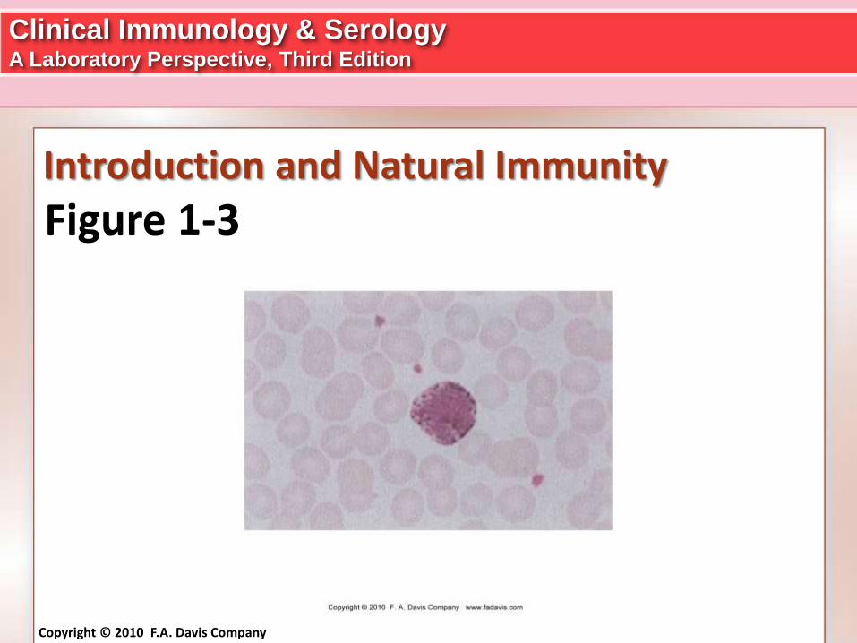
Actions of myeloid cells, including
• Neutrophils (See Figure 1-1)
• Basophils (See Figure 1-3)
• Eosinophils (See Figure 1-2)
• Monocytes & Macrophages (See Figure 1-5)
• Mast Cells (See Figure 1-4)
• Dendritic Cells

Clinical Immunology & SerologyA Laboratory Perspective, Third Edition
Copyright © 2010 F.A. Davis Company
Introduction and Natural Immunity Certain surface molecules are found on
human leukocytes and some nonleukocyte cell
types, and these are called Toll-like
receptors (TLRs).
The highest concentration of these receptors
occurs on monocytes, macrophages, and
neutrophils.
Each of these receptors recognizes a different
microbial product.

Clinical Immunology & SerologyA Laboratory Perspective, Third Edition
Copyright © 2010 F.A. Davis Company
Introduction and Natural Immunity See Figure 1-6
Once a receptor binds to its particular
substance, or ligand, phagocytosis may be
stimulated, or the cell produces cytokines that
enhance inflammation and eventual
destruction of the microorganism.

Clinical Immunology & SerologyA Laboratory Perspective, Third Edition
Copyright © 2010 F.A. Davis Company
Introduction and Natural Immunity
Figure 1-1

Clinical Immunology & SerologyA Laboratory Perspective, Third Edition
Copyright © 2010 F.A. Davis Company
Introduction and Natural Immunity
Figure 1-6

Clinical Immunology & SerologyA Laboratory Perspective, Third Edition
Copyright © 2010 F.A. Davis Company
Introduction and Natural Immunity
Figure 1-2
Clinical Immunology & SerologyA Laboratory Perspective, Third Edition
Copyright © 2010 F.A. Davis Company
Introduction and Natural Immunity
Figure 1-3

Clinical Immunology & SerologyA Laboratory Perspective, Third Edition
Copyright © 2010 F.A. Davis Company
Introduction and Natural Immunity
Figure 1-4

Clinical Immunology & SerologyA Laboratory Perspective, Third Edition
Copyright © 2010 F.A. Davis Company
Introduction and Natural Immunity
Figure 1-5

Clinical Immunology & SerologyA Laboratory Perspective, Third Edition
Copyright © 2010 F.A. Davis Company
Introduction and Natural Immunity
Phagocytosis consists of four main steps
1. Physical contact between the white cell and
the foreign particle
2. Formation of a phagosome
3. Fusion with cytoplasmic granules to form a
phagolysosome
4. Digestion and release of debris to the outside

Clinical Immunology & SerologyA Laboratory Perspective, Third Edition
Copyright © 2010 F.A. Davis Company
Introduction and Natural Immunity; 1-7
Figure 1-7

Clinical Immunology & SerologyA Laboratory Perspective, Third Edition
Copyright © 2010 F.A. Davis Company
Introduction and Natural Immunity Resting cells that engage in phagocytosis
normally derive their energy from anaerobic
glycolysis.
However, when phagocytosis is triggered, the
respiratory burst produces greater energy
via oxidative metabolism.
A radical known as O2– (superoxide) is
formed. Superoxide is highly toxic but can be
rapidly converted to more lethal products

Clinical Immunology & SerologyA Laboratory Perspective, Third Edition
Copyright © 2010 F.A. Davis Company
Introduction and Natural Immunity By adding hydrogen ions, the enzyme
superoxide dismutase (SOD) converts
superoxide to hydrogen peroxide or the
hydroxyl radical OH.
Its effect is potentiated by the formation of
hypochlorite ions.
This is accomplished through the action of the
enzyme myeloperoxidase in the presence of
chloride ions.

Clinical Immunology & SerologyA Laboratory Perspective, Third Edition
Copyright © 2010 F.A. Davis Company
Introduction and Natural Immunity Hypochlorite ions are powerful oxidizing
agents.
All of these substances contribute to killing
within the phagocyte.
See Figure 1-8

Clinical Immunology & SerologyA Laboratory Perspective, Third Edition
Copyright © 2010 F.A. Davis Company
Introduction and Natural Immunity
Figure 1-8

Clinical Immunology & SerologyA Laboratory Perspective, Third Edition
Copyright © 2010 F.A. Davis Company
Introduction and Natural Immunity Opsonization enhances phagocytosis.
Opsonins are serum proteins that attach to a
foreign substance and facilitate phagocytosis
by neutralizing repulsive forces on neighboring
cell membranes.
Examples of opsonins include C-reactive
protein, complement components, and
antibodies.
Clinical Immunology & SerologyA Laboratory Perspective, Third Edition
Copyright © 2010 F.A. Davis Company
Introduction and Natural ImmunityCellular defense mechanisms include the
action of
Lymphocytes
Macrophages
Mast cells
Dendritic cells

Clinical Immunology & SerologyA Laboratory Perspective, Third Edition
Copyright © 2010 F.A. Davis Company
Introduction and Natural Immunity Inflammation: Both cellular and humoral
mechanisms are involved.
The four cardinal signs / clinical symptoms of
inflammation are
• Redness
• Swelling
• Heat
• Pain
Clinical Immunology & SerologyA Laboratory Perspective, Third Edition
Copyright © 2010 F.A. Davis Company
Introduction and Natural ImmunityMajor events associated with the process of
inflammation are
Increased blood supply to the infected area
(due to vasodilation)
Increased capillary permeability
Migration of white blood cells, mainly
neutrophils to the injured area (diapedesis)
Migration of macrophages to the injured area
(chemotaxis)
See Figure 1-9

Clinical Immunology & SerologyA Laboratory Perspective, Third Edition
Copyright © 2010 F.A. Davis Company
Introduction and Natural Immunity
Figure 1-9

Clinical Immunology & SerologyA Laboratory Perspective, Third Edition
Copyright © 2010 F.A. Davis Company
Introduction and Natural Immunity Neutrophils are the primary cell involved in
the acute inflammatory response.
Neutrophil emigration may last 24 to 48 hours
and is proportional to the level of chemotactic
factors present in the area.
Migration of macrophages from surrounding
tissue and from blood monocytes occurs
several hours later and peaks at 16 to 48
hours.

Clinical Immunology & SerologyA Laboratory Perspective, Third Edition
Copyright © 2010 F.A. Davis Company
Introduction and Natural Immunity Macrophages attempt to clear the involved
area through phagocytosis, and in most cases
the healing process is completed with a return
of normal tissue structure.
Tissue damage and loss of function may result
from chronic inflammation.
C-Reactive Protein (CRP) is the most widely
monitored of the acute phase reactants and is
the best indicator of acute inflammation.


