Page 1
Change of attitude in the surgical treatment of early breast cancer
Ph.D. Dissertation
Tibor Takács, M.D.
Supervisor: György Lázár Ph.D., D.Sc.
Department of Surgery, Faculty of Medicine
University of Szeged
Szeged, Hungary
2013
Page 2
- 2 -
Contents
List of articles and abstracts related to the dissertation ............................................ - 3 -
List of full papers the dissertation is based on ................................................... - 3 -
List of full papers related to the subject of the dissertation ............................... - 4 -
List of abstracts related to the subject of the dissertation .................................. - 5 -
Abbreviations ............................................................................................................ - 6 -
Introduction ............................................................................................................... - 7 -
The purpose of our study .......................................................................................... - 8 -
Patients and methods ................................................................................................ - 9 -
1. WGL method ................................................................................................... - 9 -
2. ROLL method ................................................................................................ - 10 -
3. Histological methods .................................................................................... - 10 -
Pathological examination of the removed breast tissue ............................... - 10 -
Pathological examination of the removed SLNs ........................................... - 11 -
4. Statistical analysis ........................................................................................ - 12 -
Results ..................................................................................................................... - 13 -
Study 1: Comparing the ROLL and the GWL methods .................................... - 13 -
Study 2: Lymph node prognostic factors .......................................................... - 17 -
Study 3: Importance of SLNB in case of in situ breast carcinomas ................. - 27 -
Discussion ............................................................................................................... - 29 -
Summary and new results ....................................................................................... - 43 -
Acknowledgements ................................................................................................. - 44 -
References ............................................................................................................... - 45 -
Page 3
- 3 -
List of articles and abstracts related to the dissertation
List of full papers the dissertation is based on
I. Takács Tibor, Szentpáli Károly, Paszt Attila, Ormándi Katalin, Lázár Máté, Pálka István,
Kahán Zsuzsa, Lázár György: Az őrszem (sentinel) nyirokcsomó jelentősége in situ
emlőcarcinoma sebészi kezelésében. Magyar Onkológia 2006; 50:247–51.
II. Tibor Takács, Attila Paszt, Károly Szentpáli, Katalin Ormándi, Máté Lázár, István Pálka,
Zsuzsanna Kahán, György Lázár: Importance of Sentinel Lymph Node Biopsy in Surgical
Therapy of in situ Breast Cancer. Pathology & Oncology Research 2009; 15:329-33.
IF: 1,152
III. Tibor Takács, Attila Paszt, Zsolt Simonka, Szabolcs Ábrahám, Bernadett Borda, Aurél
Ottlakán, Katalin Ormándi, Máté Lázár, András Vörös, Zsuzsanna Kahán, György Lázár:
Radioguided occult lesion localization versus wire-guided lumpectomy in the treatment of
non-palpable breast lesions. Pathology & Oncology Research 2013 19: 267-73. IF:1.366
Page 4
- 4 -
List of full papers related to the subject of the dissertation
I. G. Cserni, G. Boross, R. Maráz, M.H.K. Leidenius, T.J. Meretoja, P.S. Heikkila, P.
Regitnig, G. Luschin-Ebengreuth, J. Zgajnar, A. Perhavec, B. Gazic, G. Lázár, T.
Takács, A. Vörös, R.A. Audisio: Multicentre validation of different predictive tools of
non-sentinel lymph node involvement in breast cancer. Surgical Oncology-Oxford 2012;
21: 59-65. IF: 2.444
II. Gábor Cserni, Rita Bori, Róbert Maráz, Marjut H.K. Leidenius, Tuomo J. Meretoja, Paivi
S. Heikkila, Peter Regitnig, Gero Luschin-Ebengreuth, Janez Zgajnar, Andraz Perhavec,
Barbara Gazic, György Lázár, Tibor Takács, András Vörös, Riccardo A. Audisio: Multi-
Institutional Comparison of Non-sentinel Lymph Node Predictive Tools in Breast Cancer
Patients with High Predicted Risk of Further Axillary Metastasis Pathololgy & Oncology
Research 2013; 19: 95-101. IF:1.366
III. Tuomo J. Meretoja, Marjut H.K. Leidenius, Päivi S. Heikkilä, Gábor Boross, István
Sejben, Peter Regitnig, Gero Luschin-Ebengreuth, Janez Zgajnar, Andraz Perhavec,
Barbara Gazic, György Lázár, Tibor Takács, András Vörös, Zuhair A. Saidan, Rana M.
Nadeem, Isabella Castellano, Anna Sapino, Simonetta Bianchi, Vania Vezzosi,
Emmanuel Barranger, Ruben Lousquy, Riccardo Arisio, Maria Pia Foschini, Shigeru
Imoto, Hiroshi Kamma, Tove F. Tvedskov, Niels Kroman, May-Brit Jensen, Riccardo A.
Audisio, Gábor Cserni: International Multicenter Tool to Predict the Risk of Nonsentinel
Node Metastases in Breast Cancer. Journal of the National Cancer Institute 2012; 104:
1888-96. IF:13.757
IV. Tuomo J. Meretoja, R.A. Audisio, P.S. Heikkilä, R. Bori, I. Sejben, P. Regitnig, G.
Luschin-Ebengreuth, J. Zgajnar, A. Perhavec, B. Gazic, G. Lázár, T. Takács, B. Kővári,
Z.A. Saidan, R.M. Nadeem, I. Castellano, A. Sapino, S. Bianchi, V. Vezzosi, E.
Barranger, R. Lousquy, R. Arisio, M.P. Foschini, S. Imoto, H. Kamma, T.F. Tvedskov,
M.B. Jensen, G. Cserni, M.H.K. Leidenius: International multicenter tool to predict the
risk of four or more tumor-positive axillary lymph nodes in breast cancer patients with
sentinel node macrometastases. Breast Cancer Research and Treatment 2013; 138: 817-
27. IF:5.87
Page 5
- 5 -
List of abstracts related to the subject of the dissertation
1. MST kongresszus Budapest - 2006: Az őrszem (sentinel) nyirokcsomó jelentősége ductalis
in situ emlőcarcinoma sebészi kezelésében. Takács T., Szentpáli K., Paszt A., Lázár Gy.
2. II. Emlőrák szimpózium Szeged – 2007 Az őrszem (Sentinel) nyirokcsomó jelentősége in
situ emlőcarcinoma sebészi kezelésében. Takács T., Paszt A., Szentpáli K., Ormándi K.,
Lázár M., Lázár Gy.
3. MST Dél-Magyarországi Csoportjának tudományos ülése – 2008 Kecskemét Prognosztikai
faktorok szerepe invasiv emlőcarcinomák hónalji nyirokcsomó metastasis képződésében.
Takács T., Paszt A., Simonka Zs., Török K., Lázár Gy.
4. III. Emlőrák szimpózium Szeged – 2009: Prognosztikai faktorok szerepe invasiv
emlőrákok hónalji nyirokcsomó metastasis képződésében. Takács T., Paszt A., Simonka
Zs., Lázár Gy.
5. MST kongresszus – 2012: A radioizotópos és a dróthorog jelöléses lokalizálási módszer
összehasonlítása nem tapintható emlőtumorok sebészi kezelésében Takács T., Paszt A.,
Simonka Zs., Ábrahám Sz., Borda B., Ormándi K., Lázár M., Vörös A., Kahán Zs., Lázár
Gy.
Page 6
- 6 -
Abbreviations
ALND: Axillary Lymph Node Dissection
AUC: Area Under the Curve
BCS: Breast Conservative Surgery
CNB: Core-Needle Biopsy
DCIS: Ductal Cancer In Situ (of the breast)
DCISM: Ductal Cancer In Situ (of the breast) with Microinvasion
ER: Estrogen Receptor
FNAC Fine-Needle Aspiration Cytology
GWL: Guidewire Localization
HE: Hematoxylin-Eosin
HER-2: Human Epidermal Growth Factor Receptor-2
IDC: Invasive Ductal Carcinoma
IHC: Immunohistochemistry
ILC: Invasive Lobular Carcinoma
ITC: Isolated Tumor Cells
LCIS: Lobular Cancer In Situ (of the breast)
LVI: Lymphovascular Invasion
NSLN: Non-Sentinel Lymph Node
PR: Progesterone Receptor
ROLL: Radioguided Occult Lesion Localization
ROC: Receiver Operating Characteristic
RSL: Radioguided Seed Localization
SLN: Sentinel Lymph Node
SLNB: Sentinel Lymph Node Biopsy
Page 7
- 7 -
Introduction
The extensive use of mammography for screening has resulted in the recognition of
increasing numbers of malignant or malignancy-suspicious breast tumors in an early stage.
Consequently, the ratio of non-palpable lesions has increased among early stage breast
tumors; these lesions are detected during routine mammographic screening first1. In parallel
with the extensive use of mammographic screening, the surgical treatment has changed as
well: conservative breast surgery (quadrantectomy, lumpectomy, excision of the tumor) has
replaced the earlier radical breast surgery, and axillary lymph node dissection (ALND) has
likewise been replaced by sentinel lymph node biopsy (SLNB), which is currently the
established method with which to assign the axillary lymph node status in early breast
cancers.2,3
Basically, there are two methods used to localize non-palpable breast tumors. The
method of guidewire localization (GWL) described by Kopans in 1980 has been used for
decades in the preoperative localization of non-palpable breast lesions4. The radioguided
occult lesion localization (ROLL) method was developed by Luini and colleagues in the
Institute of Oncology in Milan and is used since 19965. This method has become a standard
marking procedure; however, GWL is still used by several Institutions.
For a century, ALND has been an essential component of the surgical treatment of
invasive breast cancer. Axillary node status is one of the most important prognostic indicators
in breast cancer, and of particular value in the choice of adjuvant therapy.6,7
SLNB has been
developed as a minimally invasive diagnostic procedure for the accurate preoperative staging
of the axilla and ALND could be avoided if the patients do not have metastases in the Sentinel
Lymph Nodes (SLNs). The technique was first used by Morton and colleagues with blue dye
8,9 and later by van der Veen and colleagues
10 with lymphoscintigraphy in the treatment of
patients with melanoma. Similarly, this method can be applied in the treatment of breast
cancers in early stage. If the SLN can be accurately identified, and the histological
examination reveals no metastasis in the SLNs, the other nodes (non-SLNs - NSLN) in the
axilla are unlikely to contain metastases and the unnecessary ALND can be avoided.11
Page 8
- 8 -
Current practice guidelines recommended the ALND for breast cancer patients whose
SLN contains metastatic cancer.12,13,14
It is important to know that 40% to 70% of patients
with positive SLNs are found to have no other NSLN metastases.15,16
Therefore, these patients
undergo unnecessary ALND without therapeutic benefit or additional information for the
staging. Furthermore, completion ALND is associated with substantial morbidity affecting up
to 39% of patients, with a nearly three-fold increased risk of lymphedema or regional sensory
loss.17,18
Based on these, it would be important to create a predictive model detecting patients
who might benefit from ALND in case of SLN metastasis of the invasive breast tumor and
this would provide additional information regarding tumor staging and would have additional
therapeutic benefit as well.
In addition to early stage invasive breast tumors, the importance of in situ ductal cancers
has increased. Before the widespread introduction of mammographic screening, only 1-2% of
the recognized breast cancers comprised DCIS (Ductal Cancer in situ of the breast), but at
present, the rate of detection by mammographic screening of non-palpable breast cancers is
approximately 20%.19,20
DCIS is defined as a non-invasive breast cancer, and is widely
considered not to give metastases to the lymph nodes, so that ALND would comprise
overtreatment.20,21
Nonetheless, a number of studies have reported the detection of metastases
in SLNs in patients with DCIS, though with a very low incidence. 22,23,24,25,26,27,28,29,30
In situ
breast cancers require a novel approach in planning the surgical treatment as well, which
suggests that SLNB may be omitted in such cases.
The purpose of our study
I. Comparing the methods of localization of non-palpable breast tumors (ROLL, GWL)
(Study 1).
II. Finding factors influencing NSLN metastasis, creating predictive nomograms, and
comparing them with international nomograms (Study 2).
III. Simplifying option of the surgical treatment of in situ breast tumors (Study 3).
Page 9
- 9 -
Patients and methods
Our studies were conducted in the Department of Surgery, Faculty of Medicine,
University of Szeged.
Patients having a surgery using the GWL method (N = 69 patients) between January 1,
1997 and December 31, 2001 and the ROLL method (N = 321 patients) between January 1,
2002 and December 31, 2008 for having non-palpable unilateral malignant breast tumor and
who had primary breast conservative surgery (BCS) were enrolled in Study 1.
Patients having a surgery using the ROLL method (N = 824) for having early stage
unilateral malignant tumor between January 1, 2004 and December 31, 2011 who had primary
breast tumor removal (BCS or mastectomy) and SLNB simultaneously were enrolled in
Study 2.
Patients having a surgery using the ROLL method (N = 112) between January 1, 2002
and December 31, 2011 in case of whom the final histological examination confirmed
unilateral in situ breast tumor were enrolled in Study 3.
1. WGL method
69 patients with a non-palpable malignant breast tumor were operated on in our institute
following GWL between January 1, 1997 and December 31, 2001. During the intervention, a
hook wire was introduced by the radiologist under radiographic or ultrasound guidance
immediately before surgery. Direction of hook-wire insertion: for lesions in the upper
quadrant/central region, preference is given to the cranio-caudal direction; for lesions in the
lower quadrant, we prefer the lateral approach (these directions ensure the shortest way and at
the same time are the most appropriate for the surgical incision). Exact positioning of the
hook-wire (<5 mm) is important. The position of the wire was controlled by mammography.
During the operation, the excision involved the tumor that was marked by a wire. The level of
excision was the pectoral fascia. The excised specimen was marked with orientation stitches,
and then specimen mammographic tests were performed.4 Depending on the preoperative or
final histological results, the procedure was supplemented with ALND because at that time,
we did not use of SLNB. In the event of positive surgical margins, a supplementary operation
(re-resection or mastectomy) was performed.
Page 10
- 10 -
2. ROLL method
321 patients with non-palpable malignant breast tumor were operated on in our institute
with the application of the ROLL method between January 1, 2002 and December 31, 2008.
The ROLL technique and double marking SLNB were used simultaneously.31
One day before
the operation, 0.4 mL 99m
Tc-labeled human colloid albumin was injected into the tumor under
radiographic or ultrasound guidance. 4 hours later, it was followed by a lymphoscintigraphic
examination, and the projections of the SLNs on the skin were marked from two sides (SLN
mapping). On the following day, 10 minutes before the operation, a second marker substance
for the SLN, Patent Blue dye (2 mL) was injected into the subareolar region of the breast.
During the operation, radiocolloid activity peak was identified with a gamma probe and the
tumor was removed and the preoperative findings were also taken into account. The level of
excision was the pectoral fascia. The excised specimen was marked with orientation stitches
and controlled by specimen mammography. In patients with positive surgical margins, a
second operation (re-resection or mastectomy) was performed. If metastases were detected in
the SLNs, ALND was performed.
Radiologists recorded the time (minutes) required to localize the lesion with wire or
isotope substance. Surgeons recorded the time (minutes) required for excisions (without
axillary surgery).
3. Histological methods
Pathological examination of the removed breast tissue
During the pathological examination, the resection surface of the removed breast tissue was
stained with various substances: anterior black (Indian ink), posterior blue (Alcian Blue) and
superior red (Cadmium Red). The mass and the mediolateral, superoinferior and
anteroposterior sizes of the removed tissue were measured. During the operation, a cylinder-
form breast specimen was removed, and specimen volume could be calculated. After that,
sections were cut, and the size of the tumor and its distance from the resection surface of the
tissue were measured. During the procedure, at least 11 blocks were performed with the first
superoinferior section being the macroblock. In this way, we were able to measure the size of
the tumor and its distance from the anterior, posterior, superior and inferior resection surfaces.
Page 11
- 11 -
The second block (medial) included the medial part of the tumor from the macroblock to the
medial resection margin. With the help of this block, we were able to measure the distance
between the tumor and the medial resection margin. The third block (lateral) included the
lateral part of the tumor from the macroblock to the lateral resection margin providing the
measurement of the distance between the tumor and the lateral resection margin. The shaves
involved blocks 4–11. Besides traditional sections, we made 8 extra sections thus dividing the
external surface of the removed tissue into 8 parts (superior, superomedial, superolateral,
medial, lateral, inferomedial, inferolateral and inferior). By preparing these sections, our
investigation of the resection margins became more precise. 32
During the examination were
investigated the volume of removed specimen, the histological type of the tumor, the size of
the tumor (T stage), the presence of in situ breast cancer around the invasive component,
multifocality, the presence of lymphovascular invasion (LVI), histological grade, estrogen
(ER) and progesterone receptor (PR) status and Human Epidermal Growth Factor Receptor-2
(HER-2) gene expression. The three diameters of the removed specimen (anteroposterior,
mediolateral, superoinferior) were measured by pathologists, and the volume of the specimen
was calculated with using an equation applied in case of an elliptical tissue cylinder.
Pathologists considered multifocality where two or more invasive cancer foci could be found
in the same quadrant of the breast and where there was no contact between the invasive
focuses. Extensive in situ breast cancer around the invasive focus was defined in cases where
the proportion of intraductal component was at least 25%, and intraductal focuses were
present in the adjacent breast tissue as well. Microinvasive breast carcinoma (DCIS with
microinvasion – DCISM) was defined if the extension of cancer cells beyond the basement
membrane into the adjacent tissues is with no single focus larger than 1 mm in greatest
dimension. For hormonal receptor status, 10% staining of cells by Immunohistochemical
staining (IHC) was considered positive.
Pathological examination of the removed SLNs
SLNs were first examined using routine hematoxylin-eosin (HE) staining. If the
metastasis can be confirmed by HE staining, additional processing was not performed. If this
examination did not confirm metastasis, SLNs were evaluated in serial sections at intervals of
250 μm by means of HE. IHC staining was performed if the SLN was suspicious for
Page 12
- 12 -
metastasis but HE was not able to identify the tumor cells accurately. Negative SLNs did not
undergo IHC testing. The maximum dimension of the metastasis in each SLNs was measured
since 2008, previously were described just the types of metastases (isolated tumor cells (ITC),
micrometastasis or macrometastasis). Macrometastasis was defined if the SLN contains tumor
metastasis in higher diameter than 2 mm. Micrometastasis was defined if the measure of SLN
metastasis was between 0.2 and 2 mm. ITC metastasis in the SLN was defined if the measure
of the metastasis was smaller than 0.2 mm. All SLNs were examined for extranodal
extension. NSLNs from the ALND specimen were analyzed by routine HE staining only.
When comparing the GWL and the ROLL methods, we have taken into consideration
the preoperative localization time, the operating time, the age of the patients, the pathological
size of the tumor, the volume of the removed specimen, the ratio of the tumor size and the
removed specimen volume, the number of positive surgical margins, the subsequent
reoperations (reexcision or mastectomy) and the postoperative complications (wound
infections). Furthermore, we investigated other factors, such as the presence of an extensive in
situ breast carcinoma around the invasive cancer and the presence of multifocal tumors, as
they could have an impact on the frequency of positive resection margins.
During the examination of the predictive factors of NSLN metastasis, the following
variables were evaluated: tumor size, palpability, histological type of the tumor, grade of
differentiation of the tumor, the presence of LVI, ER status, PR status, HER-2 status, number
of removed SLNs, number of metastatic SLNs, the size of the metastasis in the SLNs, and
presence of extranodal invasion.
During the evaluation of in situ tumors, the histological type, size, and grade of
differentiation of the tumor, and the histological finding of the removed SLNs were examined.
4. Statistical analysis
For the comparison of continuous variables, t-test and one-way analysis of variance were
used, as well as the Mann-Whitney in cases of non-normality. The normal distribution of
samples was tested by using the Kolmogorov-Smirnov test. Categorical data were analyzed
by using chi-square and Fisher’s exact test. Multivariate analysis was performed by using
logistic regression. SPSS version 20.0 (© 2012 SPSS Inc.) was used for statistical analysis.
Significance was considered at p<0.05
Page 13
- 13 -
Results
Study 1: Comparing the ROLL and the GWL methods
The final histological examination revealed 69 malignant lesions in the GWL group, and
321 malignant lesions in the ROLL group. Table 1 presents the histological results of the
removed malignant breast lesions (Table 1).
Ultrasonographic guidance localization was performed in 58 cases using GWL method
and in 277 cases using ROLL method. Radiographic guidance localization was performed in
11 cases using the GWL method and in 44 cases using the ROLL method. The localization
time was significantly shorter in the ROLL group both with ultrasonographic guidance
(5.7±1.4 min vs. 21.6±2.4 min, p=0.05) and with radiographic guidance (21.8±3.1 min vs.
41.6±3.8 min, p=0.021). It must be taken into consideration, however, that every time the
GWL method was used, patients underwent mammography to verify the correct localization
of the guidewire, which of course increased the localization time in all GWL cases. There was
no significant difference in the operating time requirements (30.2±4.6 min vs. 30.7±4.7 min).
The mean age of the patients was similar in both groups (59 yrs vs. 57.7 yrs). The removed
breast specimen volume did not differ significantly between the GWL (89.5±116.3 cm3) and
the ROLL group (104.1±78.6 cm3). The pathological tumor size and ratio of tumor size and
Type of specimen GWL (N=69) ROLL (N=321)
LCIS
DCIS
Papillary in situ breast cancer
–
5 (7.3%)
–
3 (0.9%)
53 (16.5%)
3 (0.9%)
DCISM 4 (5.8%) 4 (1.3%)
IDC 54 (78.3%) 218 (67.9%)
ILC 3 (4.3%) 20 (6.3%)
Mucinous carcinoma
Tubular carcinoma
Papillary carcinoma
Phylloid carcinoma
Mixed carcinoma
Medullary carcinoma
2 (2.9%)
–
–
–
–
1 (1.4%)
1 (0.3%)
7 (2.2%)
2 (0.6%)
1 (0.3%)
9 (2.8%)
–
Table 1. Pathological features of the GWL and ROLL groups
Page 14
- 14 -
removed specimen did not show any significant difference between the GWL and the ROLL
groups. The final pathological examination revealed 16 patients (23.2%) with a positive
resection margin in the GWL group (n=69). Reoperations were performed on 14 of these
patients (20.3%); 5 patients (7.2%) underwent breast reexcision and 9 patients (13.1%)
mastectomy, and 2 patients refused consent to mastectomy. Residual tumor tissue was found
by the histological examination in 6 patients (8.7%). In the ROLL group (n=321), positive
resection margins were detected by the final pathological examination in 47 of the cases
(14.6%). Reoperations were performed on 46 of these patients (14.3%); 24 patients (7.5%)
underwent breast reexcision, and 22 patients (6.8%) underwent mastectomy. One patient
refused mastectomy. Residual tumor tissue was found by the histological examination in 25
patients (7.8%). No significant difference was detected between the GWL and the ROLL
groups in the frequency of positive resection margins. The incidence of postoperative
complications (wound infections) did not differ significantly in the two groups (Table 2).
GWL (N=69) ROLL (N=321) p value
Duration of localization (mean ±SD), min
Radiographic guidance
Ultrasound guidance
41.6±3.8
21.6±2.4
21.8±3.1
5.7±1.4
0.021
0.05
Duration of surgical excision (mean ±SD), min 30.2±4.6 30.7±4.7 NS
Mean age, yr 59 57.7 NS
Removed breast specimen volume (mean ± SD), cm3 89.5±116.3 104.1±78.6 NS
Pathological size of tumor (mean ± SD), mm 12.4±8.6 15.2±11.2 NS
Size of the tumor (cm)/specimen volume (cm3) 0.0237±0.0258 0.0181±0.0179 NS
Involved surgical margin(s), n (%) 16 (23.2%) 47 (14.6%) NS
Residual tumor, n (%) 6 (8.7%) 25 (7.8%) NS
Wound infections, n (%) 2 (2.9%) 3 (0.9%) NS
Table 2. Comparison of various factors between GWL and ROLL in malignant breast tumors
(NS-not significant)
We have also taken further factors into consideration that influenced the frequency of
positive resection margins. In the GWL group, 2 of the 69 patients (2.9%) had multifocal
breast tumor, and another 4 patients (5.8%) had extensive in situ tumor components around
Page 15
- 15 -
the invasive cancer. In the ROLL group 22 of the 321 patients (6.8%) had multifocal breast
tumors, and 37 of the 321 patients (11.5%) had extensive in situ tumor components around
the invasive cancer. The results of the histological analysis of the patients in the ROLL group
indicated, that the size of the malignant lesion (p=0.021), the presence of a multifocal tumor
(p=0.035), and the presence of an extensive in situ breast carcinoma around the invasive
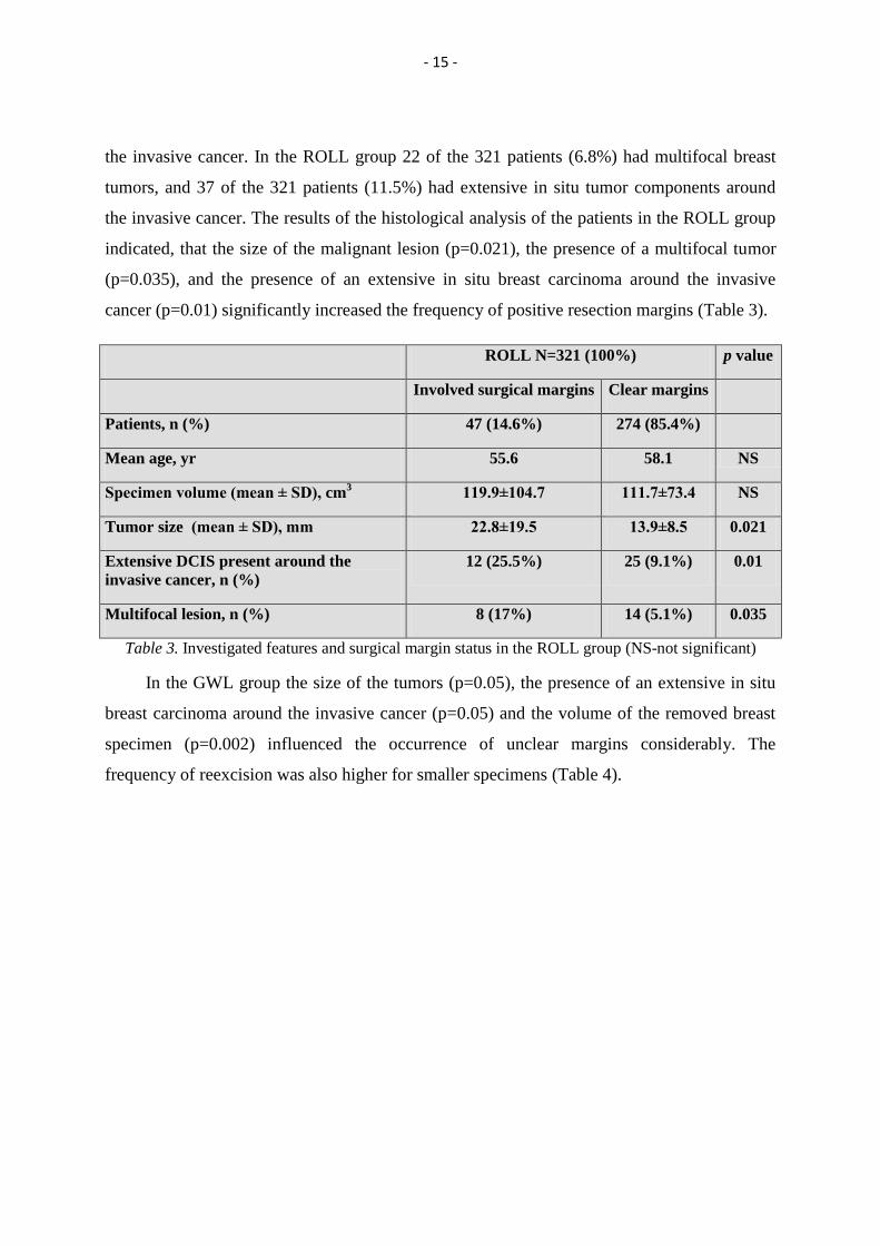
cancer (p=0.01) significantly increased the frequency of positive resection margins (Table 3).
ROLL N=321 (100%) p value
Involved surgical margins Clear margins
Patients, n (%) 47 (14.6%) 274 (85.4%)
Mean age, yr 55.6 58.1 NS
Specimen volume (mean ± SD), cm3 119.9±104.7 111.7±73.4 NS
Tumor size (mean ± SD), mm 22.8±19.5 13.9±8.5 0.021
Extensive DCIS present around the
invasive cancer, n (%)
12 (25.5%) 25 (9.1%) 0.01
Multifocal lesion, n (%) 8 (17%) 14 (5.1%) 0.035
Table 3. Investigated features and surgical margin status in the ROLL group (NS-not significant)
In the GWL group the size of the tumors (p=0.05), the presence of an extensive in situ
breast carcinoma around the invasive cancer (p=0.05) and the volume of the removed breast
specimen (p=0.002) influenced the occurrence of unclear margins considerably. The
frequency of reexcision was also higher for smaller specimens (Table 4).
Page 16
- 16 -
GWL n=69 (100%) p value
Involved surgical margins Clear margins
Patients, n (%) 16 (23.2%) 53 (76.8%)
Mean age, yr 58.4 60.6 NS
Specimen volume (mean ± SD), cm3 66.4±40.3 96±127.1 0.002
Tumor size (mean ± SD), mm 17±11.6 11.2±7.4 0.05
Extensive DCIS present around the invasive
cancer, n (%) 2 (12.5%) 2 (3.8%) 0.05
Multifocal lesion, n (%) 1 (6.3%) 1 (1.9%) NS
Table 4. Investigated features and surgical margin status in the GWL group (NS-not significant)
Page 17
- 17 -
Study 2: Lymph node prognostic factors
Between January 1, 2004 and December 31, 2011, the final histological examination
confirmed invasive carcinoma in case of 855 patients having a surgery using the ROLL
method; in case of these patients, SLNB was also planned simultaneously. In 824 cases,
marking of the SLN was successful (824/855 – 96.3%) using dual marking procedure, and
successful SLNB was performed in all 824 cases. Based on the histological findings, the
majority of the tumors was invasive ductal carcinoma (IDC) (79.1%), followed by invasive
lobular carcinoma (ILC) (9.9%) and mixed type carcinoma (4.4%) (Table 5, Figure 1).
Histology Number of cases (N) %
IDC 652 79.1
ILC 80 9.7
Mixed carcinoma 36 4.4
Other less common carcinomas 56 6.8
Tubular carcinoma
Papillary carconoma
Medullar carcinoma
Mucinous carcinoma
Aplastic carcinoma
Undifferentiated carcinoma
Carcinosarcoma
Cribriform carcinoma
Neuroendocrine carcinoma
Metaplastic carcinoma
Micropapillar carcinoma
Epithelial carcinoma
16
9
8
14
1
1
1
1
1
2
1
1
2
1.1
1
1.7
0.1
0.1
0.1
0.2
0.1
0.2
0.1
0.1
Table 5. Histological findings of invasive breasts tumors
652
8036 56
0
100
200
300
400
500
600
700
Invasive ductal
carcinoma
Invasive lobular
carcinoma
Mixed
carcinoma
Other less
common
carcinomas
Histological types
Nu
mb
er
of
cases
Figure 1. Histological types of breast tumors
Page 18
- 18 -
A total of 1328 SLNs were removed from 824 patients, which means an average of 1.6
lymph nodes per patient. SLN metastasis was not confirmed by histology in 553 cases
(553/824 – 67.1%), but in 271 cases (271/824 – 32.9%), SLN metastases were found. In 205
patients, complementary ALND was performed. In the other 66 cases, ALND was not
performed based on the decision of the oncoteam regarding the patient’s age and compliance,
the size of the metastasis (ITC or micrometastasis), and in two cases, patients did not agree to
perform the complementary surgery.
Then we studied the connection between primary tumor characteristics and NSLN
metastasis in case of SLN positivity first with one-way analysis of variance in patients having
complementary ALND (205 patients). In 70 patients (70/205 34.1%), additional metastasis
was confirmed in the axillary lymph nodes by histological examination (Figure 2).
824
271205
70
0
100
200
300
400
500
600
700
800
900
Nu
mb
er
of
cases
Successful
SLNB
Metastatic SLN Additional
ALND
Metastatic
NSLN
Figure 2. Histological finding of lymph node biopsies
Average age of the patients was significantly different between the NSLN negative and
NSLN metastatic groups (55±11.1 years vs. 58.8±10.1 years, p=0.034). 75.6% (102/135) of
NSLN negative patients had palpable tumor, while this ratio was 84.3% (59/70) in patients
with NSLN metastasis, the difference was not significant (p=0.209). The presence of
multifocal tumor did not influence the incidence of the metastasis in the NSLNs (NSLN
negative: 11/135 – 8.1%, and NSLN positive 10/70 – 14.3%; p=0.224). The tumor size
correlated with the incidence of the NSLN metastasis (NSLN negative: 18.8±8.1 mm vs.
NSLN positive: 22.6±10.6 mm; p=0.019). Comparison was also made based on histological
type. IDC was the most common (83.9%), followed by ILC (8.3%), and mixed type (lobular +
Page 19
- 19 -
ductal) carcinoma (5.9%). The incidence of less common tumors (other histological tumors)
was 2%. The histological types of the tumors did not influence the incidence of a NSLN
metastasis (p=0.375). Studying the grade of histological differentiation revealed that the ratio
of grade 1 tumors was 14.1% (19/135) in NSLN negative cases, and 8.6% (6/70) in NSLN
positive patients. The incidence of NSLN metastasis was not different in grade 2 (NSLN
negative: 64/135 - 47.4% vs. NSLN positive: 39/70 – 55.7%) or grade 3 tumors (NSLN
negative: 52/135 – 38.5% vs. NSLN positive: 25/70 – 35.7%) either (p=0.397). The presence
of LVI did not influence the incidence of additional NSLN metastasis (NSLN negative:
36/135 26.7% vs. NSLN positive: 17/70 24.3%; p=0.74). Neither ER positivity (NSLN
negative: 107/135 – 79.2% vs. NSLN positive: 51/70 – 72.9%; p=0.274), nor PR positivity
(NSLN negative: 101/135 – 74.8% vs. NSLN positive: 46/70 – 65.7%; p=0.24), nor HER-2
gene expression influenced the incidence of metastasis in the NSLN (NSLN negative: 24/135
– 17.8% vs. NSLN positive: 13/70 – 18.6%; p=0.848) (Table 6). The sizes of the metastasis in
the removed SLNs were significantly different between the NSLN negative and positive
groups (NSLN negative: 5.1±4.1 mm vs. NSLN positive: 8.1±4.6 mm, p<0.0001), however,
there were only 148 patients in the latter group regarding the fact that before 2008, the proper
size of the SLN metastasis was not routinely measured, only the type of the metastasis was
determined (ITC-, micro- or macrometastasis). These groups were examined as well, but this
comparison could be made with all 205 patients again. We found that in case of
macrometastasis, the incidence of NSLN metastasis was significantly increased (p=0.013).
Extracapsular spreading of the metastasis in the SLN did not influence the incidence of NSLN
metastases (NSLN negative: 18/135 – 13.3% vs. NSLN positive: 16/70 – 22.9% p=0.112).
The number of removed SLNs (p=0.37) and that of the SLNs containing a tumor (p=0.395)
did not increase the risk of additional NSLN metastasis (Table 6-7).
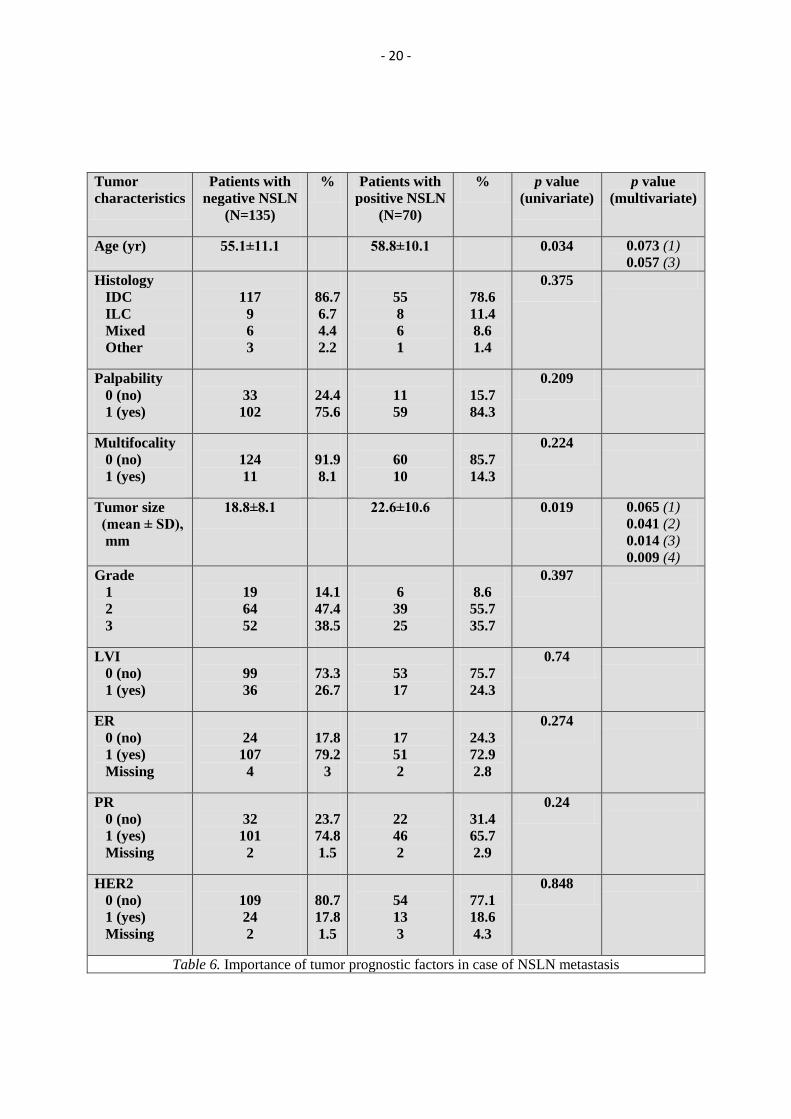
Page 20
- 20 -
Tumor
characteristics
Patients with
negative NSLN
(N=135)
% Patients with
positive NSLN
(N=70)
% p value
(univariate)
p value
(multivariate)
Age (yr) 55.1±11.1 58.8±10.1 0.034 0.073 (1)
0.057 (3)
Histology
IDC
ILC
Mixed
Other
117
9
6
3
86.7
6.7
4.4
2.2
55
8
6
1
78.6
11.4
8.6
1.4
0.375
Palpability
0 (no)
1 (yes)
33
102
24.4
75.6
11
59
15.7
84.3
0.209
Multifocality
0 (no)
1 (yes)
124
11
91.9
8.1
60
10
85.7
14.3
0.224
Tumor size
(mean ± SD),
mm
18.8±8.1 22.6±10.6 0.019 0.065 (1)
0.041 (2)
0.014 (3)
0.009 (4)
Grade
1
2
3
19
64
52
14.1
47.4
38.5
6
39
25
8.6
55.7
35.7
0.397
LVI
0 (no)
1 (yes)
99
36
73.3
26.7
53
17
75.7
24.3
0.74
ER
0 (no)
1 (yes)
Missing
24
107
4
17.8
79.2
3
17
51
2
24.3
72.9
2.8
0.274
PR
0 (no)
1 (yes)
Missing
32
101
2
23.7
74.8
1.5
22
46
2
31.4
65.7
2.9
0.24
HER2
0 (no)
1 (yes)
Missing
109
24
2
80.7
17.8
1.5
54
13
3
77.1
18.6
4.3
0.848
Table 6. Importance of tumor prognostic factors in case of NSLN metastasis
Page 21
- 21 -
Tumor characteristics Patients
with
negative
NSLN
(N=135)
% Patients
with
positive
NSLN
(N=70)
% p value
(univariate)
p value
(multivariate)
Type of SLN
metastasis
ITC
MIC
MAC
1
21
113
0.7
15.6
83.7
0
3
67
0
4.3
95.7
0.013 0.047 (3)
0.03 (4)
Size of SLN
metastasis,
(mean ± SD) mm,
(N=148)
5.1±4.1 8.1±4.6 <0.0001 0.002 (1)
0.001 (2)
Extracapsular
invasion of SLN
metastasis
0 (no)
1 (yes)
117
18
86.7
13.3
54
16
77.1
22.9
0.112
No. of removed SLNs
1
2
3
4
6
63
47
20
4
1
46.7
34.8
14.8
3
0.7
42
16
10
2
0
60
22.9
14.3
2.8
0
0.37
No. of positive SLNs
1
2
3
4
109
23
2
1
80.8
17
1.5
0.7
53
13
3
1
75.7
18.6
4.3
1.4
0.395
Table 7. Importance of SLN prognostic factors in case of NSLN metastasis
Page 22
- 22 -
Factors confirmed to be significant with one-way analysis of variance are described in
Figure 3.
p=0.034
58.8±10.155±11.1
0
10
20
30
40
50
60
70
80
NSLN negative NSLN positive
Ag
e (
yr)
p=0.019
18.8±8.1
22.6±10.6
0,0
5,0
10,0
15,0
20,0
25,0
30,0
35,0
NSLN negative NSLN pozitive
tum
or
siz
e (
mm
)
Figure A Figure B
p<0.0001
8.1±4.6
5.1±4.1
0
2
4
6
8
10
12
14
NSLN negative NSLN positive
Siz
e o
f m
eta
sta
tic S
LN
(m
m)
p=0.013
22
113
3
67
0
20
40
60
80
100
120
SLN with
micrometastasis and
ITC metastasis
SLN with
macrometastasis
SLN with
micrometastasis
SLN with
macrometastasis
NSLN negative NSLN pozitive
Nu
mb
er
of
cases
Figure C Figure D
Figure 3. Age (Figure A), tumor size (Figure B), and size of SLN metastasis (Figure C; N=148 cases),
and type of the SLN metastasis (Figure D; N=205 cases) in the NSLN negative and positive groups,
figures include mean + SD, significance values
Then factors found to be significant with one-way analysis of variance were tested with
logistic regression (age, tumor size, size, and type of the SLN metastasis). More studies were
performed. In our first study (1), if the size of the SLN metastasis was known and all three
variables were included, age (p=0.073) and tumor size (p=0.065) did not influence the
incidence of NSLN metastasis significantly, but the size of the SLN metastasis was found to
be significant (p=0.002). The second study (2) was performed omitting the age from logistic
regression. In this case, tumor size (p=0.041) and the size of the SLN metastasis (p=0.001)
significantly influenced the incidence of metastasis in NSLN. In the third study (3), if the
exact size of the SLN metastasis was not known only the type of it (macro-, or
micrometastasis) and all three variables were included, age was strictly not significant
(p=0.057), but the tumor size (p=0.014) and the type of the SLN metastasis were significantly
(p=0.047) influencing the incidence of NSLN metastasis. The fourth study (4) was performed
Page 23
- 23 -
similarly to the second one, age was not included in the analysis, in this case, tumor size
(p=0.009) and the size of the SLN metastasis (p=0.03) significantly influenced the incidence
of metastasis in NSLN (Table 6-7).
Based on our results, we tried to construct a model to predict the incidence of additional
NSLN metastasis. The probability of the metastasis can be calculated by the following
equitation if the size of the SLN metastasis is known in the first case:
PNSLNmet= )416.4s*144.0t*038.0a*034.0(e1
1
in the second case:
PNSLNmet= )562.2s*151.0t*042.0(e1
1
where: PNSLNmet – the probability of NSLN metastasis
a – age (year)
t – tumor size (mm)
s – size of the metastasis in the SLN (mm)
If the size of the SLN metastasis is not known only the type of it, the following equitation can
be used to calculate the probability of NSLN metastasis in the third case:
PNSLNmet= )701.1m*283.1t*041.0a*028.0(e1
1
in the fourth case:
PNSLNmet= )016.0m*405.1t*043.0(e1
1
where: PNSLNmet – the probability of NSLN metastasis
a – age (year)
t – tumor size (mm)
m – type of the metastasis in the SLN (mac=1, mic=2)
Page 24
- 24 -
Evaluation of the specificity and sensitivity of the first model using the Receiver Operating
Characteristic (ROC) curve showed that its predictive value – based on the AUC=0.735 (Area
Under the Curve) value is considered to be moderate, while the predictive value of the second
model using the ROC curve similarly was somewhat lower (AUC=0.717) than the first one
(Figure 4). Analysis of the third and fourth models with the ROC curve showed low
predictive values (AUC=0.66 and 0.638), it practically cannot be used to predict NSLN
metastasis (Figure 5).
Figure A
Figure B
Figure 4. Diagram presenting the reliability of the predictive models regarding specificity and
sensitivity and predicted and actual incidence of NSLN metastases (ALND or ABD met); Figure A in
case of the three-variable model (AUC=0.735), Figure B in case of the two-variable model
(AUC=0.717) if the exact size of the SLN metastasis is known
Page 25
- 25 -
Further examination of the first predictive nomogram revealed that in patients with
lower risk for NSLN metastasis, the nomogram is relatively more accurate in predicting the
NSLN metastasis. Using a cut-off value of 0.27 in case of the predictive curve, the predictive
value of the model could be increased, a sensitivity of 62.5 % and a specificity of 75 % could
be achieved, this is the most accurate range of the nomogram. We tried to improve sensitivity
Figure A
Figure B
Figure 5. Diagram presenting the reliability of predictive values of the models regarding specificity
and sensitivity and predicted and actual incidence of NSLN metastases (ALND or ABD met); Figure
A in case of the three-variable model (AUC=0.66), Figure B in case of the two-variable model
(AUC=0.638) if only the type of the SLN metastasis (macro-, or micrometastasis) is known
Page 26
- 26 -
and specificity similarly in case of the other nomograms as well; the results are seen in
Table 8.
Nomogram AUC Cut off value Sensitivity
(%)
Specificity
(%)
1. Diagram 0.735 0.27 62.5 75
2. Diagram 0.717 0.35 55.4 81.3
3. Diagram 0.66 0.34 69.6 67
4. Diagram 0.638 0.36 57.1 64.4
Table 8. Most accurate predictive ranges of the nomograms
In summary, in case of SLN positivity, the patient’s age, the tumor size, and the size
(type) of the SLN metastasis influenced the NSLN metastasis according to the one-way
analysis of variance. Multivariate analyses of cases where the size of the SLN metastasis is
known showed that only the size of the SLN metastasis was strictly significant, age and tumor
size showed only borderline significance. If the age was not included in the multivariate
analysis, the tumor size and the size of the SLN metastasis were significant factors. If only the
type of the SLN metastasis is known, multivariate analysis showed borderline significance in
case of the age, but the tumor size and the type of the SLN metastasis were still significant,
even if the age was not included in the analysis. The predictive value of the models is
moderate if the size of the SLN metastasis is known, while it is poor if only the type of the
SLN metastasis is known. In case of patients with lower risk for NSLN metastasis, the
predictive value of the nomogram is relatively more accurate.
Page 27
- 27 -
Study 3: Importance of SLNB in case of in situ breast carcinomas
Between January 1, 2002 and December 31, 2011, patients having surgery using the
ROLL method with a final histological examination confirming in situ breast tumor (N = 112
patients) were enrolled in the study. The median age of the patients was 55.2 years (range 30–
78 years). 75 patients (75/112, 67%) underwent preoperative fine-needle aspiration cytology
(FNAC) and 23 patients (23/112, 20.5%) underwent core-needle biopsy (CNB). Preoperative
histological results were not available in 13 patients. FNAC was not informative (C1) in 28 of
75 patients (28/75, 37.3%), 3 patients (3/75, 4%) had a benign breast disease (C2), 5 (5/75,
6.7%) had atypical breast disease (C3), 19 (19/75, 25.3%) gave the suspicion of malignant
disease (C4) and only in 20 (20/75, 26.7%) were malignant cells identified in the sample (C5).
Of the 23 patients investigated by CNB, malignant breast cancer (pure DCIS) was identified
in 17 (17/23 73.9%), invasive cancer in 2 (2/23, 8.7%), atypical breast disease in 2 (2/23
8.7%) and benign breast disease in 2 (2/23, 8.7%) cases (Table 9).
Results of FNAC N %
No cells detected (C1) 28 25.0
Benign disease (C2) 3 2.7
Atypical disease (C3) 5 4.4
Suspected malignancy (C4) 19 17.0
Malignant cells (C5) 20 17.8
Results of CNB
Benign disease (B2) 2 1.8
Atypical disease (B3) 2 1.8
Malignant disease pure in situ cancer (B5a) 17 15.2
Malignant disease in situ and invasive cancer (B5a+b) 2 1.8
Excision 1 0.9
Data not available 13 11.6
Table 9. Results of preoperative histological diagnosis
The final histological examination verified lobular in situ breast cancer (LCIS) in 4 of
the 112 patients (4/112, 3.6%), pure DCIS in 96 (96/112, 85.7%), papillary in situ cancer in 3
(3/112, 2.7%) and DCISM in 9 (9/112, 8%) patients (Table 10). 36 patients (36/112, 32.1%)
had palpable tumor. DCIS breast cancers have a number of histological subtypes (solid,
cribriform, papillary, micropapillary and comedo). The most important factors are the
presence of comedo necrosis and the grade of the tumor cells. In 76 of the 108 pure DCIS
patients (76/108, 70.4%), the tumor was of high grade (Grade III), in 11 (11/108, 10.2%)
Page 28
- 28 -
cases, it was of intermediate grade (Grade II), and in 21 (21/108, 19.4%) cases, it was of low
grade (Grade I).
Histological type N %
LCIS 4 3.6
DCISM 9 8.0
Pure DCIS 96 85.7
Papillary in situ cancer 3 2.7
Table 10. Final pathologic diagnosis
Simultaneous SLNB was planned in 108 of the 112 patients (108/112, 96.4%), while 4
(4/112 3.6%) patients underwent only wide excision. In 8 of 108 patients (8/108, 7.4%), SLNs
were not identified; axillary sampling or ALND was performed to remove the axillary lymph
nodes at level I-II. 100 patients underwent successful SLNB (100/108, 92.6%). In 95 cases,
SLNs were evaluated in serial sections at intervals of 250 µm with using HE, while in 5 cases
SLNs were processed only as routine axillary lymph nodes. First, during the evaluation of the
95 cases, a total of 147 sentinel lymph nodes were examined (an average of 1.5 lymph nodes
per patient, range 1–5). Metastasis was not confirmed in SLNs processed with serial
sectioning. In 11 cases, additional axillary lymph nodes were removed besides successful
SLNB; metastasis was not confirmed in the removed axillary lymph nodes. These lymph
nodes were processed with routine HE staining. In the other 13 cases (8 cases of ALND –
processed with routine HE method + 5 cases of successful SLNB, but processed with routine
HE method), metastasis was not confirmed either.
26 of the 112 (26/112, 23.2%) patients required a second complementary operation:
mastectomy in 12 cases, and reexcision in 14. Residual tumor was verified in 10 patients
treated with mastectomy and 7 patients treated with reexcision. One patient treated with
reexcision underwent mastectomy because of positive resection margin during the reexcision
(Table 11).
Complementary
surgery
Number
of cases %
Residual
tumor (N) %
Reexcision 14 12.5 7 6.3
Mastectomy 12 10.7 10 8.9
Table 11. Reoperations and results in case of patients with in situ breast cancer
Page 29
- 29 -
Discussion
The extensive use of mammography has resulted in the increased detection rate of early-
stage non-palpable malignant breast tumors. Both the GWL and the ROLL methods are
widely applied in surgical therapy to reveal and to remove non-palpable breast tumors. The
GWL method is the more widespread technique in use today despite some well-known
disadvantages: [1] radiologically, the guidewire placement is a difficult procedure to carry
out; spontaneous wire displacement, and inability to reposition can occur as well. [2] The
procedure is traumatic, causing discomfort and pain to the patient; furthermore, the wire must
remain in place until the operation. [3] The surgical excision of a wire-located lesion with
clear margins is a technically difficult procedure. There is obvious interference with the
incision line and the surgical approach, and the wire can be accidentally transected as well.
The ROLL method was developed to overcome some of the disadvantages of the GWL
technique. Its reported advantages include precise localization, accurate surgical removal,
higher rate of clear margins, reduced size of the excised specimen, better concentricity of the
lesion, less patient discomfort, shorter operating time, and reduced numbers of reoperations,
with an accompanying reduction in costs. Despite the fact that the ROLL technique has been
available for more than 10 years now, only a few studies have been published about it. The
GWL and the ROLL techniques have been compared only in 11 clinical studies and subjects
were randomized only in 4 studies (Table 12).33,34,35,36,37,38,39,40,41,42,43
Page 30
- 30 -
Authors Year Patients, N
Involved surgical
margins % p value
ROLL GWL ROLL GWL
Luini et al. 33
1999 30 30 0 0 NS
Rampaul et al.®34
2004 48 47 NA NA NA
Ronka et al. 35
2004 64 14 8 28.6 0.03
Gallegos-Hernandez et al.36
2004 65 67 16.9 35.8 0.014
Zgajnar et al.37
2005 51 92 29.4 55.2 0.005
Nadeem et al. 38
2005 65 65 17 43 0.001
Thind et al. 39
2005 70 70 16.2 40 0.002
Strnad et al. 40
2006 21 12 NA NA NA
Moreno et al.® 41
2008 61 59 10 12.5 NS
Medina-Franco et al.® 42
2008 50 50 11.1 37.5 0.04
Martinez et al. ® 43
2009 66 68 10.6 17.6 NS
Present study 2012 321 69 14.6 23.2 NS
Table 12. Comparison of international results on the complete excision rates with the
ROLL and the GWL techniques (®-randomized, NA-not available,
NS-not significant)
Altogether 6 trials have described a significant difference regarding the clear resection
margins in favor of the ROLL technique.35,36,37,38,39,42
3 studies have found that the
volume/weight ratio of the excised specimen was lower in the ROLL group than in the GWL
group.35,37,41
Several studies have confirmed the well-known advantages of the ROLL method,
such as better cosmetic results 38,39,41, less perioperative pain
34,41 and shorter localization
time.38,39,42
The newest systematic review demonstrated that radioguided localization
techniques (including ROLL and radioguided seed localization (RSL) methods) produce
lower positive resection margin rates and consequently fewer reoperations. However, this
review was limited by its small size and the quality of the randomized controlled trials.44
A
recently published multicenter, randomized, controlled trial compared the RSL method to a
standard GWL technique in the detection of non-palpable invasive and in situ breast
Page 31
- 31 -
carcinomas.45
In contrast to other trials, the positive resection margins and the reoperation
rates were similar in both techniques, but the operating time was shorter when using RSL. It is
important to know that in the RSL method, a radio-opaque titanium seed containing an 125
I-
isotope was used, therefore it is not exactly equivalent to the classic ROLL method.
In the present study, we did not find any significant differences between the two
compared methods in respect of the proportion of the average volume of the removed
specimen, the proportion of positive surgical margins, the incidence of residual tumors
(removed during a second operation), or the frequency of postoperative wound infections.
Preoperative localization time was significantly lower in the ROLL group, but there was no
significant difference in the duration of surgical excision. Nevertheless, our surgeons found
the ROLL method technically easier. International study results suggest a higher rate of
successful primary tumor excision (clear resection margins) with the use of the ROLL method
(Table 12). Higher clear resection margin rates were found in the ROLL group in our study as
well, but the difference was not significant statistically (85.4% vs. 76.8%). Although the
average removed specimen volume and the pathological tumor size were higher in the ROLL
group than in the GWL group, they were not significantly different. We did not find any
significant difference in the ratio of the tumor size and the removed specimen volume. This
indicates that a relatively smaller specimen can be removed safely using the ROLL method
for the same tumor size. Another important advantage of the ROLL technique is that it allows
concomitant removal of the invasive breast lesion and the SLN(s). Furthermore, our
investigation revealed that by applying the ROLL method, the involved surgical margin was
influenced by the tumor size, by the existence of a multifocal tumor, and by the presence of an
extensive in situ breast cancer around the invasive tumor. In case of the GWL technique, the
frequency of positive resection margins was influenced by the tumor size, by the presence of
an extensive in situ breast cancer around the invasive tumor and by a lower specimen volume.
It is important to emphasize that the size of the tumor was bigger and the specimen volume
was lower in the GWL group with positive resection margins. Therefore, the ratio of the
tumor size and the removed specimen volume are more indicative factors of the occurrence of
a positive resection margin than just the size of the tumor or the removed specimen volume
itself alone. Several studies have proved that the frequency of a positive resection margin is
Page 32
- 32 -
significantly increased by the size of the tumor46.47
, by the presence of an extensive in situ
tumor around the invasive tumor46,48
, by the presence of multifocal tumors46,49
and by the
volume of the removed specimen.46,49
Considering these facts, it is noteworthy that the most
important predictive factor of a local tumor recurrence in breast-conserving surgery is a
positive resection margin.50,51,52
If the final surgical margins are negative, the 5-year risk of
local failure is 2–7%, whereas with positive margins, this risk can rise up to 16% or even
higher53,54,55,56
. In addition to the surgical margin status, factors such as young age, large
tumor size, adjuvant chemotherapy and hormonal therapy, and positive ALNs are all
significant independent predictors of locoregional recurrence.57
International results show that both the GWL and the ROLL methods are suitable for the
localization and subsequent removal of non-palpable breast tumors. We have come to the
same conclusion in our study. However, the ROLL method has more advantages, such as
shorter localization time, more accurate surgical excisions and less discomfort to the patient.
We recommend that the ROLL method should be used for the localization of non-palpable
breast tumors if preoperative examinations prove the presence of an invasive breast cancer
and SLNB is also to be considered. We would recommend the use of the GWL technique in
cases with extensive microcalcifications and when SLNB is not going to be performed (pure
DCIS, radial scar, etc.).
Besides BCSs, SLNB, which is an accepted indicator of the axillary lymph node status
has come to the front. In accordance with previous clinical practice, complementary ALND
was routinely performed in case of metastatic SLN.12,13,14
However, several studies have
highlighted that it was unnecessary in approximately 2/3 of the cases as additional metastasis
was not detected in the removed lymph nodes.15,16
Increased risk for the occurrence of ALND
related complications, as well as the additional cost of the surgery and the treatment of
potential complications indicate the development of methods that may predict the probability
of additional axillary metastasis. This method may help in preventing or at least reducing the
number of ALNDs performed unnecessarily in case of SLN positivity. Eight NSLN
metastasis predictive models are used in the clinical practice currently.58,59,60,61,62,63,64,65
The
prospective study of Van Zee and colleagues (2003) studied the patients of the Memorial
Sloan-Kettering Cancer Center (MSKCC) in New York. 1075 patients with primary invasive
Page 33
- 33 -
breast tumor and SLN metastasis were studied in 6 years. All patients had complementary
ALND surgery. Multiparametric logistic regression was used to perform a predictive
nomogram to predict the NSLN metastasis. The nomogram is available online at
www.mskcc.org/nomograms. From the studied factors, tumor size, presence of LVI, presence
of multifocal tumor, method of detection of SLN metastasis (frozen section, routine HE, SS,
IHC), and number of positive and negative SLNs correlated with the incidence of the NSLN
metastasis. A drawback of the study is that the examination of the axillary lymph nodes was
performed with routine HE method; in case of SS, this number would have been higher. Exact
size of SLN metastasis was missing, however, the method of detection may correlate with
this. The most important disadvantage of the study is that the model does not determine when
ALND should be performed, it only predicts the probability of the metastasis.58
In a
prospective study of Hwang and colleagues (2003), 131 patients of the MD Anderson Cancer
Center, Texas were evaluated similarly. Their results showed that the tumor size, presence of
LVI, the size of metastasis in the SLN increased the occurrence of NSLN metastasis, and the
number of removed SLNs was a significant negative predictor for NSLN metastasis. The
predictive nomogram is available online as well
(http://www3.mdanderson.org/app/medcalc/bc_nomogram2/index.cfm?pagename=nsln). The
developed score system includes a positive and negative predictive value besides sensitivity
and specificity. The disadvantage of the model is that sensitivity was decreased in case of
higher values, and specificity was reduced in case of lower scores.59
Degnim and colleagues
(2005) studied 574 patients having invasive breast tumor with clinically negative axillary
status in the Mayo Clinic and University of Michigan were assessed under similar
circumstances, and age, tumor size, size of the SLN metastasis, ER positivity, extracaspular
spreading, number of positive and negative SLNs correlated statistically significantly with the
NSLN metastasis. The method is simpler and uses more easily available clinicopathological
factors compared with the MSKCC nomogram.60
Barranger and workgroup (2005) enrolled
71 patients with SLN metastasis in their analysis similarly, they evaluated the tumor size, type
of the SLN metastasis (presence of macrometastasis), and the ratio of removed positive and
negative SLNs in the final NSLN metastasis predictive model.61
Chapgar and colleagues
(2006) included 1253 patients in their multicenter, prospective database, and they found that
the tumor size (T), and the number and ratio of positive SLNs influenced the presence of
Page 34
- 34 -
additional NSLN metastasis. A novel factor was that they studied the experience of the
surgeon and differences among various regions as well. The drawback of the study was that
ER, PR, HER2 status, grade, LVI, and size of SLN metastasis were not examined.62
Khort and
colleagues published a prospective multicenter (16 institutions) study in 2008 examining 285
patients under similar circumstances and found that the tumor size, presence of LVI, and the
size of the SLN metastasis influenced metastasis formation in NSLNs. This model was the
first emphasizing synergistic interactions between factors (LVI and size of SLN metastasis, as
well as between tumor size and size of SLN metastasis). The predictive model (Stanford
nomogram) is available online as well (http://www3-hrpdcc.stanford,edu/nsln-calculator).63
In
2009, Houvenaeghel studied 909 cases in a retrospective multicenter (16 institutions) study
with similar criteria, however only cases of SLN micrometastasis were evaluated. The study
showed that the tumor size, detection method of the SLN micrometastasis, presence of LVI,
and the histological type of the tumor influenced the development of NSLN metastasis.
Omission of ALND could be recommended only in case of minimal risk for a low probability
of NSLN metastasis (<10%).64
Coufal and colleagues (2009) enrolled 330 patients in a similar
way with similar criteria to develop a predictive model which was validated in a population of
383 patients operated on for having breast tumor in the Department of Surgery in the
Kecskemét Hospital and who met the criteria. The final predictive model included tumor size,
histological type, multifocality, presence of LVI, size of SLN metastasis, extranodal
spreading, and the ratio of positive SLNs.65
The most commonly used nomograms and
evaluated and significant factors are summarized in Table 13.
Page 35
- 35 -
Nomograms
Variables MSKCC58 MDA59 Mayo60 Tennon score61 Luisville62 Stanford63 French micrometastasis64 Masaryk65
Age No No Yes No No No No No
Tumor size Yes Yes Yes Yes Yes Yes Yes Yes
Categorical No Yes Yes Yes Yes Yes Yes Yes
Continuous Yes No No No No Yes No No
Tumor type Yes No No No No No Yes Yes
Nuclear grade Yes No No No No No No No
LVI Yes Yes No No No Yes Yes Yes
ER status Yes No Yes No No No No No
Multifocality Yes No No No No No No Yes
No of pos.SLNs Yes No Yes No Yes No No No
No of neg.SLNs Yes No Yes No No No No No
No of SLNs No Yes No No No No No No
Rate of pos. SLNs No No No Yes Yes No No Yes
Detection of SLN met. Yes No No No No No Yes No
Size of SLN met. No Yes Yes Yes No Yes No Yes
Categorical No Yes Yes Yes No Yes No Yes
Continuous No No No No No No No No
Extracapsular spreading No No Yes No No No No Yes
Table 13. Variables included in the different predictive models tested
. Evaluation of the nomograms revealed that these models are not better predictive
systems in predicting NSLN metastasis in our patient population either. Based on our studies,
the most reliable methods are the Chapgar (AUC: 0.766) and MSKCC nomograms (AUC:
0.726) (Figure 6-7).
Page 36
- 36 -
MSKCC nomogram French micrometastasis nomogram
Tenon score nomogram Luisville prediction nomogram
Figure 6. Evaluation of the sensitivity and specificity of predictive nomograms in our patients 1.
Page 37
- 37 -
Mayo nomogram Masaryck nomogram
Stanford nomogram MDA nomogram
Figure 7. Evaluation of the sensitivity and specificity of predictive nomograms in our patients 2.
Cserni and colleagues (2012) studied and compared the above described 8 nomograms
and their predictive values in their multicenter study using the clinical data of the University
of Szeged as well. 200 patients having invasive breast tumor and positive SLN and in case of
whom ALND was performed were enrolled in the study from all centers (a total of 1000
patients). A low risk value for NSLN metastasis was assigned to the nomograms, and its
predictive value was studied and compared. Note that clinicopathological examination of the
SLN, or even the processing of the primary tumor may be different, therefore inter-
institutional difference was detected, which is important in identifying the low risk group as
Page 38
- 38 -
well. Therefore, validation of the selected method is recommended in an institutional level
and the most appropriate one should be used.66
In another study of Cserni and colleagues
(2012), predictive nomograms were used to identify patients with high risk for NSLN
metastasis (>50%) who would benefit from ALND.67
1000 patients having invasive breast
tumor and SLN metastasis as well as ALND were enrolled (200 from the University of
Szeged) in the study. Patients with micrometastatic SLNs were tested separately. They results
showed that identification of high risk patients is much worse than that of patients with low
risk for NSLN metastasis. There were inter-institutional differences of nomograms as well
regarding positive predictive values. Therefore, the nomograms should be validated and
selected that is most suitable for the institution. In case of SLN micrometastasis, the risk of
additional NSLN metastasis is low irrespective of the fact that the patient is in the high or low
risk group, so in such cases, ALND is not recommended. The recommendation of the 2011 St
Gallen Consensus conference is similar as well.68
Meretoja and colleagues performed a new
retrospective study with 200 cases from each 5 centers (including the University of Szeged)
examining the same factors. Their aim was to prepare a predictive model for NSLN
metastasis. After this, an internal (500 cases) and an external (1068 cases) validation was
performed. Logistic regression was used with the data of the original 1000 patients and the
probability of NSLN metastasis was determined with a mathematical model including LVI,
mutifocality, HER2 status, number of negative and positive SLNs, tumor size, size of the SLN
metastasis, and extracapsular spreading as significant variables. (The model is available
online at http://www.hus.fi/breastsurgery/prediktivemodell.)69
The gold standard in case of
SLN positivity is performing ALND, the role of ALND has recently become controversial in
selected cases. One of the most important studies examining this was the Z0011 trial
performed by an American surgeon-oncologist team and was described by Guliano and
colleagues in 2010 including patients with invasive breast tumor with T1-2, N0, M0 clinical
stage in case of whom SLNB was performed in addition to the removal of the tumor, and
macro-, or micrometastasis was found in the SLN. Pregnant women and patients receiving
neoadjuvant therapy were excluded from the study. Patients were randomized into two
groups, complementary ALND was performed in one group, and there were no additional
surgical interventions or special complementary treatments in the other group. Finally, 388
patients having ALND were compared with 425 patients who did not have ALND. The
Page 39
- 39 -
average duration of the follow-up was 6.3 years. The ratio of locoregional recurrence was
3.4% of the total patient population. There were no significant differences in local recurrence
or regional recurrence (ipsilateral axilla), or the average time until the recurrence between the
two groups. Members of both groups received systemic adjuvant oncological therapy
(hormone therapy and chemotherapy) in similar ratio. Type of chemotherapy was similar as
well. The ratio of locoregional recurrence was not significantly different in case of patients
receiving systemic adjuvant oncology treatment and patients not receiving such treatment.
Consequently, completing ALND was not beneficial in locoregional control even if tumor-
containing lymph nodes were removed.70
The effect of ALND on survival was also studied in
this patient population. 5-year survival was similar between the two groups. 5-year disease-
free survival was not significantly different either. Incidence of surgical complications
(paresthesia, wound infection, seroma, lymphedema) increased in the ALND group. 71
In our patient population, 271 patients were confirmed to have SLN metastasis, 205
ALND procedures were performed, and 70 patients in 205 had additional confirmed
metastasis. Based on our study results it can be concluded that in our patient population,
additional NSLN metastasis was influenced by the age of patients, the tumor size and the type
(size) of the SLN metastasis among the studied prognostic factors in case of SLN metastasis.
A precise predictive nomogram to predict NSLN metastasis could not be created. The
predictive value of the nomogram is better when the size of the SLN metastasis is known
compared to when only the type of the SLN metastasis is known. The predictive nomogram is
relatively more precise in identifying NSLN metastasis in patients with lower risk for NSLN
metastasis however, it is still not suitable for precise prognosis.
An increasing number of cases of malignant or malignant-suspicious non-palpable
breast disease have been recognized since the introduction of mammographic screening. The
same holds for the incidence of DCIS among early detected breast cancers.19,20
DCIS is a non-
invasive breast cancer, and is therefore not expected to give metastases. The conference
organized in the USA in 1999 accepted the suggestion that it was unnecessary to perform
ALND if the diagnosis was pure DCIS.20
However, some authors consider that SLNB in pure
DCIS is controversial, even though this might appear unnecessary. A number of studies have
been published on this issue. It was reported by the H. Lee Moffitt Cancer Center in 2000 that
Page 40
- 40 -
5 of 87 patients (5/87, 5.7%) had metastases in the SLNs.29
These results led to their proposal
to perform SLN biopsy in patients with pure DCIS. In 2003, the European Institute of
Oncology Team reported metastases in the SLNs in 7 of 223 patients with pure DCIS (7/223,
3.1%). 6 of the 7 patients underwent ALND, but other metastases were not detected.27
They
published new results in 2005, with an SLN positivity rate lower than 2 years previously
(9/508, 1.8%)72
. Results from Padova indicated that only 1 of 102 patients (1/102, 1%) had
metastasis in the SLNs and this was micrometastasis.24
Similar findings were published by the
Cleveland Clinic Breast Center (3/134, 2%), but it is important that only 41 of those patients
underwent SLNB, and the other 93 axillary sampling. 1 of the 41 patients (1/41, 2%)
exhibited SLN positivity.26
The New Orleans Ochsner Clinic Foundation investigated 44
patients with pure DCIS and found no metastasis in the SLNs.73
Other studies evaluated the
incidence of SLN metastasis in DCIS and DCISM cases as well. These results can be seen in
Table 14 as well.21,28,30,74
Thus, the rate of SLN positivity in these literature reports ranged
from 0% to 12% in patients with pure DCIS and from 10 to 16% in those with DCISM (Table
14).
Reference N SLN positivity %
Intra, M27
(DCIS)
223 7 3.1
Pendas, S29
(DCIS) 87 5 5.7
Klauber-De More,N22
(DCIS) 76 9 11.8
Veronesi, P.72
(DCIS) 508 9 1.8
Zavagno, G24
(DCIS) 102 1 1
Kelly, TA26
(DCIS) 134 3 2.2
Farkas, EA73
(DCIS) 44 0 0
Wilkie C30
(DCIS) 552 27 5
Intra28
(DCISM) 41 4 9.7
Klauber-De More, N21 (DCISM) 31 3 10
Wilkie C30
(DCISM) 51 7 13.7
Camp R74
(DCIS + DCISM) 43 7 16.3
Table 14. Literature results on SLN positivity rate in DCIS and DCISM patients
Page 41
- 41 -
How can a tumor be defined as non-invasive, which gives metastasis to the lymph
nodes? One explanation may be an inappropriate histological diagnosis. A microinvasive or
invasive focus that can give metastasis cannot be detected in the specimen besides the DCIS.
An accurate preoperative histological diagnosis is important if the patient is suspected of
having DCIS breast cancer. The main preoperative histological method in our institute is
FNAC, but this is not appropriate for the identification of DCIS preoperatively. FNAC was
not informative (C1) in 37.3% of our patients in whom in situ breast cancer was detected and
malignant cells (C5) were observed in only 26.7%, but the presence of DCIS could not be
diagnosed. CNB is a more effective method than FNAC, but FNAC is the primary
preoperative histological method in Hungary because of its cheapness.75
The literature
indicates that CNB is not a reliable method either. A group from Tampa investigated 613
DCIS patients: 290 (290/613, 47%) underwent preoperative CNB, 301 (301/613, 49%) had
excisional biopsy and 9 (9/613, 2%) had FNAC. DCISM was detected in 62 patients. 20 of
the 62 patients (20/62, 32%) underwent CNB, 40 (40/62, 65%) had excisional biopsy and 2
(2/62, 3%) had FNAC. The final histological examination indicated that 15 of the 301 patients
(15/301, 5%) with excisional biopsy had a proven invasive component besides the DCIS. The
rate in CNB was higher (38/290, 13%). The rate in preoperative DCISM patients was higher:
4 of the 40 (4/40, 10%) patients with a preoperative excisional biopsy and 6 of the 20 (6/20,
30%) patients with a preoperative CNB had a proven invasive component in the sample.30
The
reliability of CNB has likewise been investigated (Table 15). 76,77,78
These results
demonstrated that CNB is not a perfect method with which to detect pure DCIS, because there
can be an invasive component in the specimen (range 13-38%) besides the DCIS.
Reference N N (wrong) %
Wilkie C 30
290 38 13
Kurniawan E 76
375 65 17.3
Mittendorf MEA77
30 6 20
Goyal A 78
587 220 38
Table 15. Literature results on CNB reliability in DCIS patients
Another important circumstance is the pathological examination of the SLNs. In our
institute SLNs have been examined by HE serial sectioning at 250 m intervals and by IHC,
Page 42
- 42 -
which is an effective method to verify micrometastasis (<2 mm) and ITC metastasis (<0.2
mm) in the SLNs. The more detailed the preparation of the SLN, the greater the chance that
metastasis will be found in it, and this too can cause different results concerning SLN
positivity. A study from the Bethesda National Cancer Institute reported that the rate of SLN
positivity detected by IHC in the range of 2-13% when the diagnosis was high-grade DCIS,
and in the range of 8-20% when it was DCISM; 79
this was in contrast with the earlier ALND
method, which revealed an average 2% positivity in the lymph nodes. The studies from the
New York Columbia University and the Netherlands Cancer Institute furnished similar
results. These studies investigated patients with a long–term follow-up (127 months) and
found that, as compared with SLN negative patients, the survival time was not influenced by
the presence of micrometastasis or ITC metastasis in the SLNs if this was detected only by
IHC. Accordingly, these patients did not require other surgical treatment.80
It is important that
patients with SLN positivity underwent ALND, and other metastases were not detected in the
removed lymph nodes. In the majority of the SLN positive patients, only micrometastasis was
detected. These results suggest that axillary block dissection is unnecessary.
The literature and our own experience lead us not to recommend SLNB in all patients
with DCIS. SLNB can be necessary in certain circumstances: If the preoperative histological
diagnosis indicates a microinvasive focus in the sample, then SLNB is necessary
simultaneously. We suggest that, if the final histological examination indicates an invasive or
microinvasive focus, SLNB should be recommended as a second step. It is further suggested
that, if the indication is an extended DCIS tumor, and the patients must be treated with
mastectomy, then simultaneous SLNB is recommended, because an invasive or microinvasive
focus cannot be detected in the tumor and SLNB is impossible after mastectomy. If the
histological examination detects micrometastasis or ITC metastasis in the SLNs, no other
operation is necessary. A correct preoperative diagnosis is very important. If the preoperative
histological diagnosis is based only on FNAC (together with mammography and breast
ultrasound), we have two choices. We can perform an SLNB, which does not cause
significant morbidity, but is expensive, or we can base the necessity of SLNB on the final
histological diagnosis, and perform it as a second step.
Page 43
- 43 -
Summary and new results
Our results proved that both the GWL and the ROLL methods are suitable for the
localization and subsequent removal of non-palpable breast tumors. The most important
advantage of the ROLL is the shorter localization time.
We recommend that the ROLL method should be used for the localization of non-
palpable breast tumors if preoperative examinations prove the presence of an invasive
breast cancer and SLNB is also to be considered. We would recommend the use of the
GWL technique in cases with extensive microcalcifications and when SLNB is not
going to be performed (pure DCIS, radial scar, etc.).
The incidence of NSLN metastasis of invasive breast tumors was influenced
significantly only by the age of the patients, the size of the tumor, and the size (type) of
the metastasis in the SLN in case of SLN positivity.
The studied clinical and the histological characteristics cannot be used to create a
predictive nomogram that would predict the incidence of NSLN metastasis with good
specificity and sensitivity. However, the developed predictive nomograms predict the
incidence of NSLN metastasis more precisely in case of lower predictive risk; it is not
suitable to exactly predict the incidence in this range either, so the probability of NSLN
metastasis cannot be precisely predicated with the currently studied factors.
Evaluation of the eight international NSLN predictive nomograms in our patients
showed that these nomograms are not superior either and therefore cannot be used to
predict NSLN metastasis reliably.
Our studies confirm that in case of pure DCIS, there were no metastases in the SLNs,
SLNB may be avoided in such cases. In case of extensive in situ breast cancers, if the
primary surgery is mastectomy, SLNB should be performed simultaneously.
If the final histological examination indicates an invasive or microinvasive focus,
SLNB should be recommended as a second step.
Page 44
- 44 -
Acknowledgements
I am especially grateful to my supervisor, György Lázár Ph.D., D.Sc., Head of
Department of Surgery, Faculty of Medicine, University of Szeged, for the professional
advice and personal support, which helped my scientific work.
I am thankful to András Vörös from Department of Pathology, Faculty of Medicine,
University of Szeged, for making histology findings available to me, and Katalin Ormándi,
assistant professor from Euromedic Diagnostics, for supporting radiology collaboration
during my investigations.
I thank to Máté Lázár for professional advices regarding the examination of patients
with breast tumors and isotope marking.
I would like to thank Prof. Gábor Cserni, Head of Department of Pathology, Bács-
Kiskun County Teaching Hospital, Kecskemét for enabling active participation of the
Department of Surgery, University of Szeged in several multicenter studies organized by him
and for achieving several novel results together.
I thank to Krisztina Boda assistant professor from Department of Medical Physics and
Informatics and Bernadett Borda, from Department of Surgery, Faculty of Medicine,
University of Szeged for her help in statistical analyses.
I am grateful to my parents and my girlfriend for providing safe background for me with
their love and patience.
Page 45
- 45 -
References
1 Blake Cady, Michael D. Stone, John G. Schuler, et al. The New Era in Breast Cancer Invasion, Size, and Nodal
Involvement Dramatically Decreasing as a Result of Mammographic Screening Arch Surg. 1996; 131(3):301–8.
2 Rényi-Vámos F, Péley G, Bidlek M, et al. Nem tapintható emlőelváltozások radioizotópos jelöléses eltávolítása
egyidejű őrszemnyirokcsomó-biopsziával. Magyar Sebészet 2002; 56:9–15.
3 Varga L, Bende S, Botos Á, et al. Az emlősebészet változása, öt év (1998-2002) retrospektív elemzése alapján.
Magyar Sebészet; 2004; 57:13–8.
4 Kopans DB, DeLuca S. A modified needle-hook wire technique to simplify preoperative localisation of occult
breast lesions. Radiology 1980; 134(3):781.
5 Zurrida S, Galimberti V, Monti S et al. A Radioguided localisation of occult breast lesions. Breast 1998;
7(1):11–13.
6 Fisher B,Wolmark N, Bauer M, et al. The accuracy of clinical nodal staging and of limited axillary dissection
as a determinant of histological nodal status in carcinoma of the breast. Surg Gynecol Obstet 1981; 152: 765–72.
7 Consensus conference. Adjuvant chemotherapy for breast cancer. JAMA 1985; 254: 3461–3.
8 Morton D,Wen D, Cochran A. Management of early-stage melanoma by intraoperative lymphatic mapping and
selective lymphadenectomy: an alternative to routine elective lymphadenectomy or “watch and wait”. Surg
Oncol Clin N Am 1992; 1: 247–59.
9 Morton D,Wen D,Wong J, et al.Technical details of intraoperative lymphatic mapping for early stage
melanoma. Arch Surg 1992; 127: 392–9. 10
Van der Veen H, Hoekstra OS, Paul MA et al. Gamma-probe-guided sentinel node biopsy to select patients
with melanoma for lymphadenectomy. Br J Surg 1994; 81(12): 1769–70.
11
Lázár György Az emlődaganat sebészi kezelése. Emlő-és nőgyógyászati onkológia 2012; Budapest, Medicina
Könyvkiadó Zrt., ISBN 978 963 226 408 0, pp. 102-8.
12 Lyman GH, Giuliano AE, Somerfield MR et al. American Society of Clinical Oncology guideline
recommendations for sentinel lymph node biopsy in early-stage breast cancer. J Clin Oncol. 2005; 23(30):7703–
20. 13
Rubio IT, Korourian S, Cowan C et al. Sentinel lymph node biopsy for staging breast cancer. Am J Surg 1998;
176(6):532–7.
14
Carlson RW, McCormick B. Update: NCCN breast cancer Clinical Practice Guidelines. J Natl Compr Canc
Netw 2005; 3 Suppl 1:S7–11.
15
Veronesi U, Paganelli G, Galimberti V, et al. Sentinel node biopsy to avoid axillary dissection in breast cancer
with clinically negative lymph-nodes. Lancet 1997; 349:1864–7.
16
Nos C, Harding C, Freneaux P et al. Predicting of tumour in remaining axillary lymph nodes when the sentinel
node in a woman with breast cancer contains metastases. Br J Surg. 2003; 90;11:1354–60.
Page 46
- 46 -
17
Hack TF, Cohen L, Katz J et al. Physical and psychological morbidity after axillary lymph node dissection for
breast cancer. J Clin Oncol. 1999; 17(1):143–9.
18
Mansel RE, Fallowfield L, Kissin M et al. Randomized multicenter trial of sentinel node biopsy versus
standard axillary treatment in operable breast cancer: the ALMANAC Trial. J Natl. Cancer Inst 2006,
98(9):599–609.
19
Greenlee RT, Murray T, Bolden S et al. Cancer statistics, 2000. CA Cancer J Clin 2000; 50:7–33. 20
Schwartz G. F, Solin L.J, Olivotto I. A, et al. Consensus Conference on the Treatment of In Situ Ductal
Carcinoma of the Breast April 22-25. 1999; Cancer 2000; 88 (4) 946–54.
21 Stuart K, Boyages J, Brennan M, et al. Ductal carcinoma in situ – Management update. Australian Family
Physician 2005; 34:949–54.
22 Klauber-DeMore N, Tan L. K, Liberman L, et al. Sentinel Lymph Node Biopsy: Is It indicated in Patiens With
High-Risk Ductal Carcinoma-In-Situ and Ductal Carcinoma-In-Situ With Microinvasion? Annals of Surgical
Oncology 2000; 7(9): 636–42.
23 Moran CJ, Kell MR, Kerin MJ The role of sentinel lymph node biopsy in ductal carcinoma in situ. EJSO 2005;
31:1105–11.
24 Zavagno G, Carcoforo P, Marconato R, et al. Role of axillary sentinel lymph node biopsy in patient with pure
ductal carcinoma in situ of the breast. BMC Cancer 2005; 5:28–34.
25 Broekhuizen L. N, Wijsman J. H, Peterse J. L, et al. The incidence and significance os micrometastases in
lymph nodes of patients with ductal carcinoma in situ and T1a carcinoma of the breast. EJSO 2006; 32: 502–6.
26 Kelly TA, Kim JA, Patrick R, et al. Axillary lymph node metastases in patient with a final diagnosis of ductal
carcinoma in situ. Am J Surg 2003; 186:368–70.
27 Intra M, Veronesi P, Mazzarol G, et al. Axillary sentinel lymph node biopsy in patient with pure ductal
carcinoma in situ of the breast. Arch Surg 2003; 138 (3):309–13.
28 Intra M, Zurrida S, Maffini F et.al. Sentinel Lymph Node Metastasis in Microinvasive Breast Cancer 2003;
10(10): 1160–5.
29 Pendas S, Dauway E, Giuliano R, et al. Sentinel Node Biopsy in Ductal Carcinoma In Situ Patients. Annals of
Surgical Oncology 2000; 7(1):15–20.
30
Wilkie C, White L, Dupont E, et al. An update os sentinel lymph node mapping in patients with ductal
carcinoma in situ. The American Journal of Surgery 2005;190: 563–6.
31 Albertini JJ, Lyman GH, Cox C et al. Lymphatic mapping and sentinel node biopsy in the patient with breast
cancer. JAMA. 1996 Dec 11;276(22):1818–22. 32
Pathology Reporting of Breast Disease. (2005) A joint document incorporating the third edition of the NHS
Breast Screening Programme Guidelines for Pathology Reporting in Breast. Cancer Screening and the second
edition of the Royal College of Pathologists’ Minimum Dataset for Breast Cancer Histopathology. NHSBSP
Publication No 58. January
33
Luini A, Zurrida S, Paganelli G et al. Comparison of radioguided excision with wire localisation of occult
breast lesions. Br J Surg 1999; 86(4):522–5.
Page 47
- 47 -
34
Rampaul RS, Bagnall M, Burrell H et al.Randomized clinical trial comparing radioisotope occult lesion
localisation and wire-guided excision for biopsy of occult breast lesions. Br J Surg 2004; 91(12):1575–7.
35
Rönkä R, Krogerus L, Leppänen E et al. Radio-guided occult lesion localisation in patients undergoing breast-
conserving surgery and sentinel node biopsy. Am J Surg 2004; 187(4):491–6.
36
Gallegos Hernandez JF, Tanis PJ, Deurloo EE et al. Radio-guided surgery improves outcome of therapeutic
excision in non-palpable invasive breast cancer. Nucl Med Commun 2004; 25(3):227–32
37 Zgajnar J, Hocevar M, Frkovic-Grazio S et al. Radioguided occult lesion localisation (ROLL) of the
nonpalpable breast lesions, Neoplasma 2004; 51:385–9
38
Nadeem R, Chagla LS, Harris O et al. Occult breast lesions: A comparison between radioguided occult lesion
localisation (ROLL) vs. wire-guided lumpectomy (WGL). Breast 2005; 14(4):283–9.
39
Thind CR, Desmond S, Harris O et.al. Radio-guided localisation of clinically occult breast lesions (ROLL): a
DGH experience. Clin Radiol 2005; 60:681–6.
40 Strnad P, Rob L, Halaska MG et al. Radioguided occult lesion localisation in combination with detection of
the sentinel lymph node in non-palpable breast cancer tumours. Eur J Gynaecol Oncol 2006; 27(3):236–8.
41
Moreno M, Wiltgen JE, Bodanese B et al. Radioguided breast surgery for occult lesion localisation –
correlation between two methods. J Exp Clin Cancer Res 2008; 15(27):29.
42
Medina-Franco H, Abarca-Pérez L, García-Alvarez MN et al. Radioguided occult lesion localisation (ROLL)
versus wire-guided lumpectomy for non-palpable breast lesions: a randomized prospective evaluation. J Surg
Oncol 2008; 97(2):108–11.
43 Mariscal Martínez A, Solà M, de Tudela AP et al. Radioguided localisation of nonpalpable breast cancer
lesions: randomized comparison with wire localisation in patients undergoing conservative surgery and sentinel
node biopsy. Am J Roentgenol 2009; 93(4):1001–9.
44 Lovrics PJ, Cornacchi SD, Vora R et al. Systematic review of radioguided surgery for non-palpable breast
cancer. Eur J Surg Oncol 2011; 37(5):388–97.
45
Lovrics PJ, Goldsmith CH, Hodgson N et al. A Multicentered, Randomized, Controlled Trial Comparing
Radioguided Seed Localisation to Standard Wire Localisation for Nonpalpable, Invasive and in situ Breast
Carcinomas. Ann Surg Oncol 2011; 18(12):3407–14. Epub 2011 Apr 30.
46
Besic N, Zgajnar J, Hocevar M et al. Breast biopsy with wire localisation: factors influencing complete
excision of nonpalpable carcinoma. Eur Radiol 2002; 12(11):2684–9.
47
Schaefer FK, Eden I, Schaefer PJ et al. Factors associated with one step surgery in case of non-palpable breast
cancer. Eur J Radiol 2007; 64(3):426–31.
48
van Esser S, Peters NH, van den Bosch MA et al. Surgical outcome of patients with core-biopsy-proven
nonpalpable breast carcinoma: a large cohort follow-up study. Ann Surg Oncol 2009; 16(8):2252–8.
49
Jackman RJ, Marzoni FA Jr. Needle-localized breast biopsy: why do we fail? Radiology 1997; 204(3):677–84.
50
Smitt MC, Nowels KW, Zdeblick MJ et al. The importance of the lumpectomy surgical margin status in long-
term results of breast conservation. Cancer 1995; 76(2):259–67.
Page 48
- 48 -
51
Gage I, Schnitt SJ, Nixon AJ et al. Pathologic margin involvement and the risk of recurrence in patients
treated with breast-conserving therapy. Cancer 1996; 78(9):1921–8.
52
Luini A, Rososchansky J, Gatti G et al. The surgical margin status after breast-conserving surgery: discussion
of an open issue. Breast Cancer Res Treat 2009; 113(2):397–402.
53
Freedman G, Fowble B, Hanlon A et al. Patients with early stage invasive cancer with close or positive
margins treated with conservative surgery and radiation have an increased risk of breast recurrence that is
delayed by adjuvant systemic therapy Int J Radiat Oncol Biol Phys 1999; 44(5):1005–15.
54
Park CC, Mitsumori M, Nixon A et al. Outcome at 8 years after breast-conserving surgery and radiation
therapy for invasive breast cancer: influence of margin status and systemic therapy on local recurrence. J Clin
Oncol 2000; 18(8):1668–75.
55
Tartter PI, Kaplan J, Bleiweiss I et al. Lumpectomy margins, reexcision, and local recurrence of breast cancer.
Am J Surg 2000; 179(2):81–5.
56
DiBiase SJ, Komarnicky LT, Heron DE et al. Influence of radiation dose on positive surgical margins in
women undergoing breast conservation therapy. Int J Radiat Oncol Biol Phys 2002; 53(3):680–6.
57
Mirza NQ, Vlastos G, Meric F et al. Predictors of locoregional recurrence among patients with early-stage
breast cancer treated with breast-conserving therapy. Ann Surg Oncol 2002; 9(3):256–65.
58
Van Zee KJ, Manasseh DM, Bevilacqua JL et al A nomogram for predicting the likelihood of additional nodal
metastases in breast cancer patients with a positive sentinel node biopsy. AnnSurg Oncol 2003; 10:1140–51.
59
Rosa F. Hwang, MD, Savitri Krishnamurthy, MD, Kelly K. Hunt, MD et al. Clinicopathologic Factors
Predicting Involvement of Nonsentinel Axillary Nodes in Women With Breast Cancer. Annals of Surgical
Oncology 2003;, 10(3):248–54.
60
Degnim AC, Reynolds C, Pantvaidya G et al. Nonsentinel node metastasis in breast cancer patients:
assessment of an existing nd a new predictive nomogram. Am J Surg 2005 190:543–50. 61
Barranger E, Coutant C, Flahault A et al. An axilla scoring system to predict non-sentinel lymph ode status in
breast cancer patients with sentinel lymph node involvement. Breast Cancer Res Treat 2005; 91:113–9.
62
Chagpar AB, Scoggins CR, Martin RC et al. Prediction f sentinel lymph node-only disease in women with
invasive breast ancer. Am J Surg 2006; 192:882–7.
63
Kohrt HE, Olshen RA, Bermas HR et al. New models and oline calculator for predicting non-sentinel lymph
node status in sentinel lymph node positive breast cancer patients. BMC Cancer 2008; 8:66.
64
Houvenaeghel G, Nos C, Giard S et al. A nomogram predictive of non-sentinel lymph node involvement in
breast cancer patients with a sentinel lymph node micrometastasis. Eur J Surg Oncol 2009; 35:690–5.
65
Coufal O, Pavlík T, Fabian P et al. Predicting non-sentinel lymph node status after positive sentinel biopsy in
breast cancer: What model performs the best in a Czech population? Pathol Oncol Res 2009; 15:733–40.
66
G. Cserni, G. Boross, R. Maráz, et al. Multicentre validation of different predictive tools of non-sentinel
lymph node involvement in breast cancer. Surgical Oncology 2012; 21:59–65.
Page 49
- 49 -
67
G. Cserni, R Bori, R Maráz et al. Multi-Institutional Comparison of Non-sentinel Lymph Node Predictive
Tools in Breast Cancer Patients with High Predicted Risk of Further Axillary Metastasis Pathol Oncol Res 2013;
19:(1) pp. 95–101.
68 A Goldhirsch, WC Wood, AS. Coates et al. Strategies for subtypes—dealing with the diversity of breast
cancer: highlights of the St Gallen International Expert Consensus on the Primary Therapy of Early Breast
Cancer 2011. Ann Oncol 2011; 22(8):1736–47.
69 TJ Meretoja, MHK. Leidenius, PS Heikkilä et al. International Multicenter Tool to Predict the Risk of
Nonsentinel Node Metastases in Breast Cancer. J Natl Cancer Inst 2012; 19;104(24):1888–96.
70
Giuliano AE, McCall L, Beitsch P et al. Locoregional recurrence after sentinel lymph node dissection with or
without axillary dissection in patients with sentinel lymph node metastasis. Ann Surg 2010; 252:426–33.
71 Giuliano AE, Hunt KK, Ballman KV et al. Axillary dissection vs no axillary dissection in women with
invasive breast cancer and sentinel node metastasis: a randomized clinical trial. JAM. 2011; 305(6):569–75. 72
Veronesi P, Intra M, Vento AR, et al. Sentinel lymph node biopsy for localised ductal carcinoma in situ? The
Breast 2005; 14: 520–2.
73 Farkas EA, Stolier AJ, Teng SC, et al. An argument against routine sentinel node mapping for DCIS. Am Surg
2004; 70 (1):13–8.
74 Camp R, Feezor R, Kasraeian A, et al. Sentinel lymph node biopsy for ductal carcinoma in situ: an evoloving
approach at the University of Florida. Breast J 2005; 11 (6): 394–7.
75 Cserni G, Ambrózay É, Serényi P et al. A nem operatív patológiai emlődiagnosztika eredményei. Magyar
Radiológia 2005; 79 (4): 178–83.
76 Kurniawan E, Rose A, Mou A et al. Risk Factors for Invasive Breast Cancer When Core Needle Biopsy
Shows Ductal Carcinoma In Situ. Arch Surg 2010; 145 (11):1098–1104.
77 Mittendorf MEA, Arciero CA, Gutchell V et al. Core Biopsy Diagnosis of Ducta Carcinoma In situ: An
Indication for Sentinel Lymph Node Biopsy. Current Surgyery 2005; 62 (2):253–7.
78 Goyal A, Duoglas-Jones A, Monypenny I, et al. Is there a role os sentinel node biopsy in ductal carcinoma in
situ?: analysis of 587 cases. Breast Cancer Research and Treatment 2006; 98:311–4.
79 Zujewski J, Eng-Wong J. Sentinel lymph node biopsy in the management of ductal carcinomain situ. Clin
Breast Cancer 2005; 6 (3): 216–22.
80 El-Tamer M, Chun J, Gill M, et al. Incidence and Clinical Significance os Lymph Node Metastasis Detected
by Cytokeratin Immunhistochemical Staining in Ductal Carcinoma In Situ. Annals of Surgical Oncology 2005;
12 (3): 254–9.