Slouched posture is defined as a relaxed sitting posture with a flexed thoracic and lumbar spine. Such a posture is commonly encountered in our daily activities [1]. Those who use a computer often and who sit for a long time have been reported to assume this slouched posi-tion with greater frequency [2]. The posture is related to musculoskeletal pain [3], and leads to cervical and tho-racic muscles becoming overactive along with sprain of
Annals of Rehabilitation Medicine
Original Article
Ann Rehabil Med 2016;40(2):318-325pISSN: 2234-0645 • eISSN: 2234-0653http://dx.doi.org/10.5535/arm.2016.40.2.318
Received July 21, 2015; Accepted September 8, 2015Corresponding author: Kyunghoon MinDeprtment of Rehabilitation Medicine, CHA Bungdang Medical Center, CHA University, 59 Yatap-ro, Bundang-gu, Seongnam 13496, KoreaTel: +82-31-780-6287, Fax: +82-31-780-6206, E-mail: [email protected]
This is an open-access article distributed under the terms of the Creative Commons Attribution Non-Commercial License (http://creativecommons.org/licenses/by-nc/4.0) which permits unrestricted noncommercial use, distribution, and reproduction in any medium, provided the original work is properly cited.
Sang Hee Im, MD, PhD, MinYoung Kim, MD, PhD, Kyunghoon Min, MD
Department of Rehabilitation Medicine, CHA Bundang Medical Center, CHA University, Seongnam, Korea
Objective To compare quantitative muscle activation between erect and slouched sitting postures in the muscles around the scapula, and to investigate the correlation between the angle of thoracic kyphosis and the alteration of muscle activity depending on two different sitting postures.Methods Ten healthy males participated in the study. Unilateral surface electromyography (SEMG) was performed for serratus anterior, middle trapezius (MT), and lower trapezius (LT), which are scapular stabilizer muscles, as well as latissimus dorsi. Participants elevated their shoulders for 3 seconds up to 90o abduction in the scapular plane, tilting 30o anterior in the coronal plane. They were told to hold the position for 10 seconds and voluntary isometric contractions were recorded by SEMG. These movement procedures were conducted for three times each for erect and slouched sitting postures and data were averaged.Results Activities of MT and LT increased significantly more in the slouched sitting posture than in the erect one. There was no significant correlation between kyphotic angle and the area under curve of each muscle.Conclusion Because MT and LT are known as prime movers of scapular rotation, the findings of this study support the notion that slouched sitting posture affects scapular movement. Such scapular dyskinesis during arm elevation leads to scapular stabilizers becoming overactive, and is relevant to muscle fatigue. Thus, slouched sitting posture could be one of the risk factors involved in musculoskeletal pain around scapulae.
Activation of Muscles Around Scapula With Slouched Posture
319www.e-arm.org
the iliolumbar ligament [4,5].In addition, relaxed sitting has resulted in the kinemat-
ics of scapulae to become changed during arm eleva-tion, with the reduction of posterior tipping and lateral rotation of scapulae [6]. Thoracic kyphosis is caused by a slouched posture and spinal inclination, and is one of the risk factors for a limited shoulder range of motion [7]. When subjects bend forward, scapulae become more protracted and rotated downwardly, causing rotator cuff muscles to become compressed in the subacromial space. Shoulder impingement syndrome is known to be associated with inappropriate scapular positioning [8]. Briefly, shoulder elevation occurs naturally with the tho-racic spine in an extended position and slouched posture limits this motion.
Scapular position and movement to the thorax are criti-cal for normal glenohumeral function and facilitates op-timal shoulder movements [9-11]. The scapula provides a base for glenohumeral mobility and surrounding mus-cles play a role in scapula stabilization. During overhead arm elevation movements at the glenohumeral joint, it is greatly important that the scapular-stabilizing muscu-lature be strong enough to properly sustain the scapula. The muscles attached on the medial aspect of the scapula are the key muscles for stabilization. These include the middle and lower trapezius, rhomboid major and minor, and serratus anterior [12]. Previous studies have investi-gated the effect of scapular stabilizer muscles according to sitting posture [6,13,14], but no study to date reports quantitatively on muscle activities with electromyogra-phy (EMG).
We hypothesized that a slouched sitting posture may induce inappropriate activation patterns of key muscles for scapular stabilization such as the serratus anterior (SA), mid-trapezius (MT), and low-trapezius (LT). In consideration of its origin and insertion between the thoracolumbar spine and humerus, latissimus dorsi (LD) was also expected to be affected, and was therefore included in this study. This study aims to compare quan-titative activation of those four muscles between upright and slouched sitting postures, and to investigate the cor-relation between the angle of thoracic kyphosis and the alteration of muscle activities.
MATERIALS AND METHODS
Subjects A prospective cross-sectional study was conducted
from February 2015 to May 2015. Ten healthy males par-ticipated. Participants were enrolled if they were in good health and had no shoulder or neck problems. Those with a limited range of shoulder motion; surgical inter-ventions within 6 months in the dominant shoulder; complaint of pain in the neck and shoulder; and having a temporary external pacemaker, were excluded. The study protocol was approved by the Institutional Review Board of the CHA Bundang Medical Center, and all participants provided written informed consent.
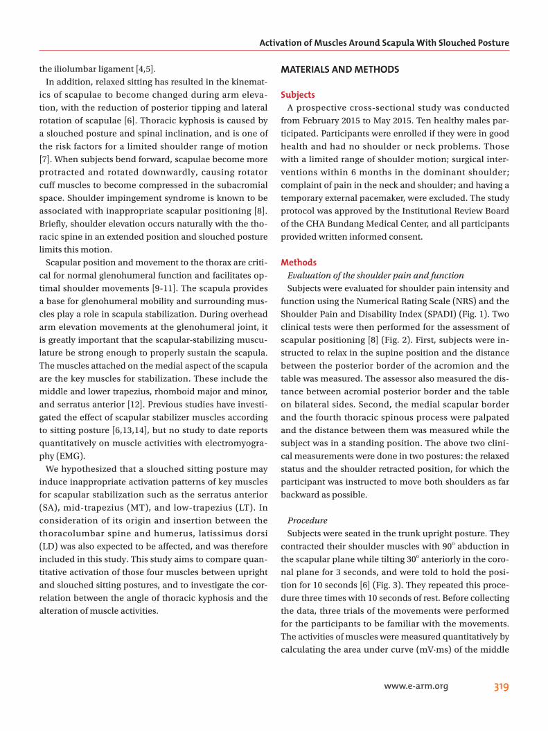
MethodsEvaluation of the shoulder pain and functionSubjects were evaluated for shoulder pain intensity and
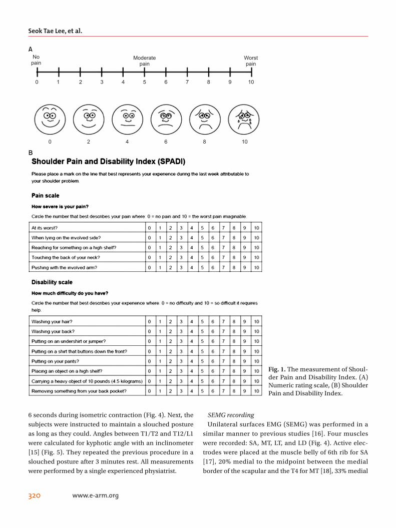
function using the Numerical Rating Scale (NRS) and the Shoulder Pain and Disability Index (SPADI) (Fig. 1). Two clinical tests were then performed for the assessment of scapular positioning [8] (Fig. 2). First, subjects were in-structed to relax in the supine position and the distance between the posterior border of the acromion and the table was measured. The assessor also measured the dis-tance between acromial posterior border and the table on bilateral sides. Second, the medial scapular border and the fourth thoracic spinous process were palpated and the distance between them was measured while the subject was in a standing position. The above two clini-cal measurements were done in two postures: the relaxed status and the shoulder retracted position, for which the participant was instructed to move both shoulders as far backward as possible.
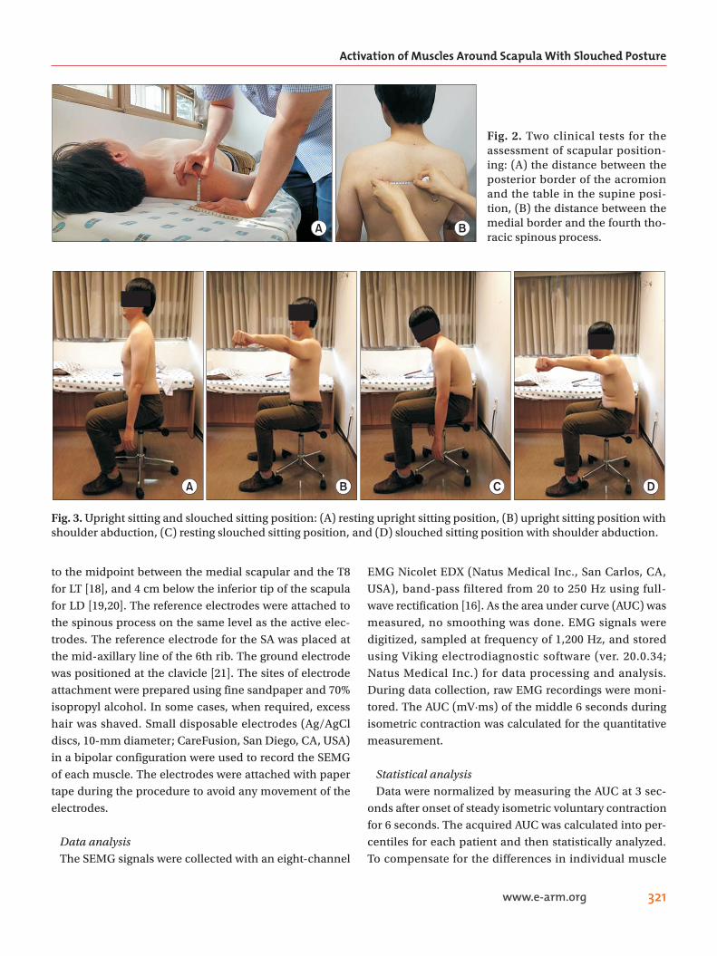
ProcedureSubjects were seated in the trunk upright posture. They
contracted their shoulder muscles with 90o abduction in the scapular plane while tilting 30o anteriorly in the coro-nal plane for 3 seconds, and were told to hold the posi-tion for 10 seconds [6] (Fig. 3). They repeated this proce-dure three times with 10 seconds of rest. Before collecting the data, three trials of the movements were performed for the participants to be familiar with the movements. The activities of muscles were measured quantitatively by calculating the area under curve (mV·ms) of the middle
Seok Tae Lee, et al.
320 www.e-arm.org
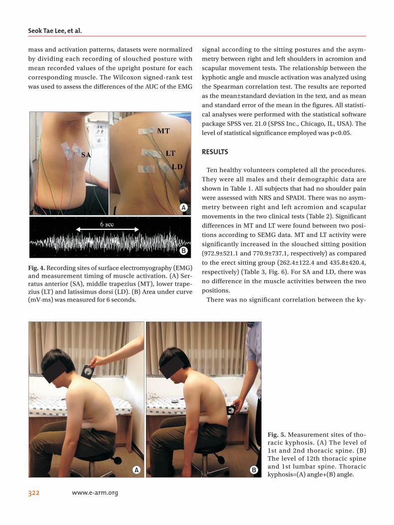
6 seconds during isometric contraction (Fig. 4). Next, the subjects were instructed to maintain a slouched posture as long as they could. Angles between T1/T2 and T12/L1 were calculated for kyphotic angle with an inclinometer [15] (Fig. 5). They repeated the previous procedure in a slouched posture after 3 minutes rest. All measurements were performed by a single experienced physiatrist.
SEMG recordingUnilateral surfaces EMG (SEMG) was performed in a
similar manner to previous studies [16]. Four muscles were recorded: SA, MT, LT, and LD (Fig. 4). Active elec-trodes were placed at the muscle belly of 6th rib for SA [17], 20% medial to the midpoint between the medial border of the scapular and the T4 for MT [18], 33% medial
A
B
0 1 2 3 4 5 6 7 8 9 10
0 2 4 6 8 10
Nopain
Moderatepain
Worstpain
Fig. 1. The measurement of Shoul-der Pain and Disability Index. (A) Numeric rating scale, (B) Shoulder Pain and Disability Index.
Activation of Muscles Around Scapula With Slouched Posture
321www.e-arm.org
to the midpoint between the medial scapular and the T8 for LT [18], and 4 cm below the inferior tip of the scapula for LD [19,20]. The reference electrodes were attached to the spinous process on the same level as the active elec-trodes. The reference electrode for the SA was placed at the mid-axillary line of the 6th rib. The ground electrode was positioned at the clavicle [21]. The sites of electrode attachment were prepared using fine sandpaper and 70% isopropyl alcohol. In some cases, when required, excess hair was shaved. Small disposable electrodes (Ag/AgCl discs, 10-mm diameter; CareFusion, San Diego, CA, USA) in a bipolar configuration were used to record the SEMG of each muscle. The electrodes were attached with paper tape during the procedure to avoid any movement of the electrodes.
Data analysisThe SEMG signals were collected with an eight-channel
EMG Nicolet EDX (Natus Medical Inc., San Carlos, CA, USA), band-pass filtered from 20 to 250 Hz using full-wave rectification [16]. As the area under curve (AUC) was measured, no smoothing was done. EMG signals were digitized, sampled at frequency of 1,200 Hz, and stored using Viking electrodiagnostic software (ver. 20.0.34; Natus Medical Inc.) for data processing and analysis. During data collection, raw EMG recordings were moni-tored. The AUC (mV·ms) of the middle 6 seconds during isometric contraction was calculated for the quantitative measurement.
Statistical analysisData were normalized by measuring the AUC at 3 sec-
onds after onset of steady isometric voluntary contraction for 6 seconds. The acquired AUC was calculated into per-centiles for each patient and then statistically analyzed. To compensate for the differences in individual muscle
A B
Fig. 2. Two clinical tests for the assessment of scapular position-ing: (A) the distance between the posterior border of the acromion and the table in the supine posi-tion, (B) the distance between the medial border and the fourth tho-racic spinous process.
C DA B
Fig. 3. Upright sitting and slouched sitting position: (A) resting upright sitting position, (B) upright sitting position with shoulder abduction, (C) resting slouched sitting position, and (D) slouched sitting position with shoulder abduction.
Seok Tae Lee, et al.
322 www.e-arm.org
mass and activation patterns, datasets were normalized by dividing each recording of slouched posture with mean recorded values of the upright posture for each corresponding muscle. The Wilcoxon signed-rank test was used to assess the differences of the AUC of the EMG
signal according to the sitting postures and the asym-metry between right and left shoulders in acromion and scapular movement tests. The relationship between the kyphotic angle and muscle activation was analyzed using the Spearman correlation test. The results are reported as the mean±standard deviation in the text, and as mean and standard error of the mean in the figures. All statisti-cal analyses were performed with the statistical software package SPSS ver. 21.0 (SPSS Inc., Chicago, IL, USA). The level of statistical significance employed was p<0.05.
RESULTS
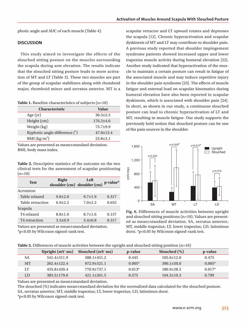
Ten healthy volunteers completed all the procedures. They were all males and their demographic data are shown in Table 1. All subjects that had no shoulder pain were assessed with NRS and SPADI. There was no asym-metry between right and left acromion and scapular movements in the two clinical tests (Table 2). Significant differences in MT and LT were found between two posi-tions according to SEMG data. MT and LT activity were significantly increased in the slouched sitting position (972.9±521.1 and 770.9±737.1, respectively) as compared to the erect sitting group (262.4±122.4 and 435.8±420.4, respectively) (Table 3, Fig. 6). For SA and LD, there was no difference in the muscle activities between the two positions.
There was no significant correlation between the ky-
A
B
Fig. 4. Recording sites of surface electromyography (EMG) and measurement timing of muscle activation. (A) Ser-ratus anterior (SA), middle trapezius (MT), lower trape-zius (LT) and latissimus dorsi (LD). (B) Area under curve (mV·ms) was measured for 6 seconds.
Fig. 5. Measurement sites of tho-racic kyphosis. (A) The level of 1st and 2nd thoracic spine. (B) The level of 12th thoracic spine and 1st lumbar spine. Thoracic kyphosis=(A) angle+(B) angle.
A B
Activation of Muscles Around Scapula With Slouched Posture
323www.e-arm.org
photic angle and AUC of each muscle (Table 4).
DISCUSSION
This study aimed to investigate the effects of the slouched sitting posture on the muscles surrounding the scapula during arm elevation. The results indicate that the slouched sitting posture leads to more activa-tion of MT and LT (Table 3). These two muscles are part of the group of scapular stabilizers along with rhomboid major, rhomboid minor and serratus anterior. MT is a
scapular retractor and LT upward rotates and depresses the scapula [12]. Chronic hyperactivation and scapular dyskinesis of MT and LT may contribute to shoulder pain. A previous study reported that shoulder impingement syndrome patients showed increased upper and lower trapezius muscle activity during humeral elevation [22]. Another study indicated that hyperactivation of the mus-cle to maintain a certain posture can result in fatigue of the associated muscle and may induce repetitive injury in the shoulder pain syndrome [23]. The effects of muscle fatigue and external load on scapular kinematics during humeral elevation have also been reported in scapular dyskinesis, which is associated with shoulder pain [24]. In short, as shown in our study, a continuous slouched posture can lead to chronic hyperactivation of LT and MT, resulting in muscle fatigue. Our study supports the previously held notion that slouched posture can be one of the pain sources in the shoulder.
Table 3. Differences of muscle activities between the upright and slouched sitting position (n=10)
Values are presented as mean±standard deviation.The slouched (%) indicates mean±standard deviation for the normalized data calculated for the slouched posture.SA, serratus anterior; MT, middle trapezius; LT, lower trapezius; LD, latissimus dorsi.*p<0.05 by Wilcoxon signed-rank test.
Table 1. Baseline characteristics of subjects (n=10)
Characteristic ValueAge (yr) 30.3±2.5
Height (cm) 176.2±4.6
Weight (kg) 73.7±9.9
Kyphotic angle difference (o ) 47.0±13.4
BMI (kg/m2) 23.8±3.1
Values are presented as mean±standard deviation.BMI, body mass index.
Table 2. Descriptive statistics of the outcome on the two clinical tests for the assessment of scapular positioning (n=10)
TestRight
shoulder (cm)Left
shoulder (cm)p-value*
Acromion
Table relaxed 9.8±2.0 9.7±1.9 0.317
Table retraction 6.9±2.2 7.0±2.2 0.655
Scapula
T4 relaxed 8.8±1.6 8.7±1.6 0.157
T4 retraction 3.3±0.9 3.4±0.8 0.317
Values are presented as mean±standard deviation.*p<0.05 by Wilcoxon signed-rank test.
SA MT LT LD
1,600
1,200
800
400
(mV
. ms)
0
UprightSlouched
p=0.445
p=0.005 p=0.013
p=0.575
* *
Fig. 6. Differences of muscle activities between upright and slouched sitting positions (n=10). Values are present-ed as mean±standard deviation. SA, serratus anterior; MT, middle trapezius; LT, lower trapezius; LD, latissimus dorsi. *p<0.05 by Wilcoxon signed-rank test.
Seok Tae Lee, et al.
324 www.e-arm.org
No difference in the activation of SA and LD was no-ticed in this study between the erect and slouched sitting positions. LD inserts along the intertubercular groove of the humerus and functionally works as a shoulder joint muscle, assisting in adduction, extension and medial rotation [25]. Considering the bridging of LD between the thoracolumbar spine and the humerus bypassing the scapula, the direct influence of different sitting positions during arm elevation on LD might not be significant. In addition, this study only assessed humeral abduction, which might have little effect on LD. As for SA, according to a previous study, there was no change in the activity of SA depending on the flexed head position, which is con-sistent with our results [26]. A different study reported that the activity of SA decreased during humeral eleva-tion with a forward head posture [27]. This difference may be due to the different study methods and small sample sizes of both studies, and they remain to be vali-dated in studies with larger samples sizes.
One limitation of the surface electrode recording tech-nique is the cross-talk phenomenon that obtains signals from proximal irrelevant muscles. In our study, the sur-face electrodes of MT and LT may have recorded signals from the rhomboid muscle. However, the majority of the signals obtained in the surface electrodes are expected to be from the most superficial muscles, MT and LT, and not the rhomboids. Further studies acquiring signals from the needle EMG during isometric contractions may fur-ther clarify the result.
As a pilot study, this study is also limited by the small sample size. Another limitation was that the muscle activity was assessed in only the arm elevation in the scapular plane. The shoulder joint shows 3-dimensional movement and arm elevation does not represent the whole picture. Furthermore, only static postures were investigated in this study. Real life postures are dynamic and may have different activation patterns. Finally, the actual clinical correlation of slouched posture to signifi-cant shoulder pain has not been well studied. Future studies involving more participants, access of all shoul-der movements, integration of dynamic components of sitting posture, and prospective cohorts of volunteers with slouched posture, may further strengthen the result and application of this study.
According to our investigation, slouched sitting posture leads MT and LT to become hyperactive. Because MT and LT are known as prime movers of scapular rotation, the findings support the notion that abnormal posture of the spine, especially a slouched sitting position, is a potential source of pain originating from scapular dys-kinesis. Therefore, spine structure needs to be evaluated in patients with shoulder and back pain, and therapeutic intervention relating to slouched posture would be help-ful for pain control.
CONFLICT OF INTEREST
No potential conflict of interest relevant to this article was reported.
REFERENCES
1. Dolan KJ, Green A. Lumbar spine reposition sense: the effect of a ‘slouched’ posture. Man Ther 2006;11:202-7.
2. Kleine BU, Schumann NP, Bradl I, Grieshaber R, Scholle HC. Surface EMG of shoulder and back muscles and posture analysis in secretaries typing at visual display units. Int Arch Occup Environ Health 1999;72:387-94.
3. Mirbagheri SS, Rahmani-Rasa A, Farmani F, Amini P, Nikoo MR. Evaluating kyphosis and lordosis in stu-dents by using a flexible ruler and their relationship with severity and frequency of thoracic and lumbar pain. Asian Spine J 2015;9:416-22.
Table 4. Correlation between kyphotic angle and muscle activation difference (n=10)
Correlation p-value*SA -0.404 0.247
MT 0.275 0.441
LT -0.440 0.203
LD -0.306 0.390
SA (%) -0.447 0.196
MT (%) -0.183 0.612
LT (%) -0.428 0.217
LD (%) -0.196 0.588
The (%) indicates mean±standard deviation for the nor-malized data calculated for the slouched posture.SA, serratus anterior; MT, middle trapezius; LT, lower tra-pezius; LD, latissimus dorsi.*p<0.05 by Spearman correlation analysis.
Activation of Muscles Around Scapula With Slouched Posture
325www.e-arm.org
4. Snijders CJ, Hermans PF, Niesing R, Jan Kleinrensink G, Pool-Goudzwaard A. Effects of slouching and mus-cle contraction on the strain of the iliolumbar liga-ment. Man Ther 2008;13:325-33.
5. Caneiro JP, O’Sullivan P, Burnett A, Barach A, O’Neil D, Tveit O, et al. The influence of different sitting pos-tures on head/neck posture and muscle activity. Man Ther 2010;15:54-60.
6. Finley MA, Lee RY. Effect of sitting posture on 3-di-mensional scapular kinematics measured by skin-mounted electromagnetic tracking sensors. Arch Phys Med Rehabil 2003;84:563-8.
7. Imagama S, Hasegawa Y, Wakao N, Hirano K, Mura-moto A, Ishiguro N. Impact of spinal alignment and back muscle strength on shoulder range of motion in middle-aged and elderly people in a prospective co-hort study. Eur Spine J 2014;23:1414-9.
8. Nijs J, Roussel N, Vermeulen K, Souvereyns G. Scapu-lar positioning in patients with shoulder pain: a study examining the reliability and clinical importance of 3 clinical tests. Arch Phys Med Rehabil 2005;86:1349-55.
10. Voight ML, Thomson BC. The role of the scapula in the rehabilitation of shoulder injuries. J Athl Train 2000;35:364-72.
11. Ludewig PM, Reynolds JF. The association of scapular kinematics and glenohumeral joint pathologies. J Or-thop Sports Phys Ther 2009;39:90-104.
12. Escamilla RF, Yamashiro K, Paulos L, Andrews JR. Shoulder muscle activity and function in common shoulder rehabilitation exercises. Sports Med 2009;39: 663-85.
13. Bullock MP, Foster NE, Wright CC. Shoulder impinge-ment: the effect of sitting posture on shoulder pain and range of motion. Man Ther 2005;10:28-37.
14. Kanlayanaphotporn R. Changes in sitting posture affect shoulder range of motion. J Bodyw Mov Ther 2014;18:239-43.
15. Lewis JS, Valentine RE. Clinical measurement of the thoracic kyphosis. A study of the intra-rater reliabil-ity in subjects with and without shoulder pain. BMC Musculoskelet Disord 2010;11:39.
16. Lee HS, Shim JS, Lee ST, Kim M, Ryu JS. Facilitating ef-
fects of fast and slope walking on paraspinal muscles. Ann Rehabil Med 2014;38:514-22.
17. Cools AM, Witvrouw EE, De Clercq GA, Danneels LA, Willems TM, Cambier DC, et al. Scapular muscle recruitment pattern: electromyographic response of the trapezius muscle to sudden shoulder movement before and after a fatiguing exercise. J Orthop Sports Phys Ther 2002;32:221-9.
18. Holtermann A, Roeleveld K, Mork PJ, Gronlund C, Karlsson JS, Andersen LL, et al. Selective activation of neuromuscular compartments within the human tra-pezius muscle. J Electromyogr Kinesiol 2009;19:896-902.
19. Park SY, Yoo WG. Differential activation of parts of the latissimus dorsi with various isometric shoulder exer-cises. J Electromyogr Kinesiol 2014;24:253-7.
20. Beaudette SM, Unni R, Brown SH. Electromyographic assessment of isometric and dynamic activation char-acteristics of the latissimus dorsi muscle. J Electro-myogr Kinesiol 2014;24:430-6.
21. Noguchi M, Chopp JN, Borgs SP, Dickerson CR. Scap-ular orientation following repetitive prone rowing: implications for potential subacromial impingement mechanisms. J Electromyogr Kinesiol 2013;23:1356-61.
22. Ludewig PM, Cook TM. Alterations in shoulder ki-nematics and associated muscle activity in people with symptoms of shoulder impingement. Phys Ther 2000;80:276-91.
23. McLean L. The effect of postural correction on muscle activation amplitudes recorded from the cervicobra-chial region. J Electromyogr Kinesiol 2005;15:527-35.
24. Poppen NK, Walker PS. Normal and abnormal motion of the shoulder. J Bone Joint Surg Am 1976;58:195-201.
25. Bagg SD, Forrest WJ. A biomechanical analysis of scapular rotation during arm abduction in the scapu-lar plane. Am J Phys Med Rehabil 1988;67:238-45.
26. Ludewig PM, Cook TM. The effect of head position on scapular orientation and muscle activity during shoulder elevation. J Occup Rehabil 1996;6:147-58.
27. Weon JH, Oh JS, Cynn HS, Kim YW, Kwon OY, Yi CH. Influence of forward head posture on scapular up-ward rotators during isometric shoulder flexion. J Bodyw Mov Ther 2010;14:367-74.


![Serratus Anterior Plane Block-An Analgesic Technique … factors in turn lead to decreased lung compliance, ventilation perfusion mismatch, hypoxemia and respiratory distress [3].](https://static.documents.pub/doc/80x56/5abfacad7f8b9aa3088e5c85/serratus-anterior-plane-block-an-analgesic-technique-factors-in-turn-lead-to.jpg)


