13
CHANGES IN THE MORPHOLOGICAL COMPOSITION OF BLOOD IN EXPERIMENTAL PLAGUE k- Translation No, iTue 1965 U. S. ARMY BIOLOGICAL TABORATORIES FORT DETRICK, FREDERICK, MARYLAND
| Date post: | 28-Jul-2018 |
| Category: |
Documents |
| Upload: | truongtram |
| View: | 227 times |
| Download: | 0 times |

CHANGES IN THE MORPHOLOGICAL COMPOSITION OF BLOOD IN EXPERIMENTAL PLAGUE
k-
Translation No,
iTue 1965
U. S. ARMYBIOLOGICAL TABORATORIES
FORT DETRICK, FREDERICK, MARYLAND

DDC AVAILABILITY NOTICE
Qualified requestors may obtain copies ofthis document from DDC.
This publication has been translated fromthe open literature and is available to thegeneral public. Non-DOD agencies may pur-chase this publication from Clearinghousefor Federal Scientific and Technical Informa-tion, U. S. Department of Commerce, Springfield,Va.
Technical Library BranchTechnical Information Division
CHANGES IN TIlE MORPHOLOGICAL COMPOSITION OF BLOOD IN EXPERIMENTAL PLAGUE
/Pollowing is the translation of an article by 1. Ye.
Kiseleva, published in the Russian-language periodicalTrudy Rostovskogo-na-Donu Gosudarstvennogo Nauchno-issledovatelskogo Protivochumnogo Instituta (Trudy ofthe Rostov on the Don State Scientific-Research Anti-plague Institute), Vol XV, 1959, pages 87--96. Trans-lation performed by Sp/ 7 Charles T. Ostertag Jr.7
Laboratory investigations of blood during the process of disease,and also during the period of treatment and recovery has occupied a stableplace in the complex of clinical examinations of a sick organism. Up untilthe present time, Soviet and foreign investigators have accumulated a vastamount of material on the changes of the morphological composition of theblood during various illnesses, infections and intoxications.
Up until now the blood picture during plague infection has beenstudied least of all. On the one hand this may be explained by the absenceof plague morbidity in a number of countries, and secondly by the difficultieswhich inevitably confront the investigator in getting hlood from plaguestriken humans or infected animals.
The data cited in literature relative to the change of the morpholog-ical composition of blood in plague stricken persons are quite diverse aud
contradictory and deal mainly with changes on the part of leukocytes.
Thus, V. K. Vysokovich (1901) points to the minor teukocytosis inpatients with the bitbonic form of plague, while H. Mueller and R. Poch,1900, detected a fluctuation in the number of leukocytes during plaguefrom 8,000 to 45,000 in I nm 3 of blood.
More than once S. I. Zlatogorov and L. V. Padlevskiy (1915) observed
the clearly expressed increase in the number of leukocytes during pestilentialpneumonia, and consider that the absence of leukocytosis may in certain casesserve as an auxiliary index for differenti-al diagno-is with croupous pneumonia.
On the contrary, D. K. Zabolotnyy (1956) observed hyperleukocytosisin the blood during all the forms of plague, and the number of leukocytesreached 20--30 thousand.
During primary pulmonary plague, G. P. Rudnev (1-940) encountered,along with minor leukocytosis, a sharply expressed hyperleukocytosis (up to20,000 and more).

H. Schar and K. Meyer, 1956, while studying the effect of the toxicfraction of P. pestia on the blood and the heinopoietic system of experimentalanimals, invariably found leukopenia and eosinopenia in white mice and ratealready in 30 minutes following the introduction of both the toxic and theatoxic fraction of the I lague ,ncrobe. These investigntors note the increase
in the number of erythrocytes up to 22--30% in white mice and rats in thepremortal condition -- in two hours following the administration of a lethaldos(e of toxin,
In the literature available to us, we have not encountered a descrip-tion of the changes in the blood picture of laboratory animals -- guinea pigsand white mice -- during the process of development of plague infection inthem following their infection with a virulent strain of the plague microbe.
Changes in the leukocytic formula of susliks during their infectionwith the plague microbe have been characterized in the work by R. F. Akulovaand G. P. Rudne,', 1930. In the case of sepsis the authors, on the basis of
Smears, ascertained the increase in the number of leukocytes by approximatelythree times in comparison with noninfected animals.
N. Ehrenkranz and L. White, 1954, refer to the changes in the number ofeosinophils during experimental pulmonary plague in monkeys.
Significant leukocytosis (up to 50,000 in 1 mm3 ) during experimental
bubonic plague in monkeys and the absence of specific changes in the numberof erythrocytes are noted by G. Hoessly, D. Walker, A. Larson and K. Meyer,1955.
In spite of the fact that the third _orrmed element of blood -- theblood platelets -- have served as the subject of investigation for more than80 years, and changes in their -umbers- hav b.en , tudid during manv acuteinfectious diseases, under the influence of ionizing radiation, canceroustumors, etc., we were not able to find a description of the change in bloodplatelets during the course of plague infection.
Investigation of the change in the number and morphology of formedelements of the blood in the dynamics of plague infection provides the basisfor the differential appraisal o4' the condition of the hemopoietic apparatusin various periods of illness.
Besides this, knowledge of the peculiarities of changes in the formedelements of the blood during plague is necessary for delimiting the action ofmedicinal preparations from the toxic factors of the plague microbe, andalso when studying the effect of vaccine strains of P. pestis on the organism.
The present work is devoted to the investigation of the above mentionedprob I em.
The tests were set up on 33 guinea pigs, weighing from 300 to 500grams, and 38 white mice.
2

7
The animals were infected with various doses (from 100 to 10,000microbes) of a virulent strain of P,. _yestis No 772 and 773, the minimumlethal dose of which comprised 100 microbes for guinea pigs and 10 forwhite mice. For infection we used a suspension of a 24 hour and a 48hour agar culture of the plague microbe, incubated at 280.
Preliminarily calculations were made of the number of erythrocytes,leukocytes and blood platelets in all the animals. These were made over aperiod of a month, in various times of the day, and on on empty stomachand after eating,
After infection the investigaticns of the blood for determining allthe formed elements were conducted daily right up until the death of theanimal.
All told 893 determinations were made.
With the white mice the batches of blood for investigation wereobtained from the blood vessels of the tail by cutting the tip of it eachtime. With the guinea pigs it was obtained from the ear vein by prickingit with a thin needle. In some cases, in the last days before the deathof the animal, it was sometimes necessary to make an incision of the conchsauriculae with the edge of a safety razor.
The diluting liquid for calculating erythrocytes was Gayem's liquid,prepared on mercuric chloride; for calculating leukocytes -- Turk's fluidin G. P. Rudnev's modification (1940). Calculation of formed elements wasmade in a Goryayev chamber.
In the first series of tests the blood was drawn up into the mixerwith the help of a syringe, connected by a thin rubber tube with the endof a blood coital pipette. Taking blood in such a manner, which guarautteedthe safety of the investigator, is not entirely convenient for carrying outthe technique, and also for transporting the blood count pipettes. There-fore in subsequent series we somewhat modified the method proposed byNikolayev (1954), which is relative to plague, and switched to taking bloodwith a graduated pipette and diluting it in test tubes. For this, in onerow of small numbered test tubes (the so-called agglutination test tubesare the most convenient) we preliminarily poured 2 ml of Gayem's liquidinto each one, and in the other row -- 0.2 ml of Turk's fluid into each one.On the distal end of a Pasteur pipette which is graduated for 10 min3 (such
a graduation for one division is easily made in the laboratory with thehelp of a chemical micropipette), a short rubber tube in the form of aballoon is attached, the pipette is led up to a drop of blood which isemerging following pricking, and by preliminarily pressing on the balloon,or sometimes by simply inclininj the pipette, the blood is collected up tothe mark which has been made. ýiien the pipette with the blood is imumersedinto a test tube with the diluting liquid, and by the pressure of the fingerson the rubber ball the blood is discharged into the test tube. It is washedseveral times with diluting liquid, withdrawn from the test tube and immersed
3

into a beaker with u disiafectanc solution. The ceot tubes are closed withrubber corks, vibrated and transferred to the laboratory.
Such a method is convenient because it makos it possible to obtainthe required solution (1:200 and 1:20) of blood in the test tube and guar-antecs safety when taking blooJ, and also when transportLing it and disin-fecting the pipettes and te!st tubes.
The bactericidal action of Gayem's and TDirk's fluids on the plaguemicrobu wau noted by itveoLigators (G. P. Rudnev, 1940; 1. S. Tinker andG. P. Rudnev, 1930) and verified by us In respect to the strain of the plaguemicrobe which we uwed in the experio•ent.
As regards the preservation of erythrocytes in Gayem's liquid, thenin the test tubes which were closed with rubber corks the number of erythro-cytes during repeated determinat ions did not chauige for us over a period oftwo months (limiting period of observation). This is testified to by thedata of E. L. Volfson (1936), who cites the case of the preservation oferythrocytes in Gayem'a liquid, nonhemiolyzed for a period of three years.
The number of blood platelets was calculated according to the Methodof A. Fonlo (1912), who used a 14-percent solution of magnesium sulfate asthe anticoagulant, and also by the method proposed by A. P. Yegorov (1939,1954) with a 4-percent solution of sodium citrate. Staining of smears wascarried out with the Giemsa-Romanovskiy stain according to the method ofPappenheym.
As the repeated investigations of the blood in healthy animals showed,the number of erythrocytes in 1 mun 3 of blood In a guinea pig was subject toconsiderable fluctuation it the course of a day.
Dependingz on the time of tht I Ou'stlont on and the phystolog'!_Iof tile animal, the number of erythrocvtes in the uame animal changed by 0.30'+ 0.24 million in I ma3 ,
Following the infection of auimols with a virulent strai|n of the plaguemicrobe, an increase was noted in the number of erythrocytes in all the daysntter inoculation (table 1).
As is apparent from the table, the greatest increase in the number oferythrocyres is reached oi, the second day following infection, exceeding theinttial value by 9.1%. In addition to the quantitative change, on the partof the blood corpuscles the presence was noted of normoblasts, anisocytosisand poikilocytosis. It must be stipulated that the uneven fortu and size ofthe erythrocytes, and also the presence of normoblasts and polychromatophiliaare observed to a somewhat lesser degree even in healthy guinea pigs.
The most expressed change was on the part of the leukocytas -- theincrease in their numbers in certain cases was by 4--5 times in comparison
4

with tile Initial number (table 2) already in 24 hours following infacLionand their was a subsequent ahatp decrease In the aumber of hLukocytea inthe days preceding the death of tho animal.
Only in two animals (No 1230 and 1476), the death of which in bothcaise set in on the fourth day following infection, the number of leukocytcawhich were measured 24 hours prior to death was increased by 17%. in thefirst case and 120% in tile second case in compariaon with thie iitial.
Along with tile change in the number of leukocytes, an increase wasalso observed in their size by approximately 1,5--2 times both duringinvestigation it) a native state and during invertigationa in stained pr-.eparat lons. Among the leukocytes, cells were sometimes detected which hada vacuolized protoplasm and sometimes cells in a condition of aecay.
Changes on the part of the blood platelets, which are presented intable 3, amounted mainly to an increase in their number in the first 3 daysafter infection, and a decrease in the number of blood platelets beginningwith the fourth day after inoculation.
The number of blood platelets In 4--5 days following infection turnedout to be sharply reduced immediately before the death of the animal, ThusIr, guinea pig No. 1237, which died on the sixth day following inoculation,the number of blood platelets in the blood, taken two minutes before thedeath of the animal, comprised 5 per 1,000 erythrocytes. At the same time,during the rapid course of the plague process, when the animals died inearly periods, the decrease in the number of blood platelets did not sur-pass the physiological fluctuations of their number in this species ofan ima I.
In considering the role of blood platelets in tile process ot formationof agglutinates with bacteria as the first response of the organism to micro-bial invasion even before the onset of phagocytosiu (Roskli. G. I., 1954:A. Copley, r. Bales, 0. Chryssostomidon, 1955; L. Duchon, 1955), we made acalculation of the blood platelets and simultaneously of the erythrocytesand leukocytes in guinea pigs 5 minutes prior to infection and in 3--10minutes after inoculation. No difference was established in the number offormed elements inl comparison with tile initial values.
The absence of thrombocytopenia in the first milutes following infection,as the above named investigators consider, points to the considerable virulenceof the microbe which was introduced.
In our tests a highly virulent strain of P. pesLtis was used, and this
in all probability explains the absence of a decrease in, tihe number uZ bloodp1 latelet:u in the first five minutes after inoculation.
The change of formed elements of blood in white mice in the process ofplague infection did not differ basically from the changes in the blood pic-ture in plague infected guinea pigs. The number of erythrocytes, measured
5

in noninfected white mice, comprised an average of 9,141,000 in I mm3 ofblood with a minimum content of 5,94.0,000 and a maximuin of 10,970,000.After Infectlon of the White mice with 1,000 microbeo (100 Dim) of the 772virulent strain of P. p)OUt in a calculation of erythrocytes shlowed an Increaseof their numbers in all days following the infection. The greatest increasein the number of erythrocytes was reached ol tLho 3rd day after infection inwhite mouse No. 2674, which died on the 4th day after inoculation. rheamount of erythrocytes in thie animal, which wao movoured twice a Iay fortwo days prior to infection, comprised 7,200,000 + 150,000 in I nmh *, intwo days following the microbial invasion, the number of erythrocyteo InI IMm3 of blood e.qualnd 9,120,000, that is, .n increase of 26". in comparisonwith the originial. In two white mice the number of erythrocytes follctd-inginfection turnod out to be decreased by 18 nnd 14.409. in coml)erison with theoriginal.
In the remaining 35 white mice the number of erythrocyteo in all daysfollowing Infeotion was tictroaued on an average by 6--107. in comparisonwith initial values.
For taking into consideration the influence of the repeated taking ofblood and, possibly, the conditions for housing the infected animals in jarscovered with gauze moistened in a solution of Lyaol, we made a calculation ofthe number of formed elements of blood not only in infected but in healthywhite mice which were housed in the Infectious-experimental division underthe same conditions as the infected animals. Tile number of erythrocytes inthese control animals during repented investigations fluctuated Insignifi-cansly both in respect to an increase and to a decrease, but did not deviateby more than 3--47. from the initial value.
The number of blood platelets in the white mIce in the first 4 daysafter infection increased. Thus, the average number of blood platelets in13 white mice which had died in 4 days following inoculation comprised 194,000"A'rfl to i-nf4cto 762 A(U)•, ,1 . .9A hnourm Irf-.*- 'f.ection, and after 3 dny• -273,000 in I nus" of blood.
Thus, ns these investtgations salowed, during experimental plague changesare ob|erved in the number of all formed elements of Mood. Erythronytostýsin plapkue is not great (up to 107% from tile initial value), however it: Inencountered with groat constancy. It muSL kie assumed that the increase inthe number of erythrocytes in I ns3 of blood which was observed by us duringplague does not reflect the true change in the number of formed elements ofbleod, but is the result of a decrease in the volume of the plasma, that ia,blood coagulation. As was shown in the investigati.ons by K. M. Mokhiln (1958),the disruption of the permeability of the vascular endothelium, which isdevelophp, In a plapue afflicted organtium, and tihe discharge of plasma intothe tissue causes blood coagulation, and by this, probably, an increase inthe number of erythrocytes,
In the plague infected organism the leukocytes are subjected to thegr atest changes. Their number increases by 3-4 times on the 2nd and 3rd
6
ay 'ol lowing Infection. Aftor 2--1 days hyperlteucocytos iin repiaced byIukuopon I an md the n ima I s usua I ly div with a decreage J number of Ieukocyt ea
ii lie peripheral bloo0d.
In contract to the ralse increase In tLhL• number of erythrocytee,leulcocytos• In plague Is undoubt.dly caused not nlly by blood coagulation,
but also by hyperpla sa of the leukopo etic part of the blood-producing
system, which is testified to by the plthu, xrphological investigantions ofS, )amberg (1926), who tudiled the p icture of the bone marrow of su iIks
in various periods following infection with plague.
The rep lacumunL of lukocytosls with eiitkopenn Indicates the loweredreact ivity of the oarn iism in the las-t period ot pla•tIue morbidity.
llasvd on (hle experimental data presented, It q pussible to explaint. hO conL rad icto'ry on firs1t appeaitrunLe reaIsul ts obta ited by various investi-
gIters when describing the blood picLure in persons sick with plague. ThuasInvestigators iAh observed the patients for b--20 hours prior to death speakof leukopenia during plague and consider it a diL-t iiguinhinig sign from
Crottpolas pneumniha (N, 1. Zlntogorov and L. V. Padlevskly, 1915). The samething was noted by pnthologoanatomtnts who were Investigating the bone nmarrowof persons who died frtom pl ague (G. S. Kul esha, 191"5). On the other hand,
Investigator, who were Istudying the blood ricture in the highest point of
the disease, noted during plague a clearly expressed leukocytosis (Zabolotnyy,
D. K., 1956; lMue llCA and p1 t-h, IQC14M).
In our investigations we also observed in the same animals, followingtheir Infection with P. peals, leiukoeytosis, hyperleukocytuiis, and leukopenta,depending on during which period of plague Infect ion the Investigation ofblood was carried out,
The increase fin the number of blood platoelets which we obsered inthe, first dnys of 1 ingtut'e in fectin I , in gn ine.•_ tgs -n ti. h!te ni.ce, ia so pnint'
to the i ncreagc.i activity of the bone marrow In 01116 period.
"The lowering of the number of blood p)latelets in 4--5 daya followingInfecton speaks in its turn of the progressing depiression of hemtatosiain tiis, the last, period of pilague i nlrc-t ion,
Conlus I Uons
1. TPhi method proposed by-US for taking bluod with a gra'duated
p i pet Ie anid dl I ut ing it in tes't tubes. JClriortlgt to &he method o4-HNtfto±rt-fvIs safe for working with plague infected animals.
2. Following the infection of guinea pigs and white mice with a
vi.rulent strain of plague microbe the number of erythrocytes in I 'mY3ofblood Increased on all the days Cotiowing infect.tl-m.
3. in the first days of pi-igue infection leukocytosis was observed in
7
the animals, sometimes hyperleukocytosis, which on the 4--5th day followinginfection !•'teepleced by leukopenia.
-4. The number of blood platelets tý increased in the first 3 daysafter infection and i-e dectreased in cases of a prolonged course of infection.
Literaturea. Volfson, E. L., 1939, Duration of Preservation of Erythrocytes in
a Fluid, Gayema Labor, praktika, No 9-10.
b Vysokovich, V. K., 1901, Plague (pestis orientalis), Kiev.
C. Damberg, S., 1926, Defensive Role of the Spleen in CombatingPlague Infection. In the book: Plague in the South-Eastern USSR andReasons for its Endemic Nature, Leningrad.
d. Yegorov, A. P., 1939, Methods for the Caiculatic-, and Differen-tiation of Thrombocytes, Labor. praktika, No 11.
e. Yegorov, A. P., 1954, Morphological Analysis of Blood, Moscow,Medgiz.
f-. Zabolotnyy, D. K,, 1956, Collected Works, Vol I, Plague, Kiev,an UkSSR Publishing House.
g. Zlatogorov, I. S. and Padlevskiy, L. B., 1915, Observations ofPatients, In the book: Pulmonary Plague in Manchuria in 1910-1911. Reportof the Russian Scientific Ex-erditin, V.l. T , Petrograd.
h. Kulesha, G. S., 1915, Concerning the Pathological Anatomy ofPulmonary Plague, Ibid, vol 2.
i. Mokhin, K. M., 1959, Peripheral Blood Formation and the Permeabil-ity of the Vascular Wall in the Dynamics- of Experimental Plague (See this samecollection).
j. Nikolayev, N. M., 1951, Application of Pipettes and Test Tubesin place of Mixers for Calculating Erythrocytes and Luekocytes, Soy Med., No 4.
k. Roskhin, G. I., 1954, Blood Platelets of Man and Mammals, Jspekhi
sovr. biol., vol 37, No 3.
1. Rudnev, G. P., 1940, Clinical Picture of Plague, Moscow, Medgiz.
m. Akulowa, R. F. and Rudnew, G. P., 1930, Das Blutbild bei experi-mentell erzeugler Pest. Zbl. f. Bakt., Bd. 119, 1 Abt., Orig., H. 1-2.
n. Copley, A. L., Balea, T., Cbryssostomidou, 0., 1955., Methodesemployees pour la determination des effets da B. C. G. et d'autres
8

mycobacteries sur Tadhesion at sur l'agglutination des plaquettes. Revinmmunol., vol 19, N3.
o. Duchon L., 1955, Du Role des plaquettes et des hemoconies dansla de'fense antimicrobienne, Ann Inst. Pasteur., vol 83, N6.
p. Ehrenkranz, N. J. and L. P. White, 1954, Hepatic function and otherphysiologic studies in monkeys with experimental pneumonic plague, J. Inf.Dis., V. 95, N6,
q. Fonio, A., 1912, Ueber ein neueus Verfahren des Blutplattchenzahlung.Dtsche Ztschr. f. Chit., Bd. 117, 11. 1-2.
r. Hoessly, G. F., Walker, D. L., Larson, A., Meyer, K. F., 1955,Experimental Bubonic Plague in Monkeys. 1. Study of the development of thedisease and the peripheral circulatore failure. Acta tropica, V. 12, N.3.
s. Mueller, H. F., and Poch, R., 1900, Die Rest. Spec. Path. uTherapie von Nothnagel. Wien.
t. Schar, M. and Meyer, K. F., 1956, Studies on Inmunncation agaiastPlague. XV. The Pathophysiologic Action of the Toxin of Pasteurella pestiGin experimental Animals. Schweizerische Ztschr. f. All Gemeine, pathologieu. Bakteriologie., V. 19, N. I.
u. Tinker, I. S. and Rudnew, G. P., 1930, Zum Studium uber dieLebensfahigkeit des Bac. pestis. Arch. f. Schiffa. u. Tropenhygiene. Bd 341
9
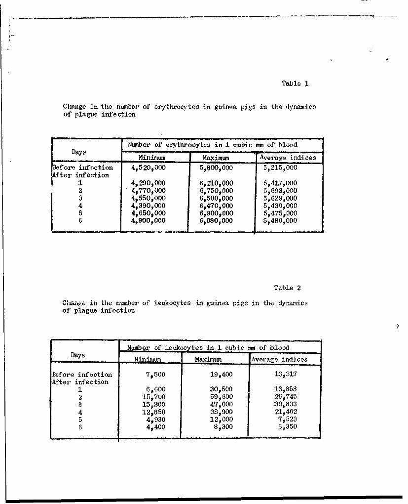
Table 1
Change in the number of erythrocytes in guinea pigs in the dynamicsof plague infe ction
Number of erythrocytes in 1 cubic mm of bloodDays Minimum Maximum Average indices
Before iifection 4,520,000 5,800,000 5,215,000(fter infection
1 49290,000 6,210,000 5,417,0002 4,770,000 6,75O,000 5,693P0003 4,550,000 6,500,000 5,629,0004 4,390,000 6,470,000 5,430,0005 4,650,000 6,900,000 5,475,0006 4,900,000 6,080,000 5,480,000
Table 2
C'aIge in the number of leukocytes in guinea pigs in the dyniunicsof plague infection
Number of leukocytes in 1 cubic mm, of bloodDays, !U.nimum Maximum Average indices
Before infection 7,500 19,400 13,317After infection
1 6,600 30,500 13,8532 15,700 59,600 26,7453 15,300 47,000 30,8334 12,850 33,900 21t4625 4,930 12,000 7,5236 4,400 8,300 6,350

Table 3
Change in the ntumber of blood platelets in guinea pigs in the dynazn2icsof plague infection
Number of platelets in I cubic mm of bloodDa~ys_____s __M__ _ininMuMr _ Aera..e indices
Before infection 256,500 650,160 472,058After infection
1 560,490 7939650 689,8342 414,990 992$250 623,0223 383,400 628,320 5029,7284 232,670 401I, 140 318t7325 117,300 399,230 255, 2076 25,000 197p600 98t493 f7
? Flaw in the original Russian version. This appears to be the nudnbr.
11.


