Chapter 1 Computer Aided Drug Design: An Overview 1 COMPUTER AIDED DRUG DESIGN: AN OVERVIEW 1.1 Abstract In this chapter a brief introduction to the computer-aided drug design (CADD) methodologies is given. The theoretical basis of CADD involves quantum mechanics and molecular modeling studies like structure based drug design; ligand-based drug design; database searching and binding affinity predictions. Finally, a brief description of the present work is given. 1.2 Introduction Drug discovery and developing a new medicine is a long, complex, costly and highly risky process that has few peers in the commercial world. This is why computer-aided drug design (CADD) approaches are being widely used in the pharmaceutical industry to accelerate the process. The cost benefit of using computational tools in the lead optimization phase of drug development is substantial. On an average, it takes 10-15 years and US $500-800 million to introduce a drug into the market, with synthesis and testing of lead analogs being a large contributor to that sum. 1 Therefore, it is beneficial to apply computational tools in hit-to-lead optimization to cover a wider chemical space while reducing the number of compounds that must be synthesized and tested in vitro. The computational optimization of a hit compound involves a structure-based analysis of docking poses and energy profiles for hit analogs, ligand-based screening for compounds with similar chemical structure or improved predicted biological activity, or prediction of favorable affinity or optimize drug metabolism and pharmacokinetics (DMPK) or absorption, distribution, metabolism, excretion, and the potential for toxicity (ADMET) properties. The comparably low cost of CADD compared with chemical synthesis and biological characterization of compounds make these methods attractive to focus, reduce, and diversify the chemical space that is explored. 2 CADD is capable of increasing the hit rate of novel drug compounds because it uses a much more targeted search than traditional high throughput screening (HTS) and combinatorial chemistry. It not only aims to explain the molecular basis of therapeutic activity but also to predict possible derivatives that would improve activity. In a drug discovery campaign, CADD is usually used for three major purposes: (1) filter large compound libraries into smaller sets of compounds that can be tested experimentally; (2)
Transcript
Chapter 1 Computer Aided Drug Design: An Overview
1
COMPUTER AIDED DRUG DESIGN:
AN OVERVIEW
1.1 Abstract
In this chapter a brief introduction to the computer-aided drug design (CADD)
methodologies is given. The theoretical basis of CADD involves quantum mechanics and
molecular modeling studies like structure based drug design; ligand-based drug design;
database searching and binding affinity predictions. Finally, a brief description of the
present work is given.
1.2 Introduction
Drug discovery and developing a new medicine is a long, complex, costly and highly risky
process that has few peers in the commercial world. This is why computer-aided drug
design (CADD) approaches are being widely used in the pharmaceutical industry to
accelerate the process. The cost benefit of using computational tools in the lead
optimization phase of drug development is substantial. On an average, it takes 10-15 years
and US $500-800 million to introduce a drug into the market, with synthesis and testing of
lead analogs being a large contributor to that sum.1 Therefore, it is beneficial to apply
computational tools in hit-to-lead optimization to cover a wider chemical space while
reducing the number of compounds that must be synthesized and tested in vitro. The
computational optimization of a hit compound involves a structure-based analysis of
docking poses and energy profiles for hit analogs, ligand-based screening for compounds
with similar chemical structure or improved predicted biological activity, or prediction of
favorable affinity or optimize drug metabolism and pharmacokinetics (DMPK) or
absorption, distribution, metabolism, excretion, and the potential for toxicity (ADMET)
properties. The comparably low cost of CADD compared with chemical synthesis and
biological characterization of compounds make these methods attractive to focus, reduce,
and diversify the chemical space that is explored.2
CADD is capable of increasing the hit rate of novel drug compounds because it
uses a much more targeted search than traditional high throughput screening (HTS) and
combinatorial chemistry. It not only aims to explain the molecular basis of therapeutic
activity but also to predict possible derivatives that would improve activity. In a drug
discovery campaign, CADD is usually used for three major purposes: (1) filter large
compound libraries into smaller sets of compounds that can be tested experimentally; (2)
Chapter 1 Computer Aided Drug Design: An Overview
2
guide the optimization of lead compounds, to increase its DMPK properties including
ADMET; (3) design novel compounds, either by "growing" starting molecules one
functional group at a time or by piecing together fragments into novel chemotypes.3
CADD can be classified into two general categories: structure-based and ligand-
based. Structure-based CADD relies on the knowledge of the target protein structure to
calculate interaction energies for all the compounds to be tested, whereas ligand-based
CADD exploits the knowledge of known active and inactive molecules through chemical
similarity searches or construction of predictive, quantitative structure-activity relationship
(QSAR) models.4 Structure based CADD is generally preferred where high-resolution
structural data of the target protein are available, i.e., for soluble proteins that can readily
be crystallized. Ligand based CADD is generally preferred when no or little structural
information is available, often for membrane protein targets. The central goal of structure
based CADD is to design compounds that bind tightly to the target, i.e., with larger
reduction in free energy, improved DMPK/ADMET properties, and are target specific, i.e.,
have reduced off-target effects.5
A successful application of these methods will result in a
compound that has been validated in vitro and in vivo and its binding location has been
confirmed, ideally through a co-crystal structure. One of the most common uses in CADD
is the screening of virtual compound libraries, also known as virtual high-throughput
screening (vHTS). Fig. 1.1 illustrates the stages in the drug discovery process6 and Fig. 1.2
explains virtual drug discovery process7.
Figure 1.1 Stages in the drug discovery process.6
Chapter 1 Computer Aided Drug Design: An Overview
3
Figure 1.2 Virtual drug discovery process.7
1.2.1 Receptor theory
The concept that therapeutic agents produce their selective action in modifying disease
symptoms by acting as "magic bullets" at discrete molecular targets within the body, is
generally attributed to Paul Ehrlich at the turn of the 19th
century as part of the now
seminal "lock and key" hypothesis.8
This hypothesis has described drugs as receptor’s
ligands or enzyme substrates that selectively modulate the function of unknown molecular
targets to produce beneficial effects. The receptor theory involves, to a very
major extent, the classical enzyme kinetic model based on the law of mass
action, which was derived by Michaelis and Menten in 1913.9 The interaction
between receptor and a ligand can be looked upon as:
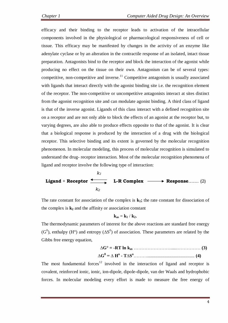
Receptor + Ligand [RL] R + Cellular Effect ... (1)
The ligand L binds to the receptor R and alters the nature of the receptor interaction with
its associated membrane components to effect a change in cellular and ultimately, tissue
function. Ligands interacting with the receptors have two intrinsic properties:10
Affinity and Efficacy. Affinity is the ability to recognize and bind to the receptor
while the ability of the ligand to effect a change in cellular processes via activation of
transmembrane transduction mechanisms involving G-protein complexes or ion channels
is a measure of efficacy. In addition to the affinity of a receptor for its ligand, the response
to the ligand is also dependent on the number of receptors on a given tissue. An additional
ligand property is that of selectivity, the degree to which the ligand interacts with the
target of choice as compared to related structural targets. The degree of selectivity
typically determines the side effect profile of a new compound, given that the targeted
mechanism itself does not produce untoward effects when stimulated beyond the
therapeutic range. Ligands may be either agonists or antagonists. Agonists have intrinsic
Chapter 1 Computer Aided Drug Design: An Overview
4
efficacy and their binding to the receptor leads to activation of the intracellular
components involved in the physiological or pharmacological responsiveness of cell or
tissue. This efficacy may be manifested by changes in the activity of an enzyme like
adenylate cyclase or by an alteration in the contractile response of an isolated, intact tissue
preparation. Antagonists bind to the receptor and block the interaction of the agonist while
producing no effect on the tissue on their own. Antagonism can be of several types:
competitive, non-competitive and inverse.11
Competitive antagonism is usually associated
with ligands that interact directly with the agonist binding site i.e. the recognition element
of the receptor. The non-competitive or uncompetitive antagonists interact at sites distinct
from the agonist recognition site and can modulate agonist binding. A third class of ligand
is that of the inverse agonist. Ligands of this class interact with a defined recognition site
on a receptor and are not only able to block the effects of an agonist at the receptor but, to
varying degrees, are also able to produce effects opposite to that of the agonist. It is clear
that a biological response is produced by the interaction of a drug with the biological
receptor. This selective binding and its extent is governed by the molecular recognition
phenomenon. In molecular modeling, this process of molecular recognition is simulated to
understand the drug- receptor interaction. Most of the molecular recognition phenomena of
ligand and receptor involve the following type of interaction:
The rate constant for association of the complex is k1; the rate constant for dissociation of
the complex is k2 and the affinity or association constant
kas = k1 / k2.
The thermodynamic parameters of interest for the above reactions are standard free energy
(G0), enthalpy (H°) and entropy (∆S
0) of association. These parameters are related by the
Gibbs free energy equation,
∆G° = -RT ln kas ……………………......…………… (3)
∆G0 = ∆ H
o - T∆S
o………......................................... (4)
The most fundamental forces12
involved in the interaction of ligand and receptor is
covalent, reinforced ionic, ionic, ion-dipole, dipole-dipole, van der Waals and hydrophobic
forces. In molecular modeling every effort is made to measure the free energy of
Chapter 1 Computer Aided Drug Design: An Overview
5
association (∆G). Various computational chemistry methods and assumptions are adopted
to arrive at a measure of association.
1.3 Molecular Modeling and Computational Chemistry
The definition currently accepted of what molecular modeling is, can be stated as this:
“molecular modeling is anything that requires the use of a computer to paint, describe or
evaluate any aspect of the properties of the structure of a molecule”.13
Methods used in the
molecular modeling arena regard automatic structure generation, analysis of three-
dimensional (3D) databases, construction of protein models by techniques based on
sequence homology, diversity analysis, docking of ligands or continuum methods. Thus,
today molecular modeling is regarded as a field concerned with the use of all sorts of
different strategies to model and deduce information of a system at the atomic level. On
the other hand, this discipline includes all methodologies used in computational chemistry,
like computation of the energy of a molecular system, energy minimization, monte Carlo
methods or molecular dynamics. In other words, it is possible to conclude that
computational chemistry is the nucleus of molecular modeling. Identification of
bimolecular moieties involved in the interaction with a specific receptor permits to
understand the molecular mechanism responsible of its specific biological activity. In turn,
this knowledge is aimed at designing new active molecules that can be successfully used
as drugs. Due to the fact that simulation accuracy is limited to the precision of the
constructed models, when it is possible, computational simulations have to be compared
with experimental results to confirm model accuracy and to modify them if necessary, in
order to obtain better representations of the system.14
1.4 Quantum Mechanics and Molecular Mechanics
There are two different approaches to compute the energy of a molecule. First, quantum
mechanics, a procedure based on first principles. In this approach, nuclei are arranged in
the space and the corresponding electrons are spread all over the system in a continuous
electronic density and computed by solving the Schrödinger equation. When chemical
reactions do not need to be simulated, classical mechanics can describe the behavior of a
bimolecular system. This mathematical model is known as molecular mechanics, and can
be used to compute the energy of systems containing a large number of atoms, such as
molecules or complex systems of biochemical and biomedical interest. In contrast to
quantum mechanics, molecular mechanics ignore electrons and compute the energy of a
system only as a function of the nuclear positions. Then, it is possible to take into account
Chapter 1 Computer Aided Drug Design: An Overview
6
in an implicit way the electronic component of the system by adequate parameterization of
the potential energy function. The set of equations and parameters which define the
potential surface of a molecule is called force field.15
1.5 Force Fields
In molecular mechanics the electrons and nuclei of the atoms are not explicitly included in
the calculations. Molecular mechanics considers a molecule to be a collection of masses
interacting with each other through harmonic forces. Thus, the atoms in molecules are
treated as ball of different sizes and flavors joined together by springs of variable strength
and equilibrium distances (bonds). This simplification allows using molecular mechanics
as a fast computational model that can be applied to molecules of any size.
In the course of a calculation the total energy is minimized with respect to the atomic
coordinates, and it consists of a sum of different contributions that compute the deviations
from equilibrium of bond lengths, angles, torsions and non-bonded interactions:
tot str bend tors vdw elecE E E E E E ... (5)
where Etot is the total energy of the molecule, Estr is the bond-stretching energy term, Ebend
is the angle-bending energy term, Etors is the torsional energy term, Evdw is the van der
Waals energy term, and Eelec is the electrostatic energy term. The equilibrium values of
bond lengths and bond angles are the corresponding force constants used in the potential
energy function in the force field and it defines a set known as force field parameters. Each
deviation from these equilibrium values will result in increasing total energy of the
molecule. So, the total energy is a measure of intramolecular strain relative to a
hypothetical molecule with an ideal geometry of equilibrium. By itself the total energy has
no strict physical meaning, but differences in total energy between two different
conformations of the same molecule can be compared.16-19
1.6 Energy-Minimizing Procedures
Energy minimization methods can be divided into different classes depending on the order
of the derivative used for locating a minimum on the energy surface. Zero order methods
are those that only use the energy function to identify the regions of low energy through a
grid search procedure. The most well-known method of this kind is the SIMPLEX method.
Within first-derivative techniques, there are several procedures like the steepest descent
method or the conjugate gradient method that make use of the gradient of the function.
Second-derivative methods, like the Newton-Raphson algorithm make use of the hessian
to locate minima.20,21
Chapter 1 Computer Aided Drug Design: An Overview
7
1.6.1 Steepest Descent Method
In the steepest descent method, the minimizer computes numerically the first derivative of
the energy function to find a minimum. The energy is calculated for the initial geometry
and then again after one of the atoms has been moved in a small increment in one of the
directions of the coordinate system. This process is repeated for all atoms which finally are
moved to a new position downhill on the energy surface. The procedure stops when a
predetermined threshold condition is fulfilled. The optimization process is slow near the
minimum, and consequently, the steepest descent method is often used for structures far
from the minimum as a first, rough and introductory run followed by a subsequent
minimization employing a more advanced algorithm like the conjugate gradient.
1.6.2 Conjugate Gradient Method
The conjugate gradient algorithm accumulates the information about the function from one
iteration to the next. With this proceeding the reverse of the progress made in an earlier
iteration can be avoided. For each minimization step the gradient is calculated and used as
additional information for computing the new direction vector of the minimization
procedure. Thus, each successive step refines the direction towards the minimum. The
computational effort and the storage requirements are greater than for steepest descent, but
conjugate gradients is the method of choice for larger systems. The greater total
computational expense and the longer time per iteration is more than compensated by
more efficient convergence to the minimum achieved by conjugate gradients.22,23
As a summary, the choice of the minimization method depends on two factors: the
size of the system and the current state of the optimization. For structures far from
minimum, as a general rule, the steepest descent method is often the best minimizer to use
for 100-1000 iterations. The minimization can be completed to convergence with
conjugate gradients.
There are several ways in molecular minimization to define convergence criteria.
In non-gradient minimizers only the increments in the energy and the coordinates can be
taken to judge the quality of the actual geometry of the molecular system. In all gradient
minimizers, however, atomic gradients are used for this purpose. The best procedure in
this respect is to calculate the root mean square gradients of the forces on each atom of a
molecule. The value chosen as a maximum derivative will depend on the objective of the
minimization. If a simple relaxation of a strained molecule is desired, rough convergence
criterions like a maximum derivative of 0.1 kcal mol-1
Å-1
is sufficient while for other cases
Chapter 1 Computer Aided Drug Design: An Overview
8
convergence to a maximum derivative less than 0.001 kcal mol-1
Å-1
is required to find a
final minimum.
1.7 Computer-aided Drug Design
Computer-aided drug design, often called structure based drug design involves using the
biochemical information of ligand-receptor interaction in order to postulate ligand
refinements. For example, if we know the binding site the steric complementarity of the
ligand could be improved to increase the affinity for its receptor. Indeed, using the crystal
structure of the complex we can target regions of the ligand that fit poorly within the
active site and postulate chemical modifications that lower the energetic potential by
making more negative van der Waals terms, thus improving complementarity with the
receptor. In a similar fashion, functional groups on the ligand can be changed in order to
augment electrostatic complementarity with the receptor.
When a target is selected for the design of new lead compounds three different
situations can be faced regarding the amount of information of the system that is available:
1) the structure of the receptor is well known and the bioactive conformation of the ligand
is not known, 2) only the bioactive conformation of the ligand is known and 3) the target
structure and the bioactive conformation of the ligand are unknown (Fig. 1.3).
The best possible starting point is an X-ray crystal structure of the target site. If the
molecular model of the binding site is precise enough, one can apply docking algorithms
that simulate the binding of drugs to the respective receptor site, like Autodock.24
In the
first step the program creates a negative image of the target site through the use of several
atom probes that determine affinity potentials for each atom type in the substrate molecule
at different points in a grid, place the putative ligands into the site and finally they evaluate
the quality of the fit. The program will try a set of different conformers of the ligand in
order to obtain the best disposition of the atoms of the molecule for maximizing the
scoring function that quantifies ligand receptor interaction.
A different strategy for obtaining new lead compounds through rational drug
design is the de novo design of ligands with the use of a builder program, like Ligbuilder.25
This program also determines the shape and the electrostatic properties of the binding site
cavity through the use of several atom probes and then it combines from a library of
chemical fragments those that better fill the cavity based on steric and electrostatic
complementarity.
Chapter 1 Computer Aided Drug Design: An Overview
9
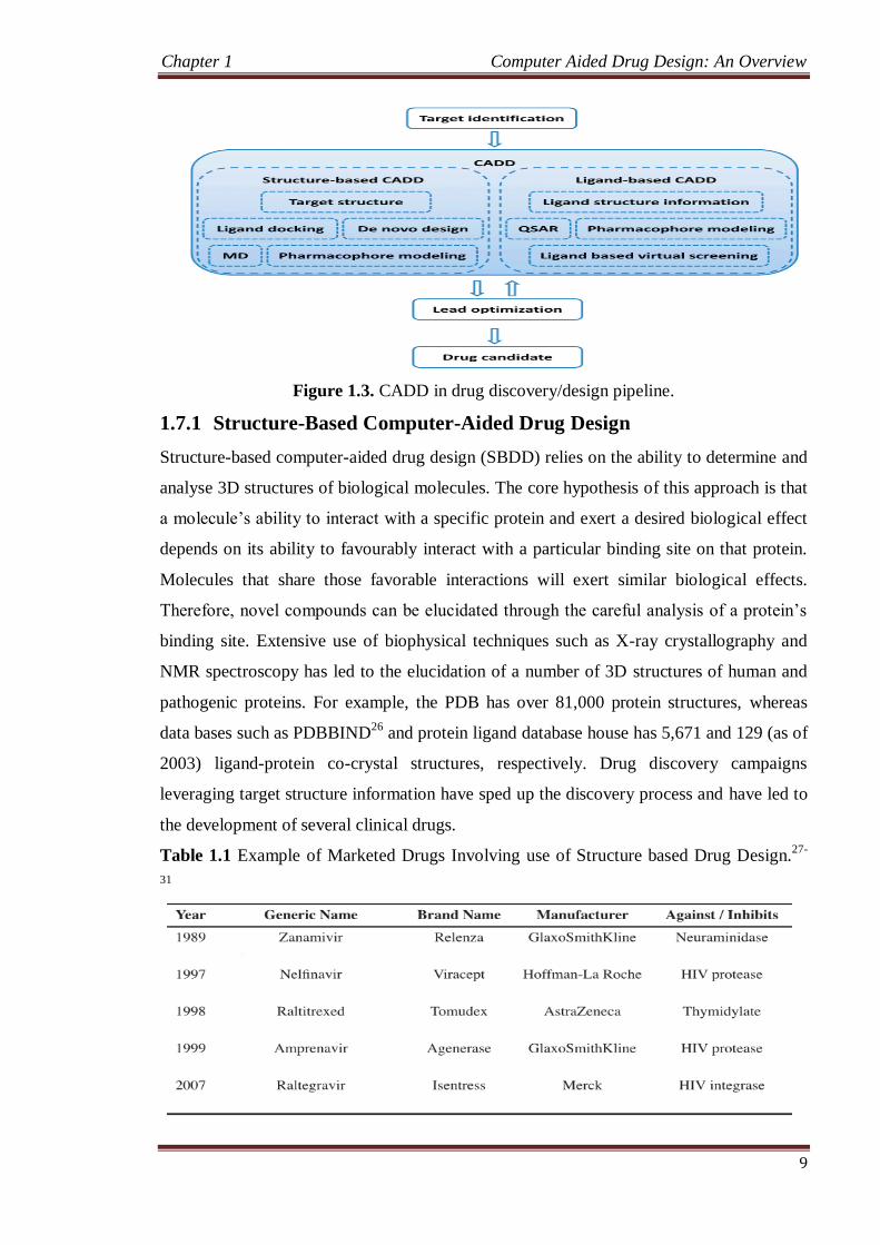
Figure 1.3. CADD in drug discovery/design pipeline.
1.7.1 Structure-Based Computer-Aided Drug Design
Structure-based computer-aided drug design (SBDD) relies on the ability to determine and
analyse 3D structures of biological molecules. The core hypothesis of this approach is that
a molecule’s ability to interact with a specific protein and exert a desired biological effect
depends on its ability to favourably interact with a particular binding site on that protein.
Molecules that share those favorable interactions will exert similar biological effects.
Therefore, novel compounds can be elucidated through the careful analysis of a protein’s
binding site. Extensive use of biophysical techniques such as X-ray crystallography and
NMR spectroscopy has led to the elucidation of a number of 3D structures of human and
pathogenic proteins. For example, the PDB has over 81,000 protein structures, whereas
data bases such as PDBBIND26
and protein ligand database house has 5,671 and 129 (as of
2003) ligand-protein co-crystal structures, respectively. Drug discovery campaigns
leveraging target structure information have sped up the discovery process and have led to
the development of several clinical drugs.
Table 1.1 Example of Marketed Drugs Involving use of Structure based Drug Design.27-
31
Chapter 1 Computer Aided Drug Design: An Overview
10
A prerequisite for the drug discovery process is the ability to rapidly determine
potential binders to the target of biological interest. Computational methods in drug
discovery allow rapid screening of a large compound library and determination of
potential binders through modeling/simulation and visualization techniques.
1.7.1.1 Preparation of a Target Structure
Success of virtual screening depends upon the amount and quality of structural
information known about both the target and the small molecules being docked. The first
step is to evaluate the target for the presence of an appropriate binding pocket.32-33
. This is
usually done through the analysis of known target-ligand co-crystal structures or using in-
silico methods to identify novel binding sites.34
A target structure experimentally determined through X-ray crystallography or
NMR techniques and deposited in the PDB is the ideal starting point for docking.
Structural genomics has accelerated the rate at which target structures are being
determined. In the absence of experimentally determined structures, several successful
virtual screening campaigns have been reported based on comparative models of target
proteins.35-37
1.7.1.2 Homology Modeling
In the absence of experimental structures, computational methods are used to predict the
3D structure of target proteins. Comparative modeling is used to predict target structure
based on a template with a similar sequence, leveraging that protein structure is better
conserved than sequence, i.e., proteins with similar sequences have similar structures.
Homology modeling is a specific type of comparative modeling in which the template and
target proteins share the same evolutionary origin. Comparative modeling involves the
following steps: (1) identification of related proteins to serve as template structures, (2)
sequence alignment of the target and template proteins, (3) copying coordinates for
confidently aligned regions, (4) constructing missing atom coordinates of target structure,
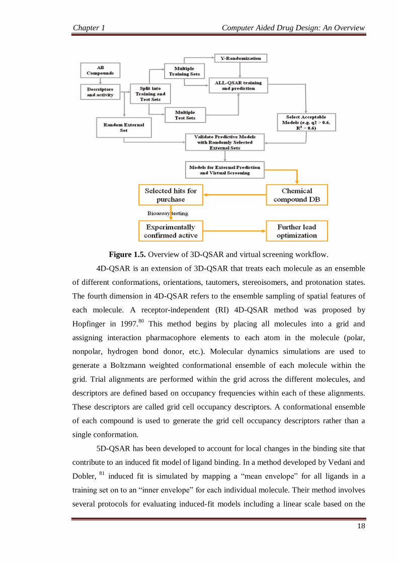
and (5) model refinement and evaluation. Fig. 1.4 illustrates the steps involved in
homology modeling. Several computer programs and web servers exist that automate the
homology modeling process e.g., PSIPRED38
and MODELER.39
1.7.1.3 Molecular dynamics-based detection
The dynamic nature of biomolecules sometimes makes it insufficient to use a single static
structure to predict putative binding sites. Multiple conformations of target are often used
to account for structural dynamics of target. Classic molecular dynamic (MD) simulations
can be used for obtaining an ensemble of target conformations beginning with a single
Chapter 1 Computer Aided Drug Design: An Overview
11
structure. The MD method uses principles of Newtonian mechanics to calculate a
trajectory of conformations of a protein as a function of time. Classic MD methods tend to
get trapped in local energy minima. To overcome this, several advanced MD algorithms
such as targeted-MD40
, conformational folding simulations41
, temperature accelerated MD
simulations42
, and replica exchange MD43
have been implemented for traversing multiple
minima energy surface of proteins.
Figure. 1.4 Steps involved in homology model building process.38-39
1.7.1.4 Small Molecules and Target Protein representation for Docking Simulations
There are three basic methods to represent target and ligand structures in silico: atomic,
surface, and grid representations.44,45
Atomic representation of the surface of the target is
usually used when scoring and ranking is based on potential energy functions. An example
is DARWIN, which uses CHARMM force-field to calculate energy.46
Surface methods
represent the topography of molecules using geometric features. The surface is represented
as a network of smooth convex, concave, and saddle shape surfaces. These features are
generated by mapping part of van der Waals surface of atoms that is accessible to probe a
sphere (Connolly, 1983).47
Docking is then guided by a complementary alignment of
ligand and binding site surfaces. Earliest implementation of DOCK48
used a set of
nonoverlapping spheres to represent invaginations of target surface and the surface of the
Chapter 1 Computer Aided Drug Design: An Overview
12
ligand. For the grid representation, the target is encoded as physicochemical features of its
surface. A grid method described by Katchalski-katzir et al49
digitizes molecules using a
3D discrete function that distinguishes the surface from the interior of the target molecule.
Molecules are scanned in relative orientation in three dimensions, and the extent of
overlap between molecules is determined using a correlation function calculated from a
Fourier transform.
1.7.1.5 Sampling Algorithms for Protein-Ligand Docking
Docking methods can be classified as rigid-body docking and flexible docking
applications depending on the degree to which they consider ligand and protein flexibility
during the docking process.50,51
Rigid body docking methods consider only static
geometric/physiochemical complementarities between ligand and target and ignore
flexibility and induced-fit51
binding models. More advanced algorithms consider several
possible conformations of ligand or receptor or both at the same time according to the
conformational selection paradigm.52
. Rigid docking simulations are generally preferred
when time is critical, i.e., when a large number of compounds are to be docked during an
initial vHTS. However, flexible docking methods are still needed for refinement and
optimization of poses obtained from an initial rigid docking procedure.
1.7.1.5.1 Systematic Methods
Systematic algorithms incorporate ligand flexibility through a comprehensive exploration
of a molecule’s degrees of freedom. In systematic algorithms, the current state of the
system determines the next state. Systematic methods can be categorized into (1)
exhaustive search algorithms and (2) fragmentation algorithms. Exhaustive searches
elucidate ligand conformations by systematically rotating all possible rotatable bonds at a
given interval. Large conformational space often prohibits an exhaustive systematic
search. Algorithms such as GLIDE53
use heuristics to focus on regions of conformational
space that are likely to contain good scoring ligand poses. Fragmentation methods sample
ligand conformation by incremental construction of ligand conformations from fragments
obtained by dividing the ligand of interest. Ligand conformations are obtained by docking
fragments in the binding site one at a time and incrementally growing them or by docking
all fragments into the binding site and linking them covalently. FLEXX54
uses the “anchor
and grow method” for ligand conformational sampling.
1.7.1.5.2 Molecular Dynamics Simulations
Molecular dynamics (MD) simulation calculates the trajectory of a system by the
application of Newtonian mechanics. However, standard MD methods depend heavily on
Chapter 1 Computer Aided Drug Design: An Overview
13
the starting conformation and are not readily appropriate for simulation of ligand-target
interactions. Because of its nature, MD is not able to cross high-energy barriers within the
simulation’s lifetime and is not efficient for traversing the rugged hyper surface of protein-
ligand interactions. Strategies like simulated annealing have been applied for more
efficient use of MD in docking.55
1.7.1.5.3 Monte Carlo Search with Metropolis Criterion (MCM) Simulations
MCM samples conformational space faster than molecular dynamics in that it requires
only energy function evaluation and not the derivative of the energy functions. Although
traditional MD drives a system toward a local energy minimum, the randomness
introduced with Monte Carlo allows hopping over the energy barriers, preventing the
system from getting stuck in local energy minima. MCM simulations have been adopted
for flexible docking applications such as in MCDOCK.56
1.7.1.5.4 Genetic Algorithms
Genetic algorithms introduce molecular flexibility through recombination of parent
conformations to child conformations. In this simulated evolutionary process, the “fittest”
or best scoring conformations are kept for another round of recombination. In this way, the
best possible set of solutions evolves by retaining favorable features from one generation
to the next. In docking, a set of values that describe the ligand pose in the protein are state
variable. State variables may include set of values describing translation, orientation,
conformation, number of hydrogen bonds, etc. The state corresponds to the genotype; the
resulting structural model of the ligand in the protein corresponds to the phenotype, and
binding energy corresponds to the fitness of the individual. Genetic operators may swap
large regions of parent’s genes or randomly change (mutate) the value of certain ligand
states to give rise to new individuals. Genetic Optimization for Ligand Docking (GOLD)57
explores full ligand flexibility with partial target flexibility using a genetic algorithm.
1.7.1.6 Scoring Functions for Evaluation of Protein Ligand Complexes
Docking applications need to rapidly and accurately assess protein-ligand complexes, i.e.,
approximate the energy of the interaction. A ligand docking experiment may generate
hundreds of thousands of target-ligand complex conformations, and an efficient scoring
function is necessary to rank these complexes and differentiate valid binding mode
predictions from invalid predictions.
1.7.1.6.1 Force-Field or Molecular Mechanics-Based Scoring Functions
Force-field scoring functions use classic molecular mechanics for energy calculations.58
These functions use parameters derived from experimental data and ab initio quantum
Chapter 1 Computer Aided Drug Design: An Overview
14
mechanical calculations. The binding free energy of protein-ligand complexes are
estimated by the sum of van der Waals and electrostatic interactions. DOCK uses the
AMBER force fields in which van der Waals energy terms are represented by the Lennard-
Jones potential function while electrostatic terms are accounted for by coulombic
interaction with a distance-dependent dielectric function.
1.7.1.6.2 Empirical Scoring Functions
Empirical scoring functions fit parameters to experimental data. An example is binding
energy, which is expressed as a weighted sum of explicit hydrogen bond interactions,
hydrophobic contact terms, desolvation effects, and entropy. Empirical function terms are
simple to evaluate and are based on approximations. The weights for different parameters
are obtained from regression analysis using experimental data obtained from molecular
data. Empirical functions have been used in several commercially available docking suits
like LUDI59
, FLEXX60
and SURFLEX.61
1.7.1.6.3 Knowledge-Based Scoring Function
Knowledge based scoring functions use the information contained in experimentally
determined complex structures. They are formulated under the assumption that interatomic
distances occurring more often than average distances represent favorable contacts. On the
other hand, interactions that are found to occur with lower frequencies are likely to
decrease affinity. Several knowledge based potentials have been developed to predict
binding affinity like potential of mean force62
, DRUGSCORE63
, SMOG64
and BLEEP.65
1.7.1.6.4 Consensus-Scoring Functions
Consensus approaches rescore predicted poses several times using different scoring
functions. These results can then be combined in different ways to rank solutions.66
Some
strategies for combining scores include (1) weighted combinations of scoring functions,
(2) a voting strategy in which cut-offs established for each scoring method is followed by
decision based on number of poses a molecule has, (3) a rank by number strategy ranks
each compound by its average normalized score values, and (4) a rank by rank method
sorts compounds based on average rank determined by individual scoring functions.67