Chapter 1 Microbial etiology of disease N. Shetty, E. Aarons, J. Andrews Microbes and their habitats have held a peculiar fascination for mankind ever since Antony van Leeuwenhoek (1632–1723) recorded some of the most important discoveries in the history of biology. Once Leeuwenhoek succeeded in creating the simple microscope, he described bacteria, free-living and parasitic creatures, sperm cells, blood cells, microscopic nematodes and much more. His publications opened up an entire world of microscopic life for scientific study. Microbes continue to excite intense research because of their virulence; their ability to cause tissue damage and death. They have been responsible for the great plagues and epidemics and have often changed the course of human history. The HIV pandemic has emerged as the single most defining occurrence in the history of infectious diseases of the late 20th and early 21st centuries. Microbes continue to baffle human ingenuity; they defy attempts at control by chemotherapeutic agents, vaccines, and the human immune system. The threat of a future pestilence is never far away. In order to study the microbial etiology of infectious disease, an understanding of the basic principles of microbiology and their interaction with the human host are essential. There are four basic groups of microbes: • Bacteria • Viruses Prokaryotic and eukaryotic cells Bacteria Sizes, shapes and arrangement of bacteria Phases of bacterial growth Viruses General properties Viral transmission Fungi Fungi of medical importance Protozoa Classification Helminths • Fungi: yeasts and molds • Protozoa. Multicellular organisms such as helminths also need to be included in the broad description of infectious disease agents. COPYRIGHTED MATERIAL
Transcript
Chapter 1
Microbial etiology of diseaseN. Shetty, E. Aarons, J. Andrews
Microbes and their habitats have held a peculiar fascination for mankind ever since Antony vanLeeuwenhoek (1632–1723) recorded some of the most important discoveries in the history of biology.Once Leeuwenhoek succeeded in creating the simple microscope, he described bacteria, free-living andparasitic creatures, sperm cells, blood cells, microscopic nematodes and much more. His publications openedup an entire world of microscopic life for scientific study. Microbes continue to excite intense research becauseof their virulence; their ability to cause tissue damage and death. They have been responsible for the greatplagues and epidemics and have often changed the course of human history. The HIV pandemic has emergedas the single most defining occurrence in the history of infectious diseases of the late 20th and early 21stcenturies. Microbes continue to baffle human ingenuity; they defy attempts at control by chemotherapeuticagents, vaccines, and the human immune system. The threat of a future pestilence is never far away.
In order to study the microbial etiology of infectious disease, an understanding of the basic principles of microbiology and their interaction with the human host are essential. There are four basic groups ofmicrobes:
• Bacteria• Viruses
Prokaryotic and eukaryotic cellsBacteria
Sizes, shapes and arrangement of bacteriaPhases of bacterial growth
VirusesGeneral propertiesViral transmission
FungiFungi of medical importanceProtozoaClassification
Helminths
• Fungi: yeasts and molds• Protozoa.
Multicellular organisms such as helminths also need to be included in the broad description ofinfectious disease agents.
ID_4_001.qxd 2/10/09 13:57 Page 3
COPYRIG
HTED M
ATERIAL
General principles of infectious diseases4
Prokaryotic and eukaryotic cells
The cell is the basic unit of life, whether it is of human or bacterial origin. Differences in bacterial (prokaryoticcells) and human (eukaryotic cells) have been exploited for diagnostic and treatment purposes. It is importantto understand what these differences are and how they contribute to disease pathogenesis (Table 1.1).
Bacteria
Sizes, shapes and arrangement of bacteria
Bacteria are unicellular organisms, ranging from 0.4 μm to 2.0 μm in size. They exist broadly in one ofthree morphological forms, spheres (cocci), rods (bacilli), or spirals. All of these forms are subject tovariation depending on existing growth conditions. The morphology of a bacterium is maintained by aunique cell wall structure and it is the chemical nature of this cell wall that is exploited by the Gram staining
Table 1.1 Differences between prokaryotic and eukaryotic cells
Cell structure
Cell wall
Cytoplasmic membrane
Nuclear body
Cytoplasmic structures
Respiratory enzymes and electron transport chains
Cell division
Organelles of locomotion
Prokaryotic
Complex cell wall containingpeptidoglycan/lipopolysaccarideSome cells have a capsule
Cytoplasmic membrane withoutcarbohydrates and usually lacking sterols.Incapable of endocytosis and exocytosis
Not bounded by a nuclear membraneUsually contains one circular chromosomecomposed of deoxyribonucleic acid (DNA). No nucleolus. May containextrachromosomal DNA – the plasmid
70S ribosomes composed of a 50S and a 30S subunit. Mitochondria, endoplasmicreticulum, Golgi apparatus, vacuoles, andlysosomes are absent. No microtubules.Contains only actin-like protein thatcontribute to cell shape. Spore formationmay occur
Located in the cell membrane
Usually by binary fission. No mitosis ormeiosis
Some have hair-like flagellae, fimbriae or pili may also be present. No cilia
Eukaryotic
Animal cells lack cell walls; plants, algae andfungi do have cell walls, but they differ incomposition from those of bacteria
Cytoplasmic membrane contains sterols andcarbohydrates and is capable of endocytosis(phagocytosis and pinocytosis) and exocytosis
Nucleus is bounded by a nuclear membrane,connecting it with the endoplasmicreticulum. Contains one or more paired,linear chromosomes composed of DNA.Nucleolus present
Ribosomes composed of a 60S and a 40Ssubunit; mitochondria, endoplasmicreticulum, Golgi apparatus, vacuoles, andlysosomes present. Mitotic spindle involvedin mitosis. Cytoskeleton with microtubules,actin, and intermediate filaments
Located in the mitochondria
By mitosis; sex cells in diploid organisms areproduced through meiosis
May have flagella or cilia: organellesinvolved in locomotion; consist of a distinctarrangement of sliding microtubulessurrounded by a membrane
ID_4_001.qxd 2/10/09 13:57 Page 4
Microbial etiology of disease 5
technique (see Chapter 4). The Gram stain remains the single most important diagnostic test in the study ofinfection – dividing bacteria into two basic groups: Gram positive bacteria and Gram negative bacteria –thereby influencing the all too important decision: which antibiotic does the clinician use immediately andempirically before full microbiological results are available.
With the help of the Gram stain and a microscope it is possible to visualize the size (relative to a human redor white cell), the shape, the arrangement (if distinctive), and the Gram reaction of the bacterial cell. All theabove features are important clues that help identify the infectious agent from a patient’s clinical specimen.
Cocci are spherical or oval bacteria having one of several distinct arrangements based on their planes of division:
1 Division in one plane produces either a diplococcus (paired; Figure 1.1) or streptococcus (chain)arrangement.If you were to Gram stain a smear of the specimen containing a putative diplococcus you would be able to see a Gram positive (purple) or -negative (pink) coccus in pairs; note the size relative to apolymorphonuclear leukocyte (Figure 1.2a and b). Streptococci, including medically important ones such as Streptococcus pyogenes, are Gram positive (Figure 1.3). The streptococci can be arranged in pairs(e.g. Streptococcus pneumoniae, Figure 1.2a) or in chains (e.g. S. pyogenes, Figure 1.3).
2 Division in random planes produces a staphylococcus arrangement. Note the Gram stained smear of apus sample showing numerous polymorphs ‘pus cells’ and staphylococci: cocci in irregular, grape-like clusters(Figure 1.4). Ordered division in two or three planes can result in sarcinial arrangements (tetrads) respectively.
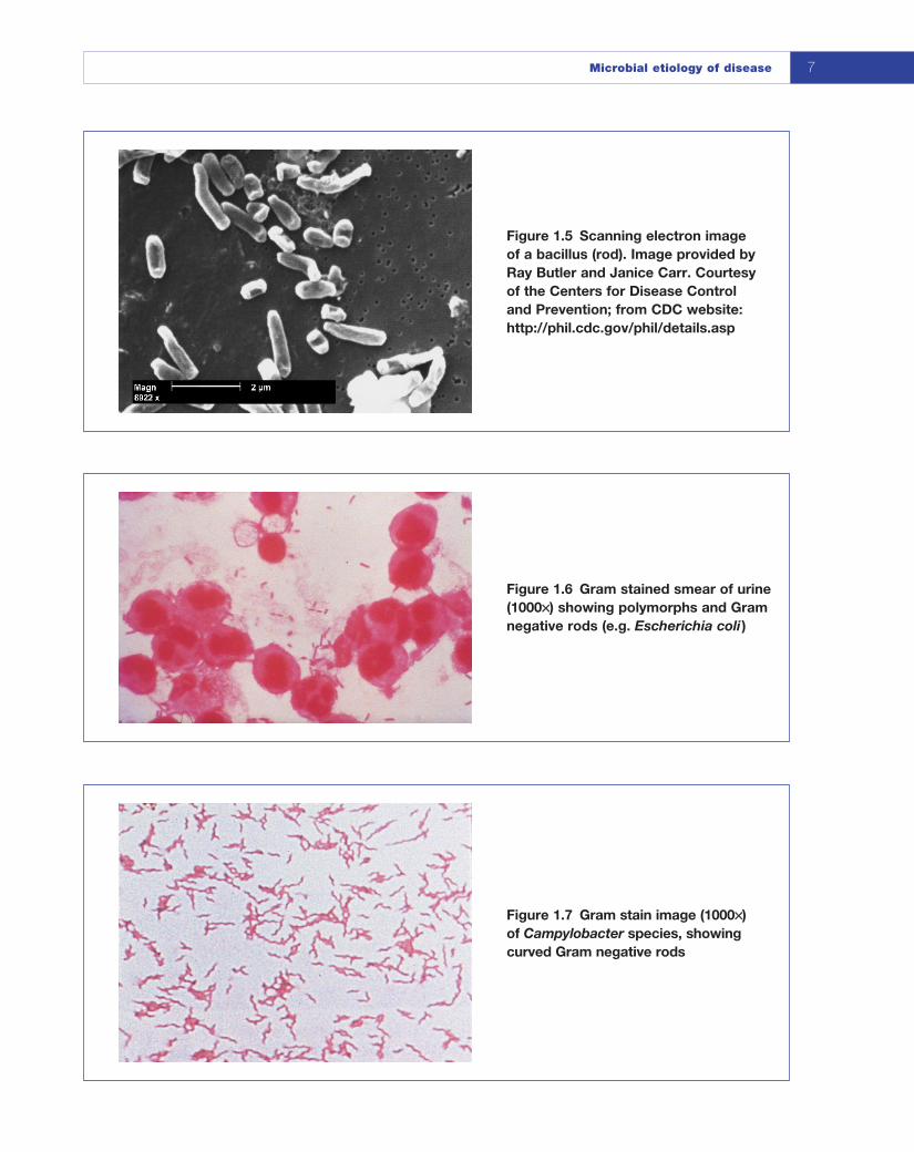
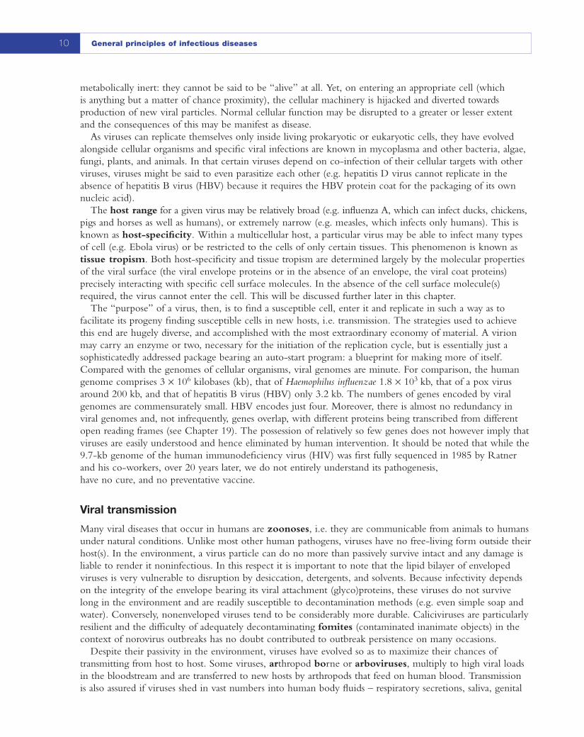
Bacilli are rod-shaped bacteria (Figure 1.5). Bacilli divide in one plane and are arranged singly or in chainsas in Bacillus anthracis. For many clinically important bacilli the arrangement is not distinctive; some bacillimay be rounded off looking more coccoid; they are often called cocco-bacillary forms. Bacilli, like cocci,can be Gram positive or -negative (Figure 1.6)
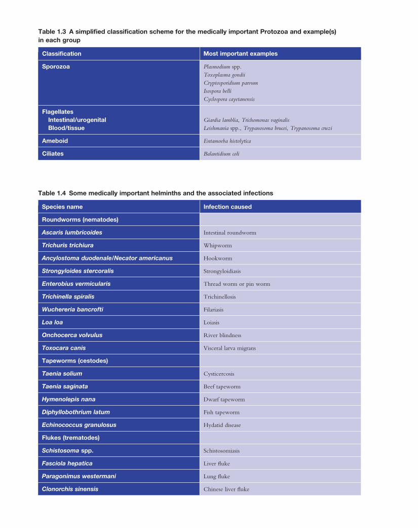
Other common shapes of bacteria are: curved bacteria as in Campylobacter (Figure 1.7) and Vibrio species;Spirillum species have thick rigid spirals; and spirochaete forms such as Leptospira species have flexible spirals(Figure 1.8). Spirals range in size from 1 μm to over 100 μm in length.
It is worth remembering that not all bacteria stain with the Gram stain. In Chapter 2 we will discussorganisms that do not take up the Gram stain readily, those that do not have typical bacterial cell wallstructures or arrangements, and those that are obligate intracellular microorganisms.
Figure 1.1 Scanning electronmicrograph of a diplococcus. Imageprovided by Dr Richard Facklam.Courtesy of the Centers for DiseaseControl and Prevention; from CDCwebsite: http://phil.cdc.gov/phil/details.asp
Figure 1.3 Gram positive cocci inchains: the streptococci
Figure 1.4 Gram stain of a smear(1000××) from pus showing numerouspolymorphs and Gram positive cocci inclusters (e.g. Staphylococcus aureus)
ID_4_001.qxd 2/10/09 13:58 Page 6
Microbial etiology of disease 7
Figure 1.5 Scanning electron image of a bacillus (rod). Image provided byRay Butler and Janice Carr. Courtesyof the Centers for Disease Control and Prevention; from CDC website:http://phil.cdc.gov/phil/details.asp
Figure 1.6 Gram stained smear of urine(1000××) showing polymorphs and Gramnegative rods (e.g. Escherichia coli )
When an organism is inoculated into suitable media such as a liquid culture medium in the laboratory or if it were to encounter a susceptible human/animal host it will exhibit a growth curve (Figure 1.9).
In the lag phase the microorganism adapts to a new and often more favourable environment. During this phase, there is a marked increase in enzymes and intermediates, in preparation for active growth. The lag phase is a period of adjustment necessary for the accumulation of metabolites until they are present inconcentrations that permit cell division to resume.
In the exponential or logarithmic phase, cells are in a state of balanced growth. The cells increase innumber and there is a logarithmic expansion of mass and volume. In other words imagine a single celldividing into two, each further divides in a binary manner and two becomes four, four to eight, eight tosixteen, and so on. A steady state is reached where one of many factors come into play; either essentialnutrients become exhausted, there is accumulation of waste products, change in pH, induction of hostimmune mechanisms and other obscure factors exert a deleterious effect on the culture, and growth isprogressively slowed.
During the stationary phase, accumulation of toxic products or exhaustion of nutrients causes netgrowth to cease. The viable cell count remains constant. The formation of new organisms equals the deathof organisms in the system. The stationary phase is important to the clinical microbiologist as microbialtoxins, antimicrobial substances and other proteins such as bacteriocins and lysins accumulate to significantlevels at the end of the stationary phase. Thus, they affect not only growth in the laboratory but alsopathogenesis of disease in the host.
As factors detrimental to the bacteria accumulate, more bacteria are killed than are formed. During thephase of decline there is a negative exponential phase, which results in a decrease in the numbers of viablebacteria within the system.
Figure 1.8 Scanning electronmicrograph of Leptospira species.Image provided by Rob Weyant.Courtesy of the Center for Disease Control and Prevention; from CDC website:http://phil.cdc.gov/phil/details.asp
Log numberof cells
Time
a
b
c
d
Figure 1.9 The bacterial growth curveshowing the four phases of growth. (a) The lag phase; (b) the exponentialphase; (c) the stationary phase; (d) thephase of decline
ID_4_001.qxd 2/10/09 13:58 Page 8
Microbial etiology of disease 9
Viruses
General properties
Of all the agents infectious to man, and indeed to other living things, viruses are the smallest. (See Box 1.1for a description of prions.) An individual infectious unit, comprising a nucleic acid genome, packagedinside a protein coat with or without a surrounding lipid-containing envelope membrane, is known as a viralparticle or virion (Figure 1.10).
Only the very largest of these, the poxviruses measuring up to 400 nm in their longest dimension and the even larger mimivirus, can be visualized with a light microscope. Cell-free, intact virions are entirely
Box 1.1 Prions
Even smaller infectious agents have been identifiedthat, amazingly, lack a nucleic acid genome. Itwould appear that the protein alone is theinfectious agent. This infectious agent has beencalled a prion, short for proteinaceous infectiousparticle. They are unique structures as they lackDNA or RNA (the very code for life in a livingmicroorganism) and they resist all conventionalattempts at inactivation. The discovery thatproteins alone can transmit an infectious diseasehas led to much debate and controversy in thescientific community.
Prion diseases target the brain and are oftencalled spongiform encephalopathies. The post
mortem appearance of the brain is characteristic,with large vacuoles in the cortex and cerebellum.Many mammalian species develop these diseases.Specific examples include:
• Scrapie in sheep• TME (transmissible mink encephalopathy)
in mink• CWD (chronic wasting disease) in muledeer
and elk• BSE (bovine spongiform encephalopathy)
in cows; this led to banning of British beefworldwide (commonly known as mad cowdisease).
(a) (b)
Figure 1.10 Electron photomicrograph of: (a) a nonenveloped virus, the adenovirus; (b) anenveloped virus, the herpes virus. Courtesy of: (a) Dr G. William Gray Jr; (b) Dr Fred Murphy; fromCDC website: http://phil.cdc.gov/phil/details.asp
ID_4_001.qxd 2/10/09 13:58 Page 9
General principles of infectious diseases10
metabolically inert: they cannot be said to be “alive” at all. Yet, on entering an appropriate cell (which is anything but a matter of chance proximity), the cellular machinery is hijacked and diverted towardsproduction of new viral particles. Normal cellular function may be disrupted to a greater or lesser extent and the consequences of this may be manifest as disease.
As viruses can replicate themselves only inside living prokaryotic or eukaryotic cells, they have evolvedalongside cellular organisms and specific viral infections are known in mycoplasma and other bacteria, algae,fungi, plants, and animals. In that certain viruses depend on co-infection of their cellular targets with otherviruses, viruses might be said to even parasitize each other (e.g. hepatitis D virus cannot replicate in theabsence of hepatitis B virus (HBV) because it requires the HBV protein coat for the packaging of its ownnucleic acid).
The host range for a given virus may be relatively broad (e.g. influenza A, which can infect ducks, chickens,pigs and horses as well as humans), or extremely narrow (e.g. measles, which infects only humans). This isknown as host-specificity. Within a multicellular host, a particular virus may be able to infect many typesof cell (e.g. Ebola virus) or be restricted to the cells of only certain tissues. This phenomenon is known astissue tropism. Both host-specificity and tissue tropism are determined largely by the molecular propertiesof the viral surface (the viral envelope proteins or in the absence of an envelope, the viral coat proteins)precisely interacting with specific cell surface molecules. In the absence of the cell surface molecule(s)required, the virus cannot enter the cell. This will be discussed further later in this chapter.
The “purpose” of a virus, then, is to find a susceptible cell, enter it and replicate in such a way as tofacilitate its progeny finding susceptible cells in new hosts, i.e. transmission. The strategies used to achievethis end are hugely diverse, and accomplished with the most extraordinary economy of material. A virionmay carry an enzyme or two, necessary for the initiation of the replication cycle, but is essentially just asophisticatedly addressed package bearing an auto-start program: a blueprint for making more of itself.Compared with the genomes of cellular organisms, viral genomes are minute. For comparison, the humangenome comprises 3 × 10⁶ kilobases (kb), that of Haemophilus influenzae 1.8 × 10³ kb, that of a pox virusaround 200 kb, and that of hepatitis B virus (HBV) only 3.2 kb. The numbers of genes encoded by viralgenomes are commensurately small. HBV encodes just four. Moreover, there is almost no redundancy inviral genomes and, not infrequently, genes overlap, with different proteins being transcribed from differentopen reading frames (see Chapter 19). The possession of relatively so few genes does not however imply thatviruses are easily understood and hence eliminated by human intervention. It should be noted that while the9.7-kb genome of the human immunodeficiency virus (HIV) was first fully sequenced in 1985 by Ratnerand his co-workers, over 20 years later, we do not entirely understand its pathogenesis, have no cure, and no preventative vaccine.
Viral transmission
Many viral diseases that occur in humans are zoonoses, i.e. they are communicable from animals to humansunder natural conditions. Unlike most other human pathogens, viruses have no free-living form outside theirhost(s). In the environment, a virus particle can do no more than passively survive intact and any damage isliable to render it noninfectious. In this respect it is important to note that the lipid bilayer of envelopedviruses is very vulnerable to disruption by desiccation, detergents, and solvents. Because infectivity dependson the integrity of the envelope bearing its viral attachment (glyco)proteins, these viruses do not survivelong in the environment and are readily susceptible to decontamination methods (e.g. even simple soap andwater). Conversely, nonenveloped viruses tend to be considerably more durable. Caliciviruses are particularlyresilient and the difficulty of adequately decontaminating fomites (contaminated inanimate objects) in thecontext of norovirus outbreaks has no doubt contributed to outbreak persistence on many occasions.
Despite their passivity in the environment, viruses have evolved so as to maximize their chances oftransmitting from host to host. Some viruses, arthropod borne or arboviruses, multiply to high viral loadsin the bloodstream and are transferred to new hosts by arthropods that feed on human blood. Transmission is also assured if viruses shed in vast numbers into human body fluids – respiratory secretions, saliva, genital
ID_4_001.qxd 2/10/09 13:58 Page 10
Microbial etiology of disease 11
secretions, urine, and stool. Human behavior takes care of the rest: face to face conversation, sexual activity,use of the hands for eating as well as for toileting facilitates transmission and continued survival. Certainviruses, influenza and rotavirus for example, go even further by causing the volume of the infectious fluid to be considerably increased and literally sprayed into the environment by sneezing or explosive, waterydiarrhea respectively. Mother-to-child transmission (also known as vertical transmission) is usually anincidental means of transmission rather than the predominant one. The transmission of blood-borneviruses through parenteral exposure (blood transfusion, contaminated surgical implements, tattooing, sharing of equipment for IV drug use, etc.) is of course an artifact of very recent (in evolutionary terms)human behavior, where the major means of transmission is mucosal or skin lesion contact with blood orgenital secretions. In the absence of universal immunization, knowledge of the route by which a viral disease is transmitted is essential to the control of that infection in human populations.
FungiFungi are an extremely diverse group of organisms, ubiquitous in the environment. They are found as twomain forms, yeasts and molds. They are nonphotosynthetic organisms with the ability to absorb solublenutrients by diffusion from living or dead organic matter. Molds consist of branching filaments (hyphae),which interlace to form a mycelium. The hyphae of the more primitive molds remain aseptate (withoutwalls) whereas those of the more developed groups are septate with a central pore in each cross wall. Yeastsare unicellular organisms consisting of separate round or oval cells. They do not form a mycelium, althoughthe intermediate yeast-like fungi form a pseudomycelium consisting of chains of elongated cells.
Many fungi, including some of clinical importance, can exist in both forms dependent on temperatureand other environmental conditions. These are known as dimorphic fungi.
Like mammalian cells, fungi are eukaryotes (Table 1.1) with DNA organized into chromosomes withinthe cell nucleus. Fungi also have distinct cytoplasmic organelles including Golgi apparatus, mitochrondria,and storage vacuoles. Homology with mammalian cells also extends to biosynthesis, where fungi sharesimilar pathways for both protein synthesis and DNA replication.
A formal classification scheme of fungi has little medical relevance so a simplified clinical classification for pathogenic fungi, based on initial site of infection, is more commonly used (Table 1.2).
Cutaneous superficial fungal infections are very common. The majority are caused by three groups offungi: mold dermatophytes such as Microsporium spp. and Trichophyton spp., Candida albicans, and Malasseziaspp. Keratin-containing structures such as hair shafts, nails, and skin are affected. Dermatophyte skininfection (sometimes called ringworm) is commonly named after the area affected, for example tinea capitis(head) or tinea corporis (body).
The systemic fungi include Coccidioides immitis, Paracoccidioides braziliensis, and Histoplasma capsulatum.These are thermally dimorphic fungi, meaning they have both yeast-like and filamentous forms. They are environmental organisms, which enter the body usually via inhalation. Infection is geographically
Table 1.2 Classification of fungi associated with human infection (mycoses)
circumscribed and often clinically mild. Severe disseminated disease can occur, however, particularly inimmunocompromised patients.
The main fungi that cause disease in immunocompromised patients are the yeasts C. albicans and relatedspecies such as Candida krusei. Aspergillus species are important environmental filamentous fungi, which maycause pulmonary or disseminated infection. The yeast-like fungi Cryptococcus neoformans can cause chronicmeningitis in patients with HIV infection.
Fungi of medical importance
Over 200 000 species of fungi have been described although only about 200 have been associated withhuman disease. With a few exceptions, fungal infections of humans originate from an exogenous source inthe environment and are acquired through inhalation, ingestion, or traumatic implantation.
A few species of fungi are capable of causing significant disease in otherwise normal individuals. Manymore are only able to produce disease when the host has some aspect of impaired immunity e.g. HIVseropositivity, or during treatment for malignancy. With the increasing number of immunocompromisedpatients, fungi previously considered to be nonpathogenic are being recognized as the cause of sporadicinfections. Any fungus capable of growing at the temperature of the human host (37°C) must now beregarded as a potential human pathogen.
Recent DNA sequencing work has shown that Pneumocystis jiroveci, long believed to be a protozoon, isin fact also a fungus. This organism is an important pathogen in immunocompromised patients, especiallythose with AIDS.
Protozoa
Protozoa are unicellular microorganisms, which are found in almost every type of environment. Almost two-thirds of the world population live in conditions in which infection with protozoa or helminths (seelater) are thought to be unavoidable. Protozoan lifecycles are extremely diverse and can be complex and thusthey display a much wider range of morphology than bacteria or viruses. Many species are parasites of higherplants and animals but this chapter will deal only with those that cause disease in humans.
Protozoans such as Plasmodium spp., Leishmania spp., and Entamoeba histolytica are important causes ofmorbidity and mortality in the tropics but can be seen in the developed world in the returning traveler.Pathogens such as Cryptosporidium parvum and Giardia lamblia are important causes of morbidity in both the developed and developing world. Protozoan organisms including Toxoplasma gondii, Leishmania spp., andC. parvum have been shown to cause particularly severe disease in patients with AIDS. With increased levelsof international travel and the immunosuppressive effects of infection with HIV, one needs to have a raisedawareness of diseases caused by protozoa.
Classification
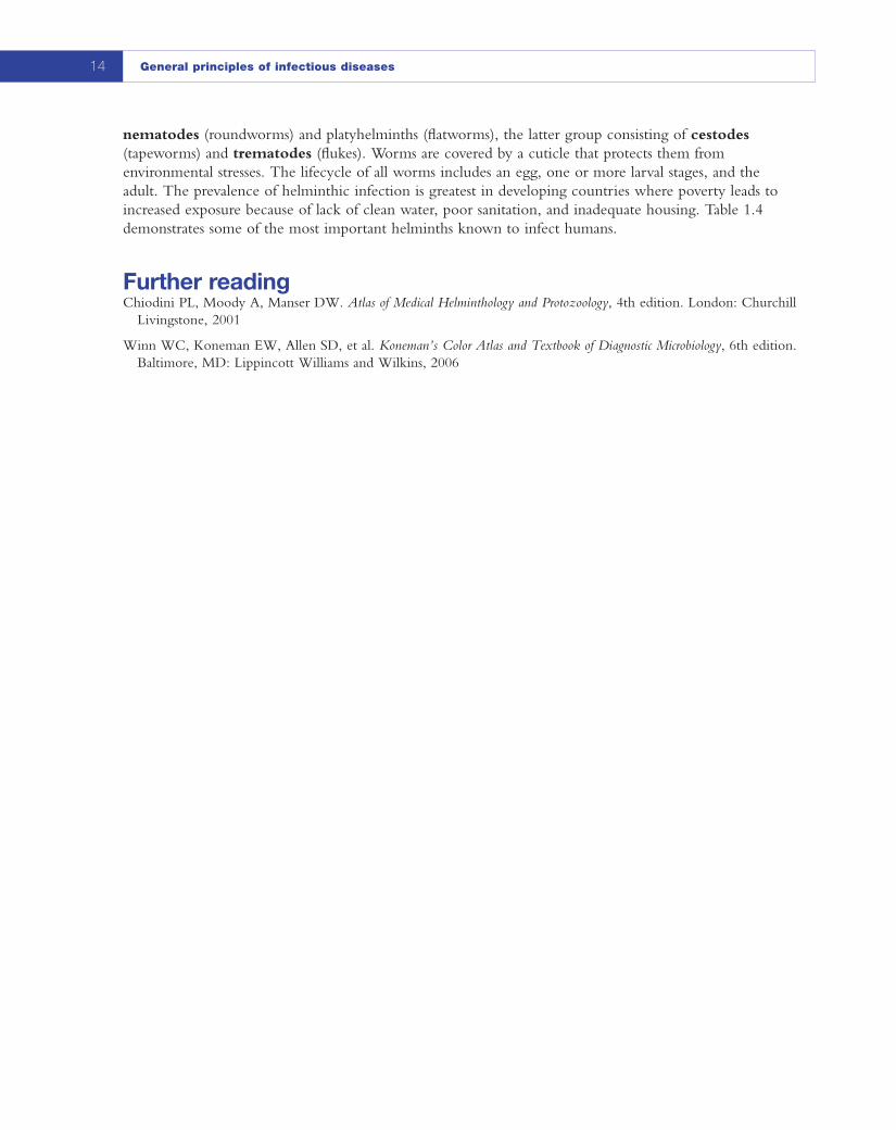
The classification of protozoa that are medically important can be simplified by subdivision into four maingroups, which in part relate to the method of locomotion (Table 1.3). These distinctions are by no meansabsolute, for example some organisms may be flagellate or amoeboid at different stages of their lifecycle.Flagellates are often divided into organisms causing intestinal or urogenital infection such as Giardia lambliaor Trichomonas vaginalis, and flagellates found in the blood or tissues (hemoflagellates) such as Leishmania spp.or Trypansoma spp.
HelminthsFor completeness a short section on helminths has been included here. Helminths are multicellular organismsthat range from less than 1 cm to more than 10 m in length. The helminths that infect man include the
ID_4_001.qxd 2/10/09 13:58 Page 12
Table 1.3 A simplified classification scheme for the medically important Protozoa and example(s) in each group
Classification
Sporozoa
FlagellatesIntestinal/urogenitalBlood/tissue
Ameboid
Ciliates
Table 1.4 Some medically important helminths and the associated infections
nematodes (roundworms) and platyhelminths (flatworms), the latter group consisting of cestodes(tapeworms) and trematodes (flukes). Worms are covered by a cuticle that protects them fromenvironmental stresses. The lifecycle of all worms includes an egg, one or more larval stages, and the adult. The prevalence of helminthic infection is greatest in developing countries where poverty leads toincreased exposure because of lack of clean water, poor sanitation, and inadequate housing. Table 1.4demonstrates some of the most important helminths known to infect humans.
Further readingChiodini PL, Moody A, Manser DW. Atlas of Medical Helminthology and Protozoology, 4th edition. London: Churchill
Livingstone, 2001
Winn WC, Koneman EW, Allen SD, et al. Koneman’s Color Atlas and Textbook of Diagnostic Microbiology, 6th edition.Baltimore, MD: Lippincott Williams and Wilkins, 2006