38
Chapter 10-11 Review
| Date post: | 28-Dec-2015 |
| Category: |
Documents |
| Upload: | kimberly-henry |
| View: | 216 times |
| Download: | 0 times |

Chapter 10-11 Review
The Heart: Heart WallThe Heart: Heart Wall
Slide 11.4Copyright © 2003 Pearson Education, Inc. publishing as Benjamin Cummings
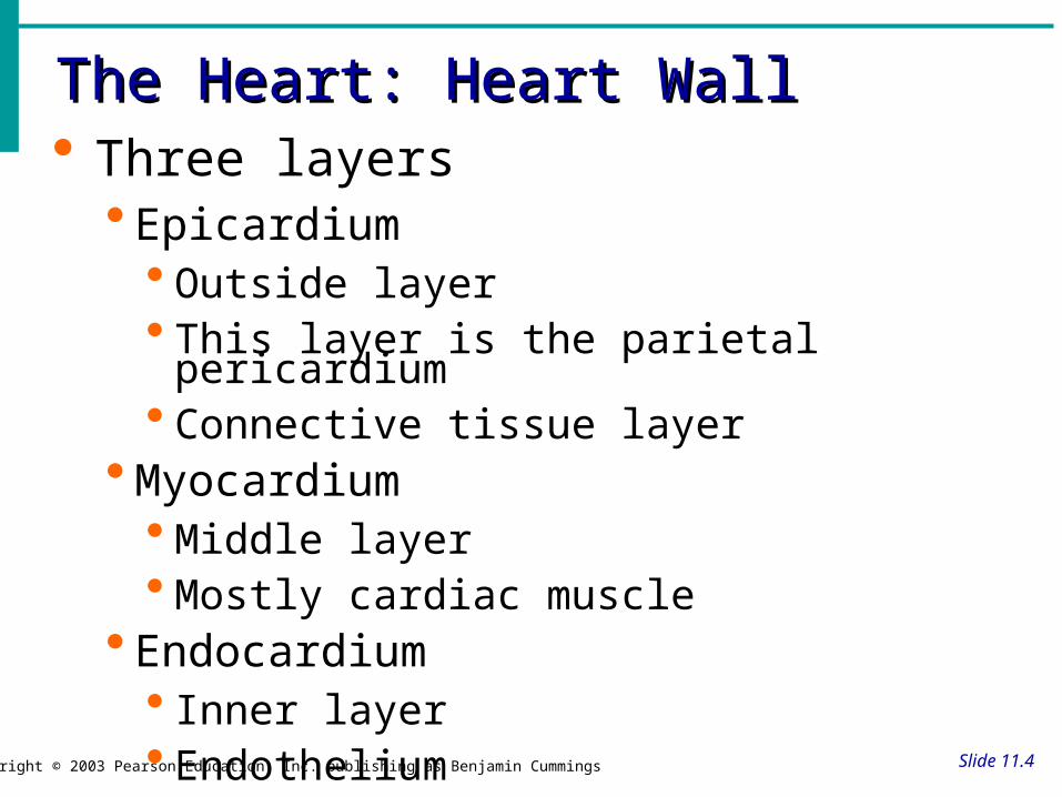
Three layers Epicardium
Outside layer This layer is the parietal pericardium Connective tissue layer
Myocardium Middle layer Mostly cardiac muscle
Endocardium Inner layer Endothelium

The Heart: CoveringsThe Heart: Coverings
Slide 11.3Copyright © 2003 Pearson Education, Inc. publishing as Benjamin Cummings
Pericardium – a double serous membrane Visceral pericardium
Next to heart
Parietal pericardium
Outside layer
Serous fluid fills the space between the layers of pericardium

The Heart: ChambersThe Heart: Chambers
Slide 11.6Copyright © 2003 Pearson Education, Inc. publishing as Benjamin Cummings
Right and left side act as separate pumps Four chambers
Atria Receiving chambers (thinner walls)
Right atrium Left atrium
Ventricles Discharging chambers
Right ventricle (2nd thickest) Left ventricle (thickest)

The Cardiovascular System2. Blood Vessels -A network of tubes
–Arteriesarterioles move away from the heart•Elastic Fibers•Circular Smooth Muscle
–Capillaries – where gas exchange takes place.•One cell thick•Serves the Respiratory System
–VeinsVenules moves towards the heart•Skeletal Muscles contract to force blood from legs back to heart•One way leaflet valves
– When the valves no longer meet properly (valvular incompetence) varicose veins form b/c of pooling blood/ backflow
6
Regulation of the Heart• Intrinsic regulation: Results from normal functional
characteristics—initiated by heart’s own pacemaker nodes, not on neural or hormonal regulation– Starling’s law of the heart--stroke volume of the heart increases in
response to an increase in the volume of blood filling the heart (the end diastolic volume) when all other factors remain constant (more blood in the heart=stronger heart muscular contraction)
• Extrinsic regulation: Involves neural and hormonal control– Parasympathetic stimulation (by medulla oblongata)
• Supplied by vagus nerve, decreases heart rate, acetylcholine secreted
– Sympathetic stimulation• Supplied by cardiac nerves, increases heart rate and force of contraction,
epinephrine and norepinephrine released
Heart Homeostasis• Effect of blood pressure
– Baroreceptors monitor blood pressure
• Effect of pH, carbon dioxide, oxygen– Chemoreceptors monitor
• Effect of extracellular ion concentration– Increase or decrease in extracellular K+ decreases heart rate
• Effect of body temperature– Heart rate increases when body temperature increases, heart rate
decreases when body temperature decreases


9
Arteries: Built for their job• Arteries
– blood flows away from heart– thicker walls
• provide strength for high pressure pumping of blood
– elastic & stretchable • maintains blood
pressure even when heart relaxes

10
Major arteries
pulmonaryartery
pulmonaryartery =to lungs
aorta carotid = to headto brain & left arm to right arm
coronary arteries
to body

Veins: Built for their job• Veins
– blood returns back to heart– thinner-walled
• blood travels back to heart at low speed & pressure
• why low pressure?– far from heart
• blood flows because muscles contract when we move – squeeze blood through veins
– valves in large veins• in larger veins one-way valves
allow blood to flow only toward heart
Open valve
Blood flowstoward heart
Closed valve
12
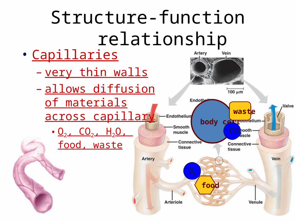
Structure-function relationship• Capillaries
– very thin walls – allows diffusion of
materials across capillary• O2, CO2, H2O,
food, wastebody cell
O2
food
waste
CO2

13
Blood Vessels: Anatomy
Slide 11.25Copyright © 2003 Pearson Education, Inc. publishing as Benjamin Cummings
Three layers (tunics)Tunic intima
Endothelium
Tunic media
Smooth muscle
Controlled by sympathetic nervous system
Tunic externa
Mostly fibrous connective tissue
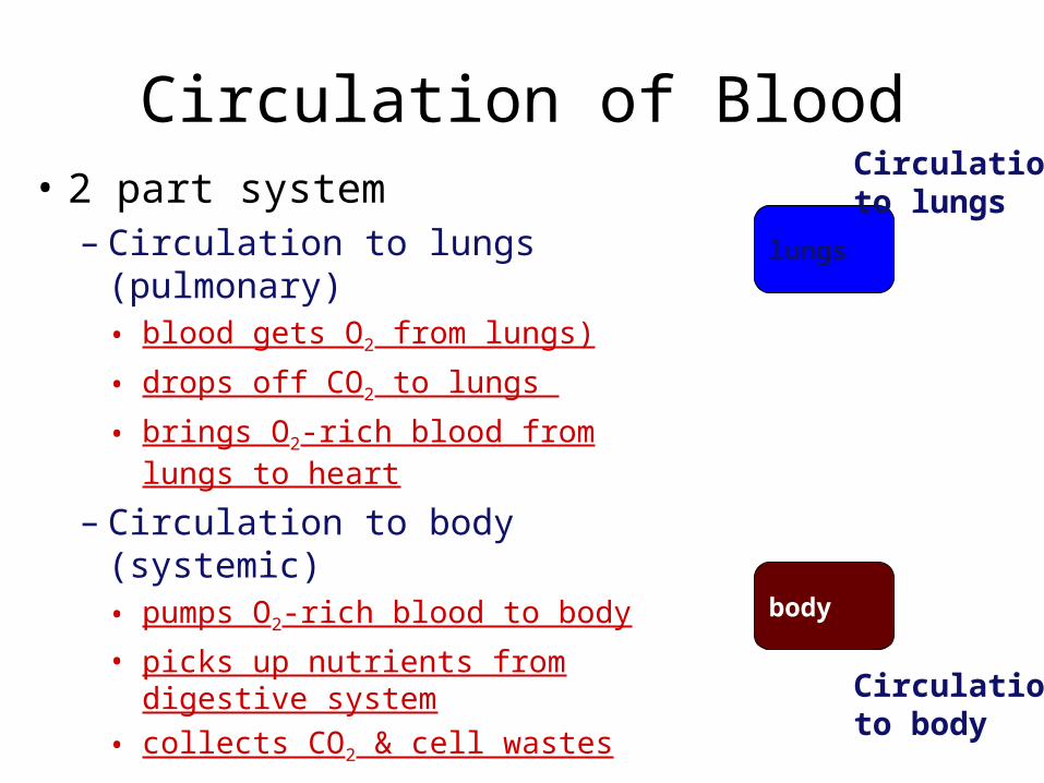
• 2 part system– Circulation to lungs
(pulmonary)• blood gets O2 from lungs)
• drops off CO2 to lungs
• brings O2-rich blood from lungs to heart
– Circulation to body (systemic)• pumps O2-rich blood to body
• picks up nutrients from digestive system
• collects CO2 & cell wastes
Circulation of Blood
lungs
body
Circulationto lungs
Circulationto body
15
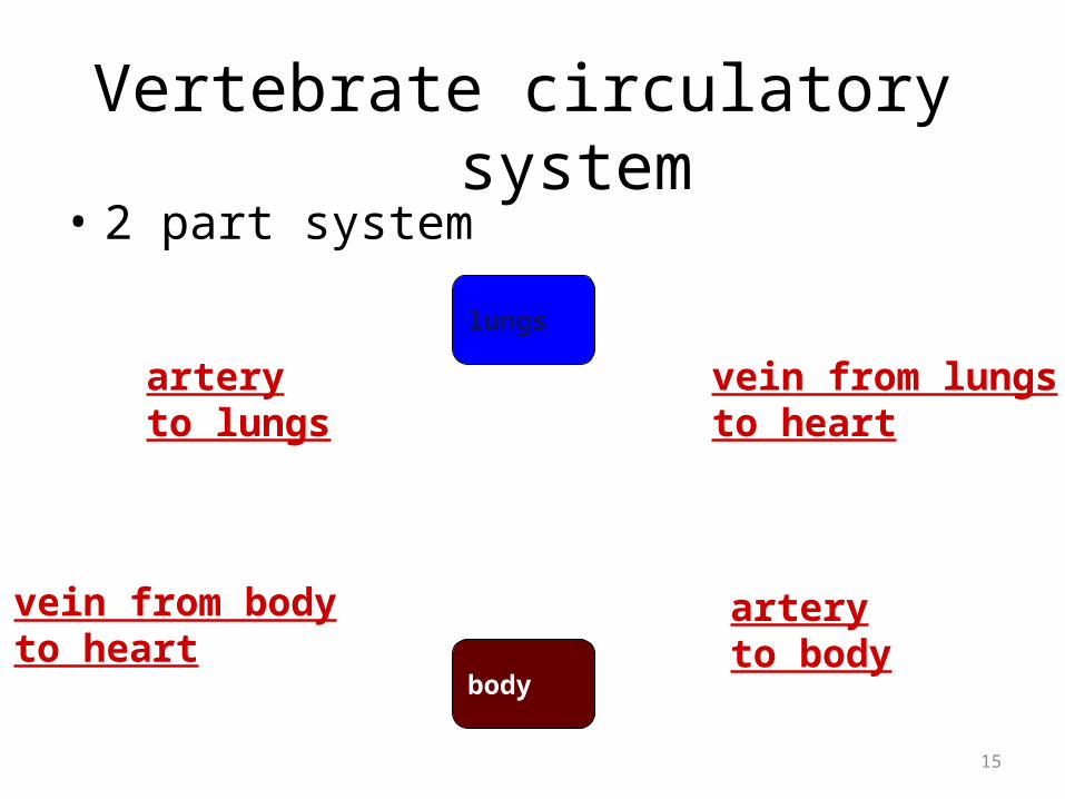
Vertebrate circulatory system
lungs
body
• 2 part system
arteryto body
arteryto lungs
vein from lungsto heart
vein from bodyto heart
16
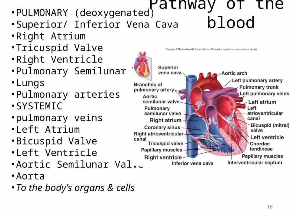
Pathway of the blood•PULMONARY (deoxygenated)•Superior/ Inferior Vena Cava•Right Atrium•Tricuspid Valve•Right Ventricle•Pulmonary Semilunar Valve•Lungs•Pulmonary arteries•SYSTEMIC •pulmonary veins•Left Atrium•Bicuspid Valve•Left Ventricle•Aortic Semilunar Valve•Aorta•To the body’s organs & cells

17
Cardiac Muscle
• Elongated, branching cells containing 1-2 centrally located nuclei• Contains actin and myosin myofilaments • Intercalated disks: Specialized cell-cell contacts• Desmosomes hold cells together and gap junctions allow action potentials• Electrically, cardiac muscle behaves as single unit

Conducting System of Heart
Refractory Period
• Absolute: Cardiac muscle cell completely insensitive to further stimulation
• Relative: Cell exhibits reduced sensitivity to additional stimulation
• Long refractory period prevents tetanic contractions
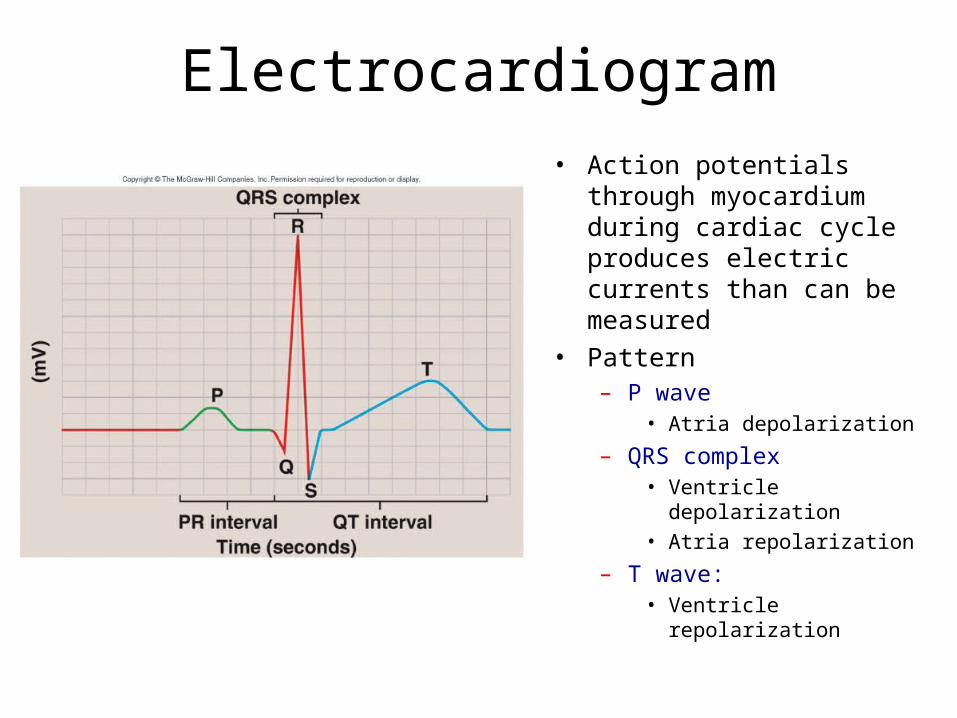
Electrocardiogram• Action potentials through
myocardium during cardiac cycle produces electric currents than can be measured
• Pattern– P wave
• Atria depolarization
– QRS complex• Ventricle depolarization• Atria repolarization
– T wave: • Ventricle repolarization

EKG Interpretation
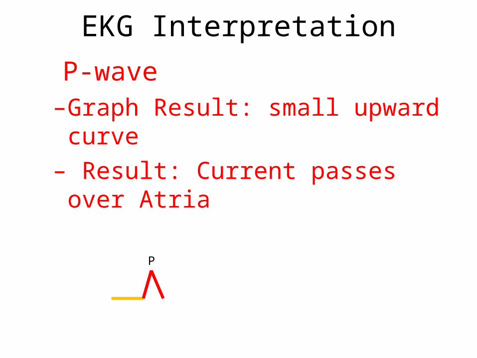
• P-wave– SA node sends signal through atria muscle
–Atria depolarize (becomes more positive)
P

Conducting System of Heart
EKG Interpretation
• P-wave–Graph Result: small upward curve
– Result: Current passes over Atria
P

EKG Interpretation
• P Q segment–Graph Result: Flat line –Response: Atrial contraction time
P
Q
EKG Interpretation
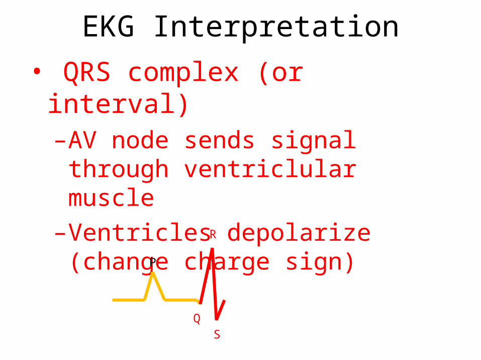
• QRS complex (or interval)–AV node sends signal through ventriclular muscle
–Ventricles depolarize (change charge sign)
Q
R
S
P
EKG Interpretation
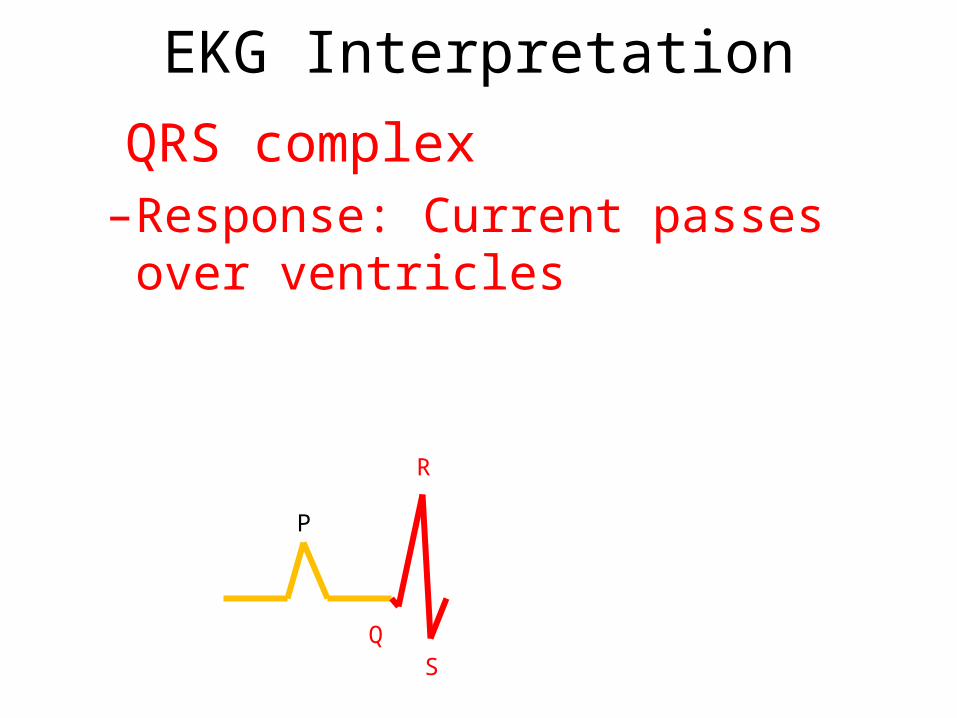
• QRS complex–Response: Current passes over ventricles
Q
R
S
P

EKG Interpretation
• S T segment–Graph: rise, flat line and bump–Response: Ventricular contraction time
Q
R
S
PT
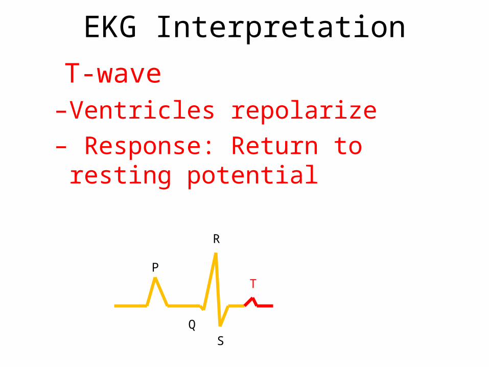
EKG Interpretation
• T-wave–Ventricles repolarize– Response: Return to resting potential
Q
R
S
PT

EKG Interpretation
• U-wave (if present)–Repolarization of papillary muscles
Q
R
S
PT U
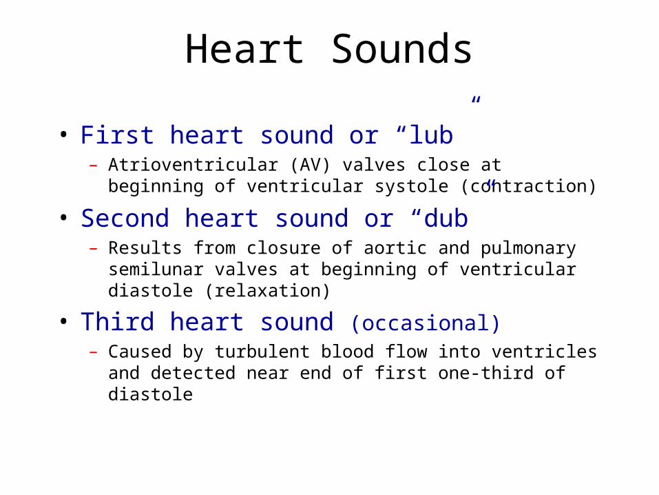
Heart Sounds
• First heart sound or “lub”– Atrioventricular (AV) valves close at beginning of ventricular
systole (contraction)
• Second heart sound or “dub”– Results from closure of aortic and pulmonary semilunar valves at
beginning of ventricular diastole (relaxation)
• Third heart sound (occasional)– Caused by turbulent blood flow into ventricles and detected near
end of first one-third of diastole
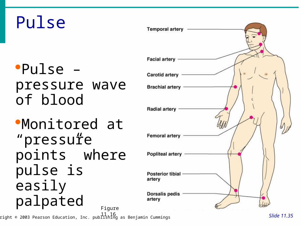
Pulse
Slide 11.35Copyright © 2003 Pearson Education, Inc. publishing as Benjamin Cummings
Pulse – pressure wave of blood
Monitored at “pressure points” where pulse is easily palpated
Figure 11.16

Blood Vessels: Anatomy
Slide 11.25Copyright © 2003 Pearson Education, Inc. publishing as Benjamin Cummings
Three layers (tunics)Tunic intima
Endothelium
Tunic media
Smooth muscle
Controlled by sympathetic nervous system
Tunic externa
Mostly fibrous connective tissue

Plasma Proteins
Slide 10.4Copyright © 2003 Pearson Education, Inc. publishing as Benjamin Cummings
Albumin – regulates osmotic pressure
Clotting proteins – help to stem blood loss when a blood vessel is injured
Antibodies – help protect the body from antigens
Platelets
Slide 10.13Copyright © 2003 Pearson Education, Inc. publishing as Benjamin Cummings
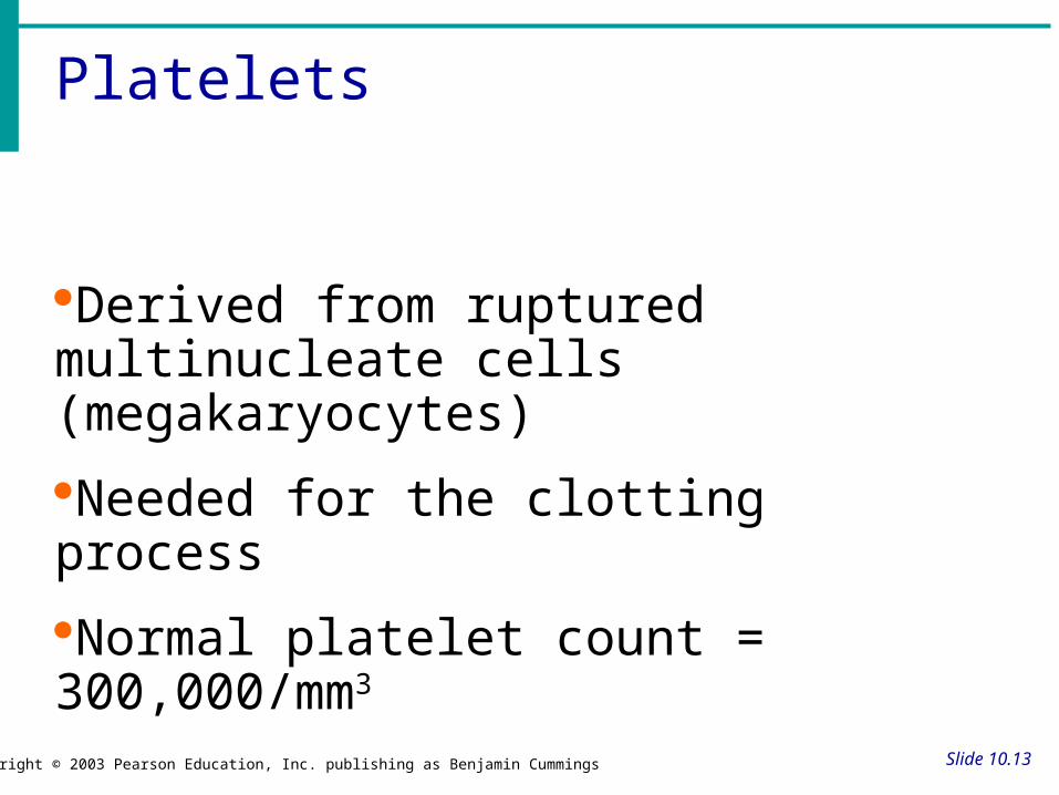
Derived from ruptured multinucleate cells (megakaryocytes)
Needed for the clotting process
Normal platelet count = 300,000/mm3
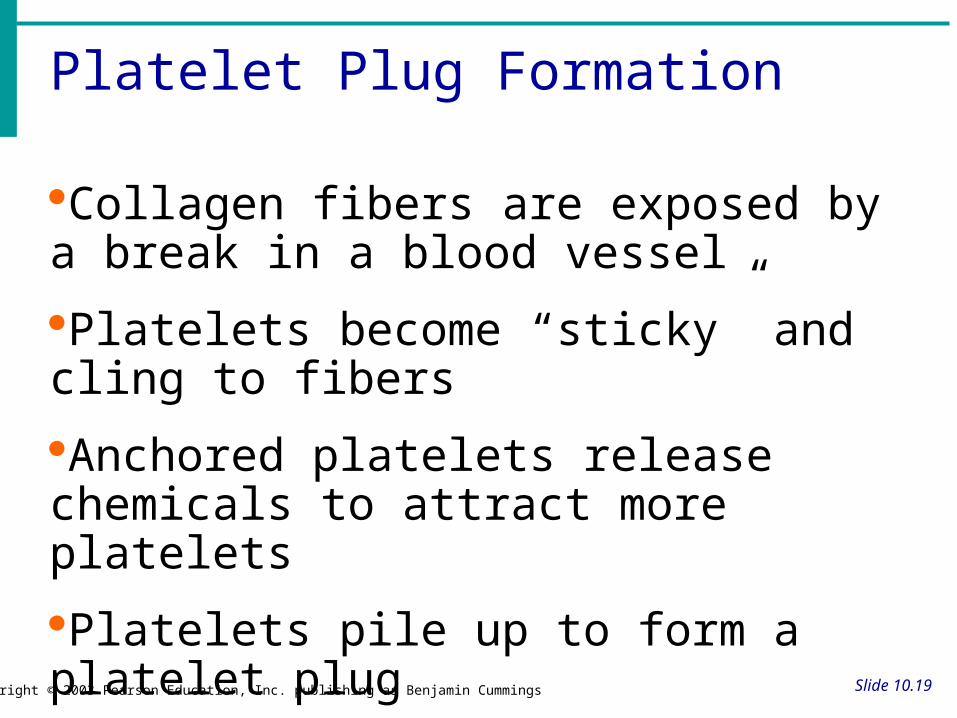
Platelet Plug Formation
Slide 10.19Copyright © 2003 Pearson Education, Inc. publishing as Benjamin Cummings
Collagen fibers are exposed by a break in a blood vessel
Platelets become “sticky” and cling to fibers
Anchored platelets release chemicals to attract more platelets
Platelets pile up to form a platelet plug

pump (peak pressure)_________________fill (minimum pressure)
Cardiac CycleHow is this reflected in blood pressure measurements?
chambers fill
ventriclesfill
ventriclespump
systolic________diastolic
110________80

37
Cardiac Cycle
• Heart is two pumps that work together, right and left half
• Repetitive contraction (systole) and relaxation (diastole) of heart chambers
• Blood moves through circulatory system from areas of higher to lower pressure.– Contraction of heart produces the pressure

Cardiac Cycle


