23
CHAPTER 10 Gaseous Exchange Animation 10 : Gaseous Exchange Source & Credit: Wikispaces
CHAPTER
10 Gaseous Exchange
Animation 10 : Gaseous ExchangeSource & Credit: Wikispaces
2
10. Gaseous Exchange eLearn.Punjab
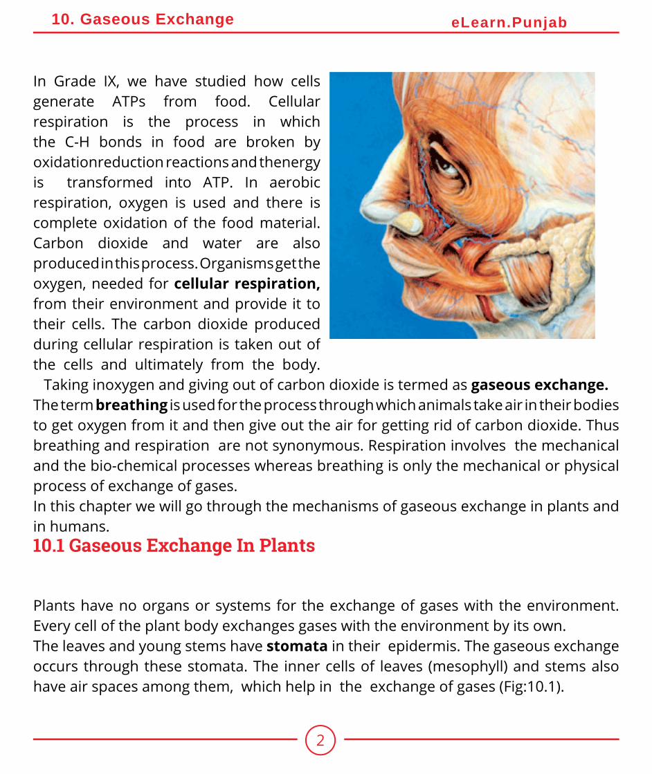
In Grade IX, we have studied how cells generate ATPs from food. Cellular respiration is the process in which the C-H bonds in food are broken by oxidationreduction reactions and thenergy is transformed into ATP. In aerobic respiration, oxygen is used and there is complete oxidation of the food material. Carbon dioxide and water are also produced in this process. Organisms get the oxygen, needed for cellular respiration, from their environment and provide it to their cells. The carbon dioxide produced during cellular respiration is taken out of the cells and ultimately from the body.
Taking inoxygen and giving out of carbon dioxide is termed as gaseous exchange.The term breathing is used for the process through which animals take air in their bodies to get oxygen from it and then give out the air for getting rid of carbon dioxide. Thus breathing and respiration are not synonymous. Respiration involves the mechanical and the bio-chemical processes whereas breathing is only the mechanical or physical process of exchange of gases.In this chapter we will go through the mechanisms of gaseous exchange in plants and in humans.10.1 Gaseous Exchange In Plants
Plants have no organs or systems for the exchange of gases with the environment. Every cell of the plant body exchanges gases with the environment by its own.The leaves and young stems have stomata in their epidermis. The gaseous exchange occurs through these stomata. The inner cells of leaves (mesophyll) and stems also have air spaces among them, which help in the exchange of gases (Fig:10.1).
3
10. Gaseous Exchange eLearn.Punjab
Recalling
Organisms need energy in the form of ATP for their activities.
In young stems and leaves, some gaseous exchange also occurs through the cuticle which is present over their epidermis.
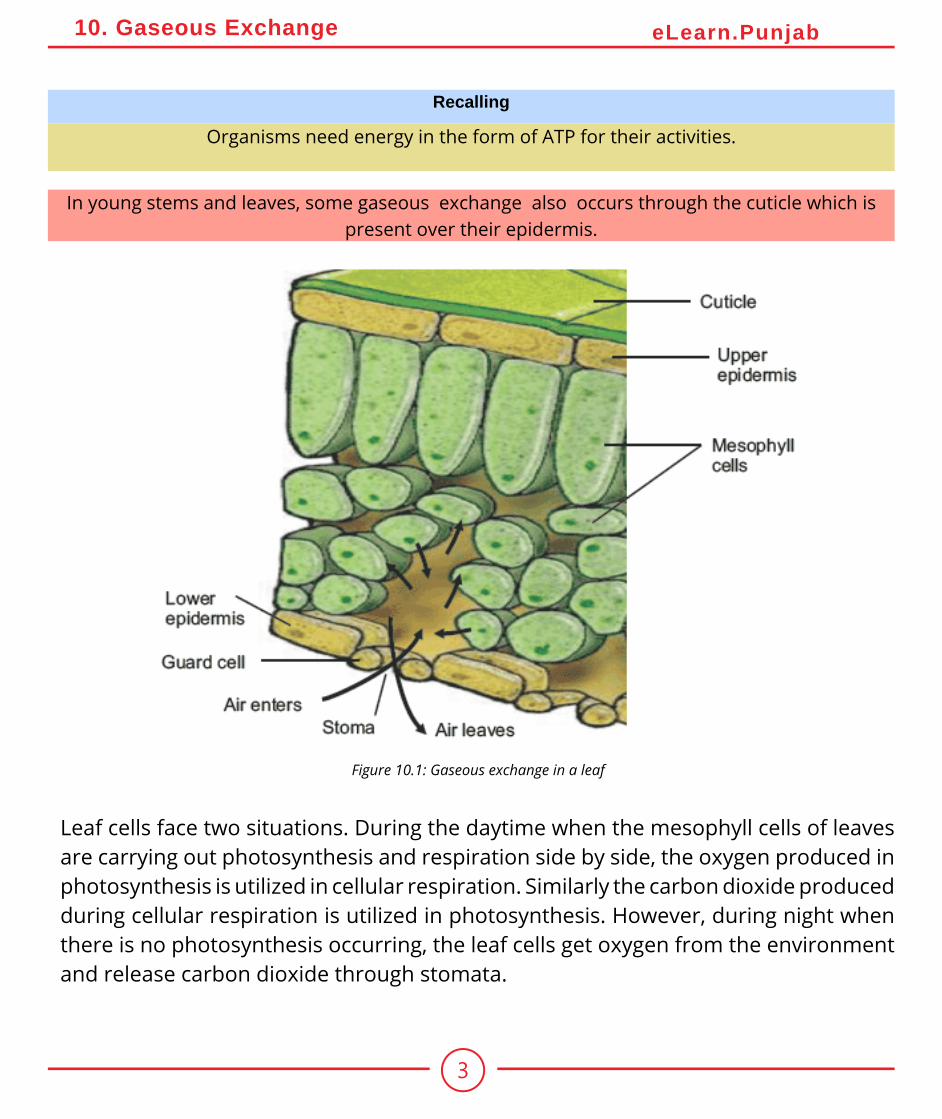
Figure 10.1: Gaseous exchange in a leaf
Leaf cells face two situations. During the daytime when the mesophyll cells of leaves are carrying out photosynthesis and respiration side by side, the oxygen produced in photosynthesis is utilized in cellular respiration. Similarly the carbon dioxide produced during cellular respiration is utilized in photosynthesis. However, during night when there is no photosynthesis occurring, the leaf cells get oxygen from the environment and release carbon dioxide through stomata.
4
10. Gaseous Exchange eLearn.Punjab
Analyzing and InterpretingDraw diagram of stomata of a leaf indicating the movement of gases.
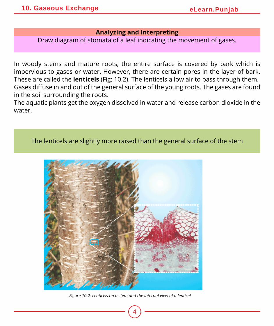
In woody stems and mature roots, the entire surface is covered by bark which is impervious to gases or water. However, there are certain pores in the layer of bark. These are called the lenticels (Fig: 10.2). The lenticels allow air to pass through them.Gases diffuse in and out of the general surface of the young roots. The gases are found in the soil surrounding the roots.The aquatic plants get the oxygen dissolved in water and release carbon dioxide in the water.
The lenticels are slightly more raised than the general surface of the stem
Figure 10.2: Lenticels on a stem and the internal view of a lenticel
5
10. Gaseous Exchange eLearn.Punjab
Practical Work:Investigate the effect of light on the net gaseous exchange from leaf Stomata are the microscopic pores in the epidermis of leaves. They are the passageways for gases and water vapours. Opening and closing of stomata controls the gaseous exchange.Problem: What is the net gaseous exchange from leaves during day and night times?Apparatus required: Petri dish, water, glass slides and cover slips, methylene blue, light microscope
10.2 Gaseous Exchange In Humans
In humans and other higher animals the exchange of gases is carried out by the respiratory system. We can divide the respiratory system in two parts i.e. the air passageway and the lungs.
Background information:• A stoma is an opening through which leaves exchange gases.• The cells of leaves carry out photosynthesis during daytime only.• The cells of leaves carry out respiration all the times.Procedure:1. Take a thick leaf and peel off a thin layer (epidermis) from its surface.2. Place the thin layer in water in a Petri dish.3. Cut a piece of the peeled off epidermis and place it in a drop of water on a glass slide.4. Pour a drop of methylene blue and place a cover slip on the material.5. Observe under the low and high powers of the microscope.6. Perform the same steps by taking the epidermis of leaf at night time.Observation: Observe both epidermis (upper and lower) and point out the stomata. Count the number of open stomata in both and compare their numbers. Draw your observation on the notebook.Evaluation:1. How many stomata did you observe?2. What is the structure of guard cells and how does it helps in the opening and closing of stomata?
6
10. Gaseous Exchange eLearn.Punjab
10.2.1 The Air Passageway
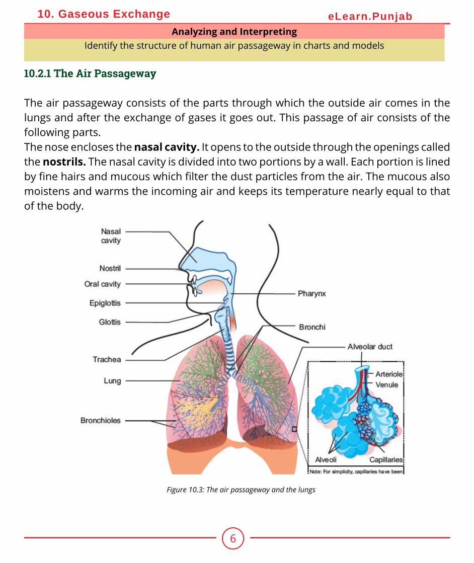
The air passageway consists of the parts through which the outside air comes in the lungs and after the exchange of gases it goes out. This passage of air consists of the following parts.The nose encloses the nasal cavity. It opens to the outside through the openings called the nostrils. The nasal cavity is divided into two portions by a wall. Each portion is lined by fine hairs and mucous which filter the dust particles from the air. The mucous also moistens and warms the incoming air and keeps its temperature nearly equal to that of the body.
Analyzing and InterpretingIdentify the structure of human air passageway in charts and models
Figure 10.3: The air passageway and the lungs
7
10. Gaseous Exchange eLearn.Punjab
The nasal cavity opens into the pharynx by means of two small openings called internal nostrils. Pharynx is a muscular passage and is common to both food and air.It extends to the opening of the oesophagus and the larynx. The air goes from the pharynx into the larynx. We know that glottis is a narrow opening at the floor of pharynx which leads into larynx.The larynx is a box, made of cartilage. It is present between pharynx and trachea. It is also called the voice box. Two pairs of fibrous bands called vocal cords are stretched across the larynx. The vocal cords vibrate when the air passes through them. This vibration produces sounds.Larynx continues to the trachea, which is also called the windpipe. It is about 12 cm long tube which lies in front of the oesophagus. There are C-shaped cartilagenous rings in the wall of trachea. The cartilages keep the trachea from collapsing even when there is no air in it.
RecallingThe glottis is guarded by a flap of tissue called the epiglottis.
8
10. Gaseous Exchange eLearn.Punjab
On entering the chest cavity, the trachea divides into two smaller tubes called bronchi (Singular: bronchus). The bronchi also have cartilagenous plates in their walls. Each bronchus enters into the lung of its side and then divides into smaller branches.The bronchi continue dividing in the lungs until they make several fine tubes called bronchioles. The bronchioles progressively lose the cartilages as they become narrower. The bronchioles end as fine tubules called the alveolar ducts. Each alveolar duct opens into a cluster of pouches called alveoli. The alveoli form the respiratory surface in human body. Each alveolus is a sac-like structure lined by a single layer of epithelial cells. It is bound on the outside by a network of capillaries (Fig: 10.3).The pulmonary artery from the heart containing deoxygenated blood enters the lungs and branches into arterioles and then into capillaries which surround the alveoli. These then join together to form the venules which form pulmonary vein. The pulmonary vein carries the oxygenated blood back to the heart.
The vibrations in vocal cords and the movements of lips, cheeks, tongue and jaws produce specific sounds which result in speech. Speech is an ability that only humans are gifted with and
this is one of the characteristics which has put human beings superior to all.
The trachea and the bronchi are also lined with ciliated and glandular cells. The glandular cells secrete mucus which moistens the air and also traps any fine particles of dust or bacteria that have escaped from the nasal cavity. The cilia beat with an upward motion so that the foreign
particles along the mucus are sent to the oral cavity from where it may be either swallowed or coughed out.
10.2.2 The Lungs
All the alveoli on one side constitute a lung. There is a pair of lungs in the thoracic cavity. The chest wall is made up of 12 pairs of ribs and the rib muscles called intercoastal muscles. A thick muscular structure, called diaphragm, is present below the lungs.
9
10. Gaseous Exchange eLearn.Punjab
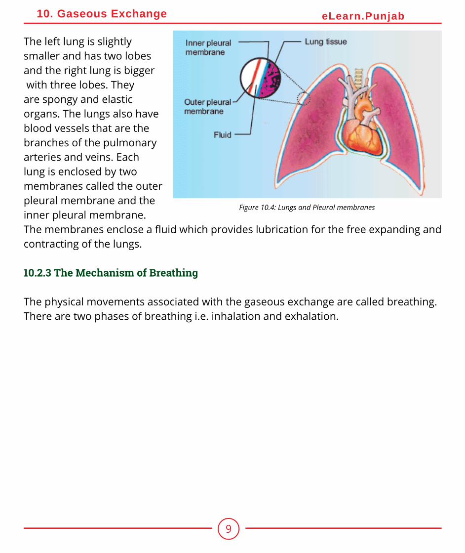
The left lung is slightly smaller and has two lobes and the right lung is bigger with three lobes. They are spongy and elastic organs. The lungs also have blood vessels that are the branches of the pulmonary arteries and veins. Each lung is enclosed by two membranes called the outer pleural membrane and the inner pleural membrane. The membranes enclose a fluid which provides lubrication for the free expanding and contracting of the lungs.
10.2.3 The Mechanism of Breathing
The physical movements associated with the gaseous exchange are called breathing. There are two phases of breathing i.e. inhalation and exhalation.
Figure 10.4: Lungs and Pleural membranes
10
10. Gaseous Exchange eLearn.Punjab
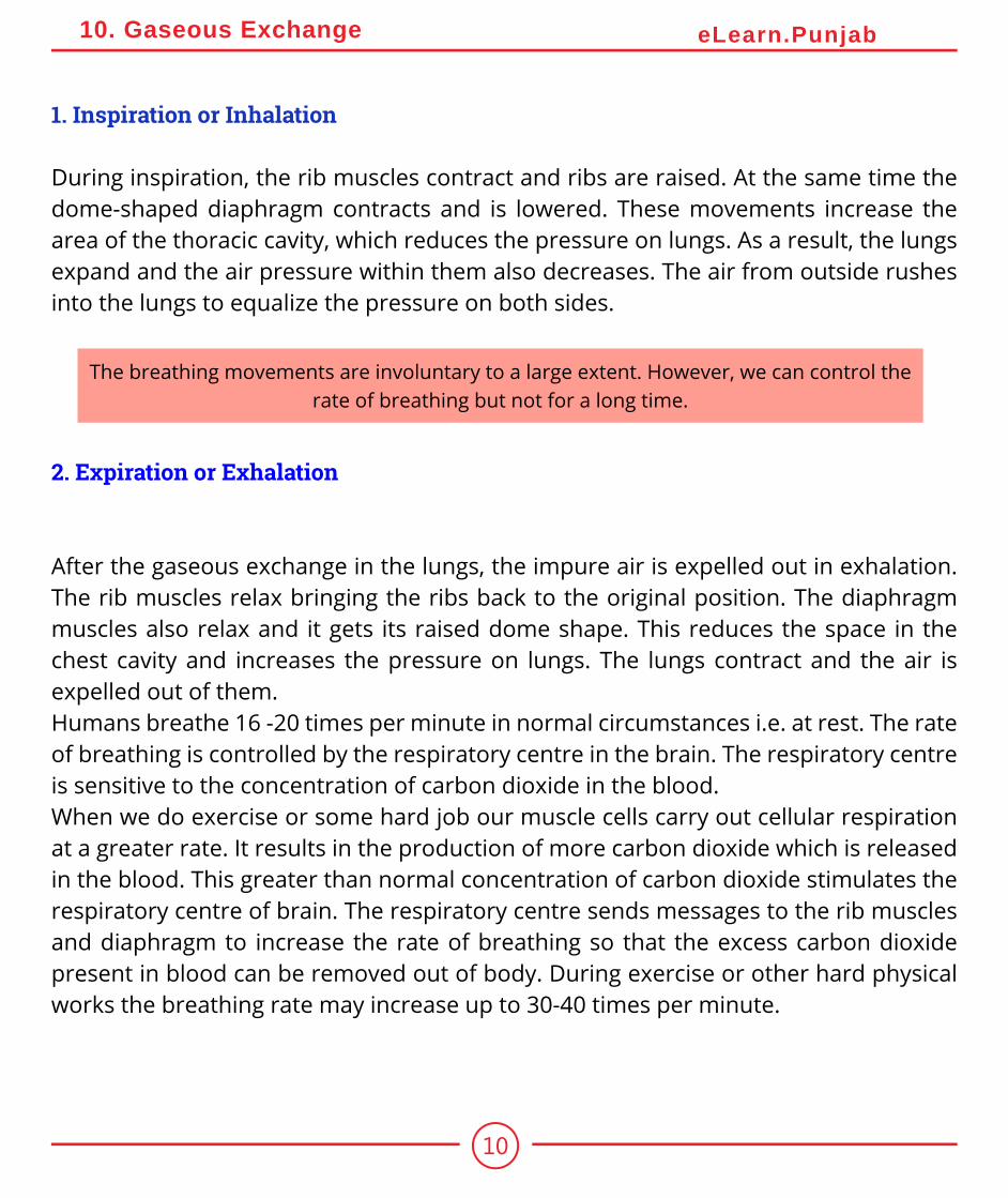
1. Inspiration or Inhalation
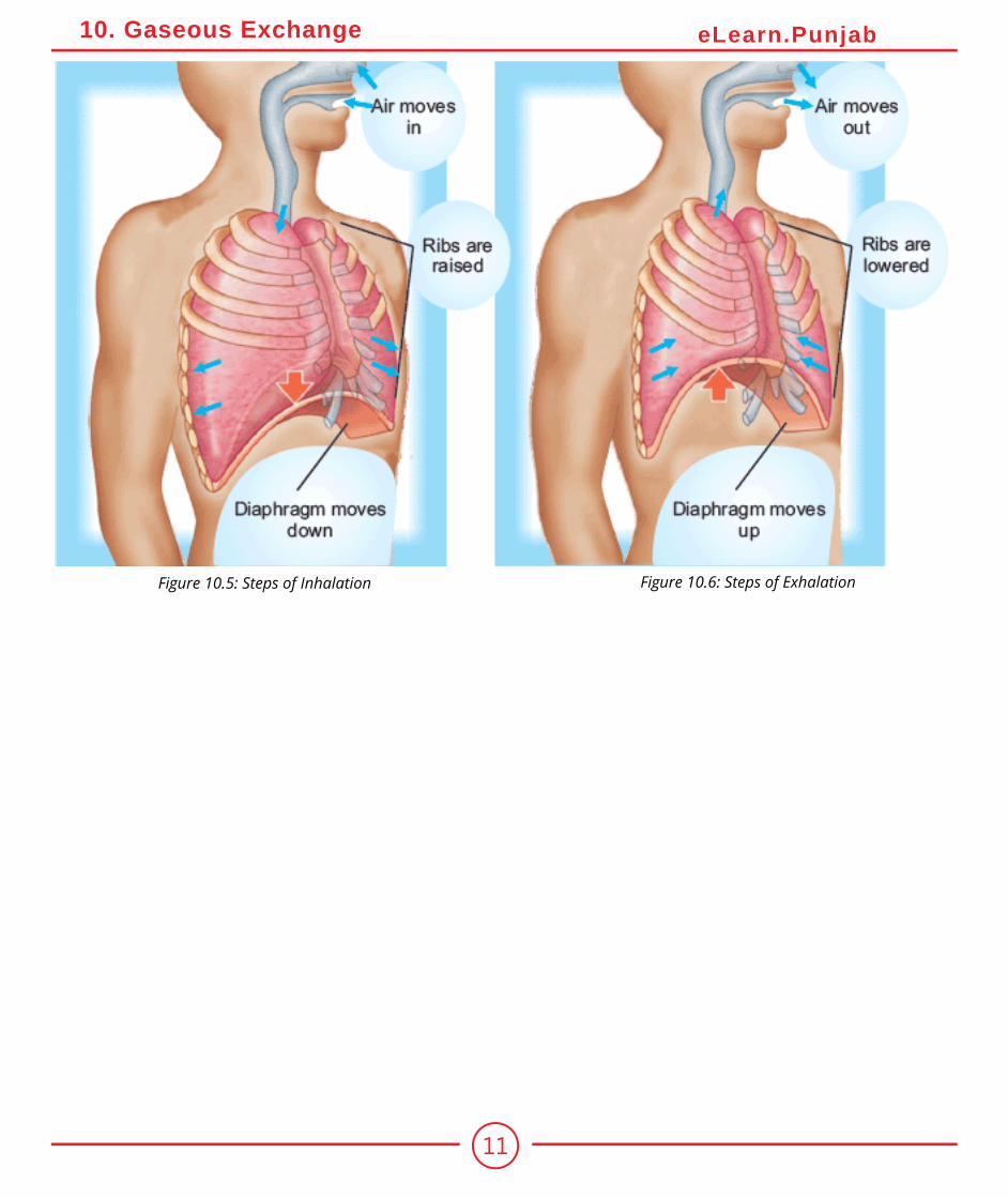
During inspiration, the rib muscles contract and ribs are raised. At the same time the dome-shaped diaphragm contracts and is lowered. These movements increase the area of the thoracic cavity, which reduces the pressure on lungs. As a result, the lungs expand and the air pressure within them also decreases. The air from outside rushes into the lungs to equalize the pressure on both sides.
The breathing movements are involuntary to a large extent. However, we can control the rate of breathing but not for a long time.
2. Expiration or Exhalation
After the gaseous exchange in the lungs, the impure air is expelled out in exhalation. The rib muscles relax bringing the ribs back to the original position. The diaphragm muscles also relax and it gets its raised dome shape. This reduces the space in the chest cavity and increases the pressure on lungs. The lungs contract and the air is expelled out of them.Humans breathe 16 -20 times per minute in normal circumstances i.e. at rest. The rate of breathing is controlled by the respiratory centre in the brain. The respiratory centre is sensitive to the concentration of carbon dioxide in the blood.When we do exercise or some hard job our muscle cells carry out cellular respiration at a greater rate. It results in the production of more carbon dioxide which is released in the blood. This greater than normal concentration of carbon dioxide stimulates the respiratory centre of brain. The respiratory centre sends messages to the rib muscles and diaphragm to increase the rate of breathing so that the excess carbon dioxide present in blood can be removed out of body. During exercise or other hard physical works the breathing rate may increase up to 30-40 times per minute.
11
10. Gaseous Exchange eLearn.Punjab
Figure 10.5: Steps of Inhalation Figure 10.6: Steps of Exhalation
12
10. Gaseous Exchange eLearn.Punjab
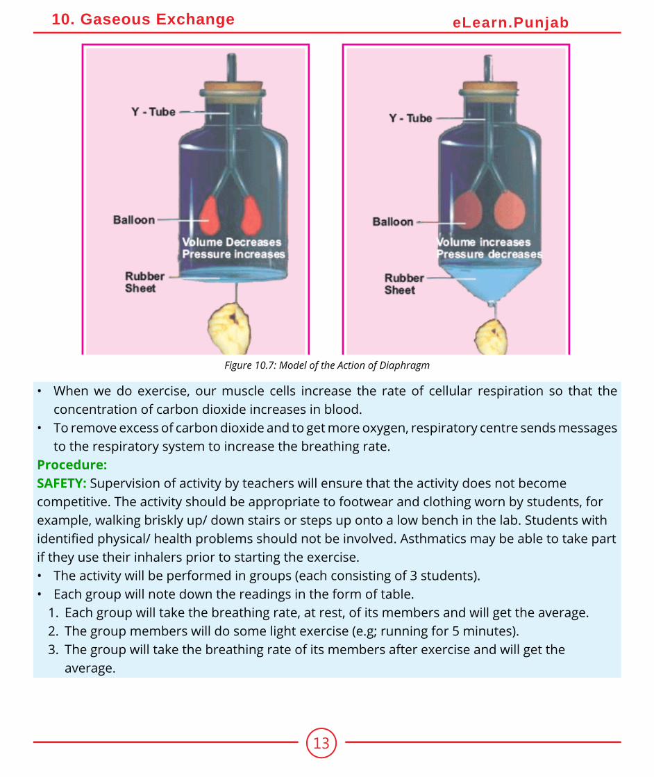
A model to show the action of diaphragmApparatus: a bell jar, ‘Y’ shaped glass tube, two balloons, rubber sheetProcedure:• Take a bell jar. Fix a ‘Y’ shaped glass tube towards its rounded end, as shown in the figure. Tie
a balloon on the open ends of the two branches of glass tube.• Tie a thin rubber sheet on the open end of the jar. The cavity of the bell jar acts as the thoracic
cavity, the “Y” shaped tube as the trachea that branches into bronchi. The rubber sheet acts as the diaphragm and the balloon act as the lungs.
• To demonstrate inspiration, pull the rubber sheet down. The balloons get inflated. This shows how the lungs are filled with air when the diaphragm moves down.
• To demonstrate expiration, the rubber sheet is allowed to go back to its original position. The balloons get deflated. This shows how the lungs are deflated when the diaphragm comes back to its original position.
Practical Work:Investigate the breathing rate at rest and after exerciseThe activity involves students exerting themselves in light exercise and monitoring their breathing rate for a period afterwards.Problem: What is the effect of exercise on the breathing rate?Apparatus required: Stopwatch or wristwatchBackground information:• The autonomic nervous system is specialized for controlling our automatic responses, for
example breathing rate, heart rate and digestion. These are the processes that we do without conscious thought.
• The respiratory centre in the brain is sensitive for the blood carbon dioxide concentration.
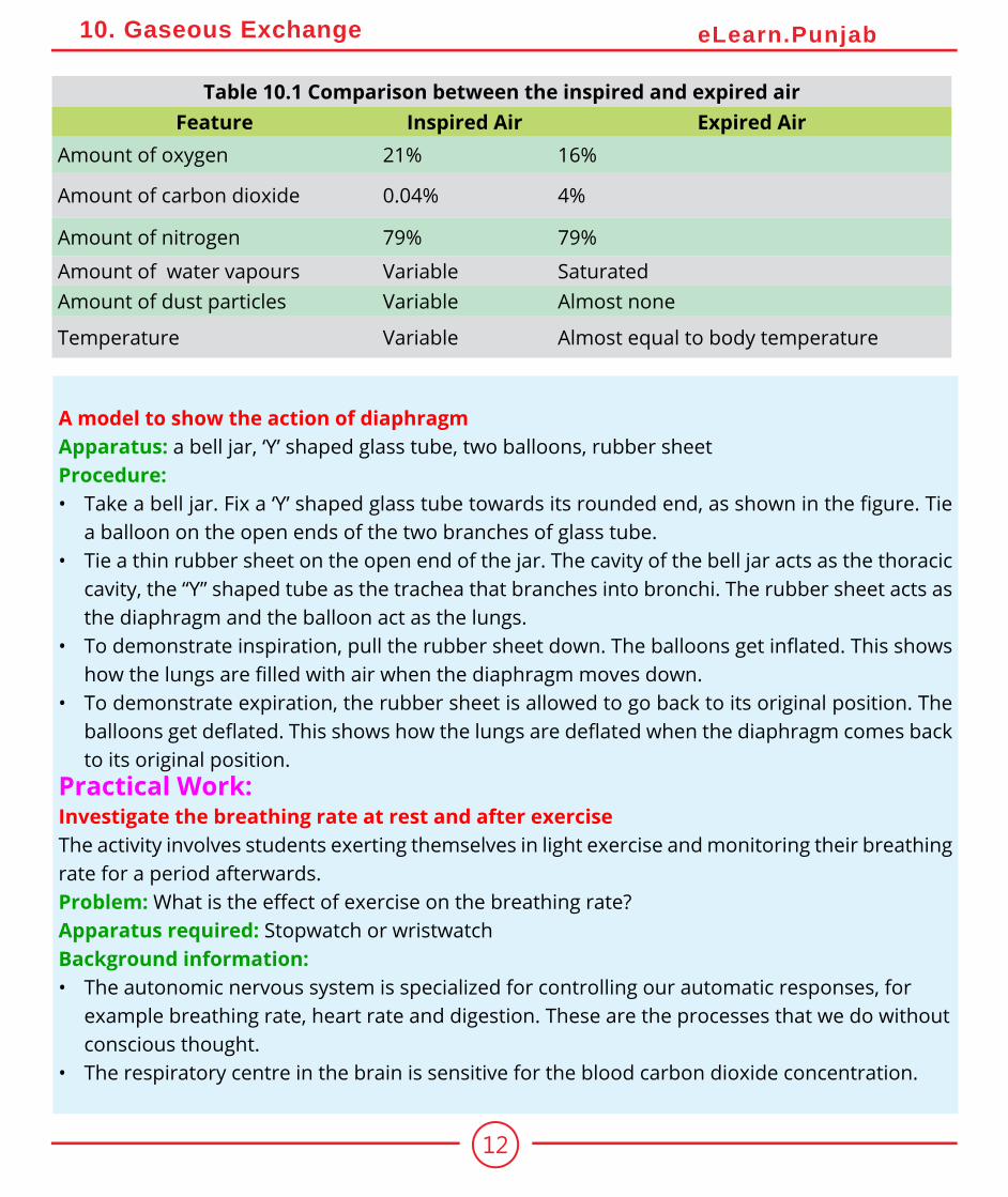
Table 10.1 Comparison between the inspired and expired airFeature Inspired Air Expired Air
Amount of oxygen 21% 16%
Amount of carbon dioxide 0.04% 4%
Amount of nitrogen 79% 79%
Amount of water vapours Variable SaturatedAmount of dust particles Variable Almost none
Temperature Variable Almost equal to body temperature
13
10. Gaseous Exchange eLearn.Punjab
• When we do exercise, our muscle cells increase the rate of cellular respiration so that the concentration of carbon dioxide increases in blood.
• To remove excess of carbon dioxide and to get more oxygen, respiratory centre sends messages to the respiratory system to increase the breathing rate.
Procedure:SAFETY: Supervision of activity by teachers will ensure that the activity does not become competitive. The activity should be appropriate to footwear and clothing worn by students, for example, walking briskly up/ down stairs or steps up onto a low bench in the lab. Students with identified physical/ health problems should not be involved. Asthmatics may be able to take part if they use their inhalers prior to starting the exercise.• The activity will be performed in groups (each consisting of 3 students).• Each group will note down the readings in the form of table.
1. Each group will take the breathing rate, at rest, of its members and will get the average.2. The group members will do some light exercise (e.g; running for 5 minutes).3. The group will take the breathing rate of its members after exercise and will get the
average.
Figure 10.7: Model of the Action of Diaphragm
14
10. Gaseous Exchange eLearn.Punjab
4. The members will do more hard exercise (running for 10 minutes).5. The group will take the breathing rate of its members after hard exercise and will get the
average.Evaluation:• What was the average breathing rate at rest?• What was the average breathing rate after light exercise?• After which exercise, the breathing rate showed more increase?• Why did the breathing rate increase during exercise?
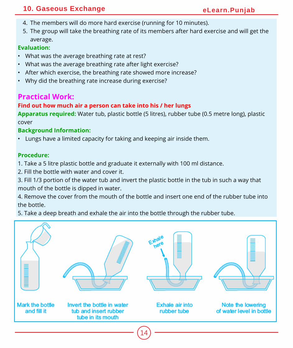
Practical Work:Find out how much air a person can take into his / her lungsApparatus required: Water tub, plastic bottle (5 litres), rubber tube (0.5 metre long), plastic coverBackground Information:• Lungs have a limited capacity for taking and keeping air inside them.
Procedure:1. Take a 5 litre plastic bottle and graduate it externally with 100 ml distance.2. Fill the bottle with water and cover it.3. Fill 1/3 portion of the water tub and invert the plastic bottle in the tub in such a way that mouth of the bottle is dipped in water.4. Remove the cover from the mouth of the bottle and insert one end of the rubber tube into the bottle.5. Take a deep breath and exhale the air into the bottle through the rubber tube.
15
10. Gaseous Exchange eLearn.Punjab
Observation:• Note the lowering of the water level in the bottle.Result:• The water level lowers when the exhaled air goes in the bottle. The volume of water that leaves
the bottle is equal to the volume of the air exhaled from the lungs.Evaluation:• What does the lowering of water level in the bottle indicate?
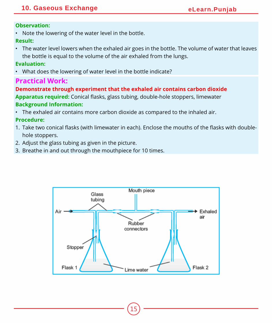
Practical Work:Demonstrate through experiment that the exhaled air contains carbon dioxideApparatus required: Conical flasks, glass tubing, double-hole stoppers, limewaterBackground Information:• The exhaled air contains more carbon dioxide as compared to the inhaled air.Procedure:1. Take two conical flasks (with limewater in each). Enclose the mouths of the flasks with double-
hole stoppers.2. Adjust the glass tubing as given in the picture.3. Breathe in and out through the mouthpiece for 10 times.
16
10. Gaseous Exchange eLearn.Punjab
10.3 Respiratory Disorders
There are a number of respiratory disorders which affect people. The percentage of such disorders is particularly high in Pakistan. It is due to the more concentration of air pollutants not only in the urban but also in the rural atmosphere. Some of the important respiratory disorders are described next.1. BronchitisBronchitis is the inflammation of the bronchi or bronchioles. It results in excessive secretions of mucus into the tubes, leading to the swelling of tubular walls and narrowing of tubes (Fig. 10.8). It is caused by viruses, bacteria or exposure to chemical irritants (e.g. tobacco smoke).
There are two major types of bronchitis i.e. acute and chronic. The acute bronchitis usually lasts about two weeks and patients recover with no permanent damage to the bronchi or bronchioles. In chronic bronchitis, the bronchi develop chronic inflammation. It usually lasts for three months to two years. Symptoms of bronchitis include a cough, mild wheezing, fever, chills and shortness of breath (especially when doing hard job).
Observation:• Observe the colour of the limewater after a few minutes.• Look for differences in the cloudiness of the limewater in the two flasks.Results:• Conclude why the limewater in flask 2 turned more cloudy than in flask 1.
17
10. Gaseous Exchange eLearn.Punjab
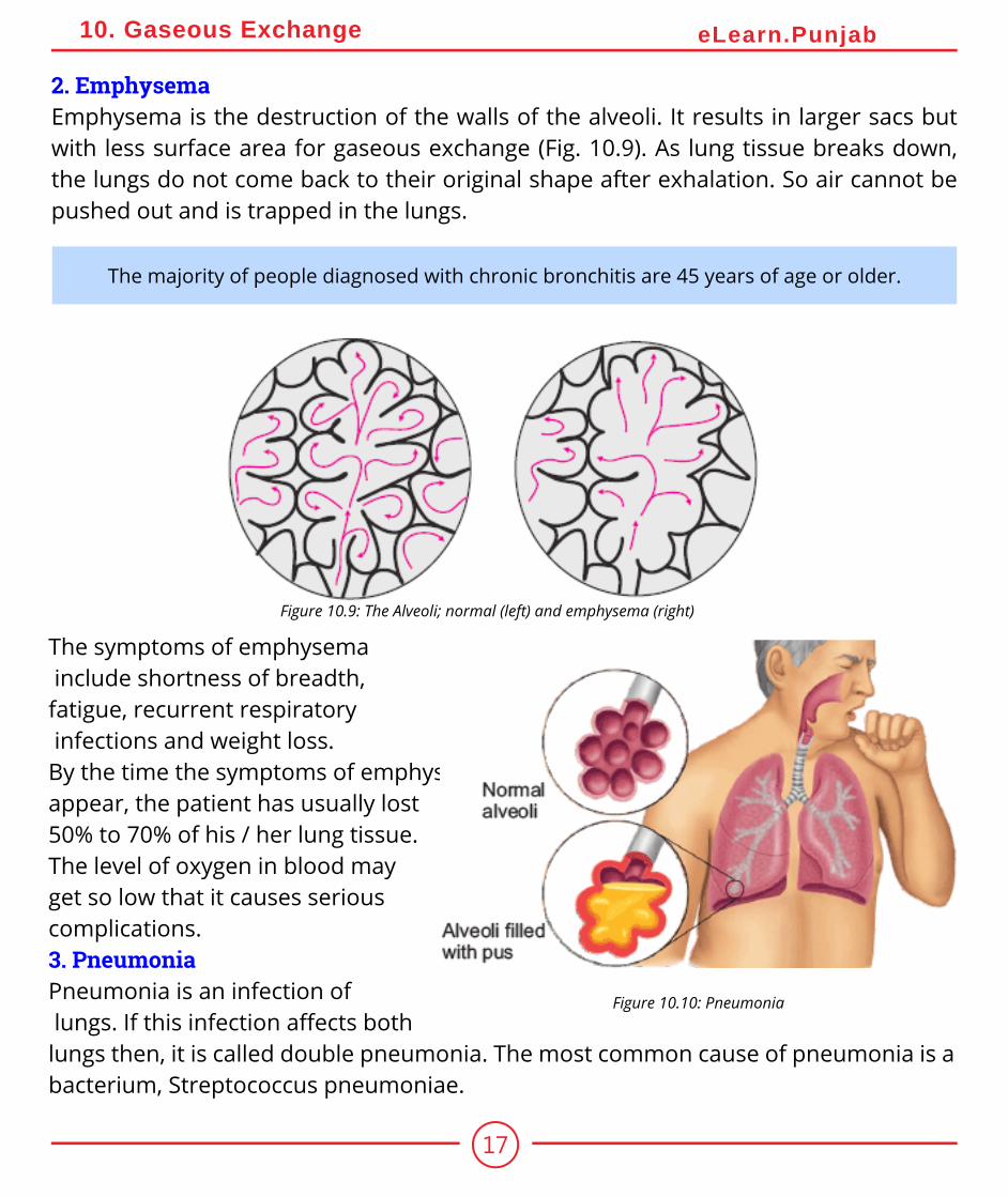
2. EmphysemaEmphysema is the destruction of the walls of the alveoli. It results in larger sacs but with less surface area for gaseous exchange (Fig. 10.9). As lung tissue breaks down, the lungs do not come back to their original shape after exhalation. So air cannot be pushed out and is trapped in the lungs.
Figure 10.9: The Alveoli; normal (left) and emphysema (right)
The symptoms of emphysema include shortness of breadth, fatigue, recurrent respiratory infections and weight loss. By the time the symptoms of emphysema appear, the patient has usually lost 50% to 70% of his / her lung tissue. The level of oxygen in blood may get so low that it causes serious complications.3. PneumoniaPneumonia is an infection of lungs. If this infection affects both lungs then, it is called double pneumonia. The most common cause of pneumonia is a bacterium, Streptococcus pneumoniae.
Figure 10.10: Pneumonia
The majority of people diagnosed with chronic bronchitis are 45 years of age or older.
18
10. Gaseous Exchange eLearn.Punjab
Some viral (influenza virus) and fungal infections may also lead to pneumonia. When the causative organisms enter the alveoli, they settle there and grow in number. They break the lung tissues and the area becomes filled with fluid and pus. The symptoms of pneumonia include a cold that is followed by a high fever, shivering, and a cough with sputum production. Patient may become short of breath. The patient’s skin colour may change and become dusky or purplish. It is due to poor oxygenation of blood.Vaccines are available to prevent pneumonia caused by S. pneumoniae. Antibiotics are used in the treatment of this type of pneumonia.
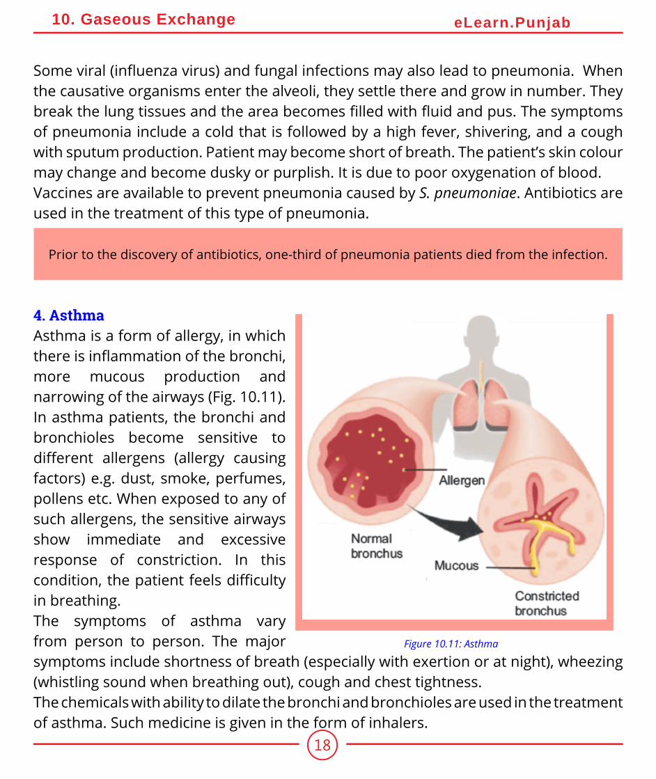
4. AsthmaAsthma is a form of allergy, in which there is inflammation of the bronchi, more mucous production and narrowing of the airways (Fig. 10.11). In asthma patients, the bronchi and bronchioles become sensitive to different allergens (allergy causing factors) e.g. dust, smoke, perfumes, pollens etc. When exposed to any of such allergens, the sensitive airways show immediate and excessive response of constriction. In this condition, the patient feels difficulty in breathing.The symptoms of asthma vary from person to person. The major symptoms include shortness of breath (especially with exertion or at night), wheezing (whistling sound when breathing out), cough and chest tightness.The chemicals with ability to dilate the bronchi and bronchioles are used in the treatment of asthma. Such medicine is given in the form of inhalers.
Figure 10.11: Asthma
Prior to the discovery of antibiotics, one-third of pneumonia patients died from the infection.
19
10. Gaseous Exchange eLearn.Punjab
5. Lung Cancer
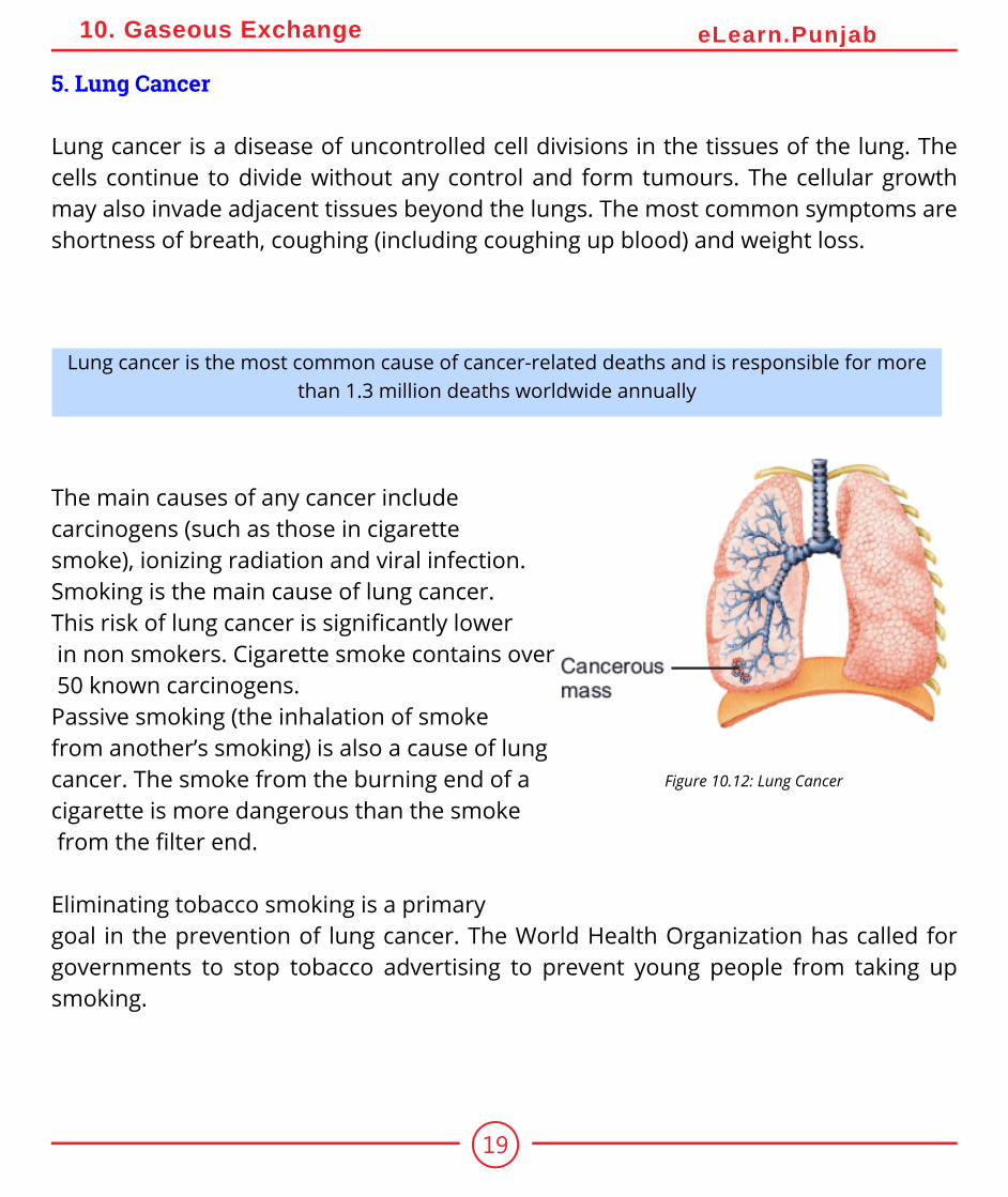
Lung cancer is a disease of uncontrolled cell divisions in the tissues of the lung. The cells continue to divide without any control and form tumours. The cellular growth may also invade adjacent tissues beyond the lungs. The most common symptoms are shortness of breath, coughing (including coughing up blood) and weight loss.
Lung cancer is the most common cause of cancer-related deaths and is responsible for more than 1.3 million deaths worldwide annually
The main causes of any cancer include carcinogens (such as those in cigarette smoke), ionizing radiation and viral infection.Smoking is the main cause of lung cancer. This risk of lung cancer is significantly lower in non smokers. Cigarette smoke contains over 50 known carcinogens.Passive smoking (the inhalation of smoke from another’s smoking) is also a cause of lung cancer. The smoke from the burning end of a cigarette is more dangerous than the smoke from the filter end.
Eliminating tobacco smoking is a primary goal in the prevention of lung cancer. The World Health Organization has called for governments to stop tobacco advertising to prevent young people from taking up smoking.
Figure 10.12: Lung Cancer
20
10. Gaseous Exchange eLearn.Punjab
10.3.1 Bad Effects of Smoking
Smoking is harmful due to the chemicals in cigarettes and smoke. Tobacco smoke contains over 4,000 different chemicals, out of which at least 50 are carcinogens and many are poisonous.Many people think that lung cancer is the only smoking-related disease and it is the number one cause of death among smokers. But it is not right. Cigarette smoke affects the body from head to toe. Smokers have a much higher risk of developing a number of life threatening diseases.
Smoking may also lead to the cancers in kidneys, oral cavity, larynx, breast, bladder and pancreas etc. Many chemicals in tobacco smoke damage the air passageway, which leads to emphysema and other respiratory disorders.
The World No Tobacco Day is celebrated on the 31st of May every year
Nicotine is a powerful poison and was widely used as an insecticide in the past. When inhaled through tobacco smoking, it reaches our circulatory system and not only hardens the walls of
the arteries but also damages the brain tissues.
If a person stops smoking, the chance to develop cancer decreases as damage to the lungs is repaired and contaminant particles are gradually removed.
According to the WHO, the rates of smoking have declined in the developed world. In the developing world, however, it is rising by 3.4% per year as of 2002.
21
10. Gaseous Exchange eLearn.Punjab
Non-smokers who are exposed to second-hand smoke (passive smoke) at home or work increase their heart disease risk by 25-30% and their lung cancer risk by 20-30%
Smoking also affects the social life of a person. Smokers may face social un-acceptance because other people may not want to be exposed to other’s smoke.
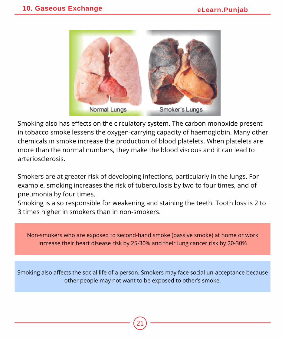
Smoking also has effects on the circulatory system. The carbon monoxide present in tobacco smoke lessens the oxygen-carrying capacity of haemoglobin. Many other chemicals in smoke increase the production of blood platelets. When platelets are more than the normal numbers, they make the blood viscous and it can lead to arteriosclerosis.
Smokers are at greater risk of developing infections, particularly in the lungs. For example, smoking increases the risk of tuberculosis by two to four times, and of pneumonia by four times.Smoking is also responsible for weakening and staining the teeth. Tooth loss is 2 to 3 times higher in smokers than in non-smokers.
22
10. Gaseous Exchange eLearn.Punjab
UNDERSTANDING THE CONCEPT1. How do the different parts of the plant body exchange gases with the environment?2. Write down the steps of inhalation and exhalation.3. State the signs and symptoms, causes and treatments of bronchitis, emphysema and
pneumonia.4. How does the tobacco smoke damage the respiratory system?
SHORT QUESTIONS1. Differentiate between breathing and cellular respiration.2. Trace the path of air from the nasal cavity to the alveoli.3. How will you differentiate between a stoma and a lenticel?
THE TERMS TO KNOW
Alveolar ductAlveolusAsthma
BreathingBronchioles
BronchusBronchitis
DiaphragmEmphysemaExhalation
Gaseous exchangeInhalation
LarynxLenticels
Nasal cavityNostril
PneumoniaTrachea
Vocal cords
ACTIVITIES1. Investigate the effect of light on the net gaseous exchange from leaf, by using bicarbonate as
the indicator.2. Investigate the breathing rate at rest and after exercise.3. Find out how much air a person can take into his lungs.4. Demonstrate through experiment that carbon dioxide is exhaled during respiration.
23
10. Gaseous Exchange eLearn.Punjab
ON-LINE LEARNING
1. en.wikipedia.org/wiki/Respiratory_system2. www.biotopics.co.uk/humans/resyst.html3. www.who.int/respiratory/4. www.tutorvista.com › Science › Science II › Respiration
SCIENCE, TECHNOLOGY AND SOCIETY1. Evaluate the effects of tilling on roots for better exchange of gases with the soil air.2. Outline the concept of Artificial Ventilator for artificial breathing in patients.3. Interpret the dangers of breathing in exhausts of fossil fuels (Petrol and others)4. Rationalize the importance of cross ventilation in homes.5. Assess the adverse effects associated with smoking on health.6. Point out bad social aspects of smoking.


![7[1].3 the concept of gaseous exchange](https://static.documents.pub/doc/80x56/5454c1cdaf795946778b63dd/713-the-concept-of-gaseous-exchange-5584af30d86e0.jpg)
