18
Biochemistry 3100 Lecture 8 Slide 1 Chapter 10: Chapter 10: Hemoglobin Hemoglobin Voet & Voet: Voet & Voet: Pages 320-353 Pages 320-353
Biochemistry 3100Lecture 8 Slide 1
Chapter 10:Chapter 10:HemoglobinHemoglobin
Voet & Voet: Voet & Voet: Pages 320-353Pages 320-353
Biochemistry 3100Lecture 8 Slide 2
Hemoglobin FunctionHemoglobin Function
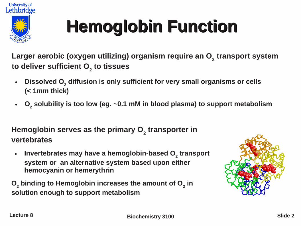
Larger aerobic (oxygen utilizing) organism require an O2 transport system
to deliver sufficient O2 to tissues
● Dissolved O2 diffusion is only sufficient for very small organisms or cells
(< 1mm thick)
● O2 solubility is too low (eg. ~0.1 mM in blood plasma) to support metabolism
Hemoglobin serves as the primary O2 transporter in
vertebrates
● Invertebrates may have a hemoglobin-based O2 transport
system or an alternative system based upon either hemocyanin or hemerythrin
O2 binding to Hemoglobin increases the amount of O
2 in
solution enough to support metabolism
Biochemistry 3100Lecture 8 Slide 3
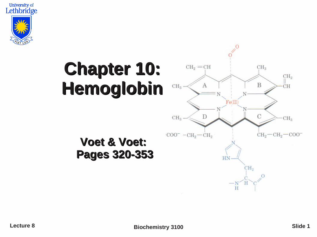
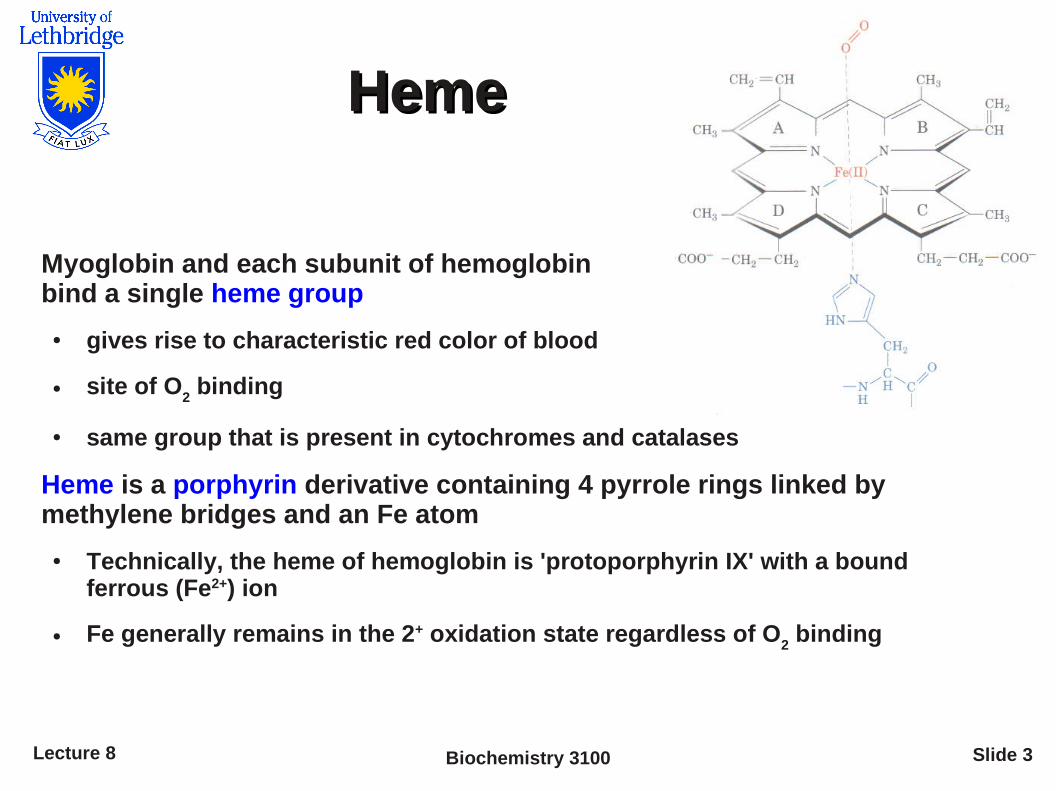
HemeHeme
Myoglobin and each subunit of hemoglobin bind a single heme group
● gives rise to characteristic red color of blood
● site of O2 binding
● same group that is present in cytochromes and catalases
Heme is a porphyrin derivative containing 4 pyrrole rings linked by methylene bridges and an Fe atom
● Technically, the heme of hemoglobin is 'protoporphyrin IX' with a bound ferrous (Fe2+) ion
● Fe generally remains in the 2+ oxidation state regardless of O2 binding

Biochemistry 3100Lecture 8 Slide 4
Quantifying Binding
Reversible binding of a protein (P) and ligand (L) can be described by an equilibrium expression characterized by an equilibrium association constant, K
a(M-1)
● In cells, the [ligand] is typically far larger than [protein]
P + L PL K a=[PL ]
[P ][L ]
Equilibrium can be expressed as the fraction of ligand binding sites occupied by ligand
=binding sites occupied
total binding sites=
[PL ]
[PL ][P ]
Substituting Ka [L][P] for [PL] and rearranging
=K a [L ][P ]
K a [L ][P ][P ]=
K a [L ]
K a [L ]1=
[L ]
[L ]1
K a

Biochemistry 3100Lecture 8 Slide 5
Determining Ka (or K
d)
Plotting θ versus [L] yields a hyperbolic curve
● At θ = 0.5; the equilibrium equation yields [L] = (1/Ka)
θ
0
0.5
1.0
[L]1/Ka
θ can be followed spectroscopically by detecting conformation differences associated with ligand binding
Kd; the dissociation constant is the reciprocal of K
a and is often used in its place
Why? a) the equilibrium equation is (slightly) simplifiedb) units are concentration
=[L ]
[L ]K d

Biochemistry 3100Lecture 8 Slide 6
Oxygen binding to myoglobin
Must modify (slightly) equilibrium binding equation as O2 is a gas
● Partial oxygen pressure (pO2) is easier to measure than dissolve oxygen
concentration ([O2])
● P50
is the concentration at which half the binding sites are filled (Kd)
=[L ]
[L ]K d =
pO2
pO2K d =
pO2
pO2P50
θ
0
0.5
1.0
pO2 (kPa)
P50
1
Partial pressure of O2 is pressure of O
2 above solution
O2 binds tightly to myoglobin with a P
50 of 0.26 kPa
(Note: oxygen is ~21% of the 101.3 kPa gas pressure)

Biochemistry 3100Lecture 8 Slide 7
Hemoglobin
● Myoglobin: Comparatively insensitive to small changes in physiological [O
2]
– Suited to storage
– O2 bound under physiological conditions
● Hemoglobin (Hb) is an α2β
2 oligomer and a
homologue of myoglobin
– It carries almost all oxygen in animals
● Interaction between the Hb subunits modulate its binding affinity allowing it to respond to small changes in physiological [O
2]
– Suited for oxygen transport
– Binds/Releases O2 under physiological conditions
Biochemistry 3100Lecture 8 Slide 8
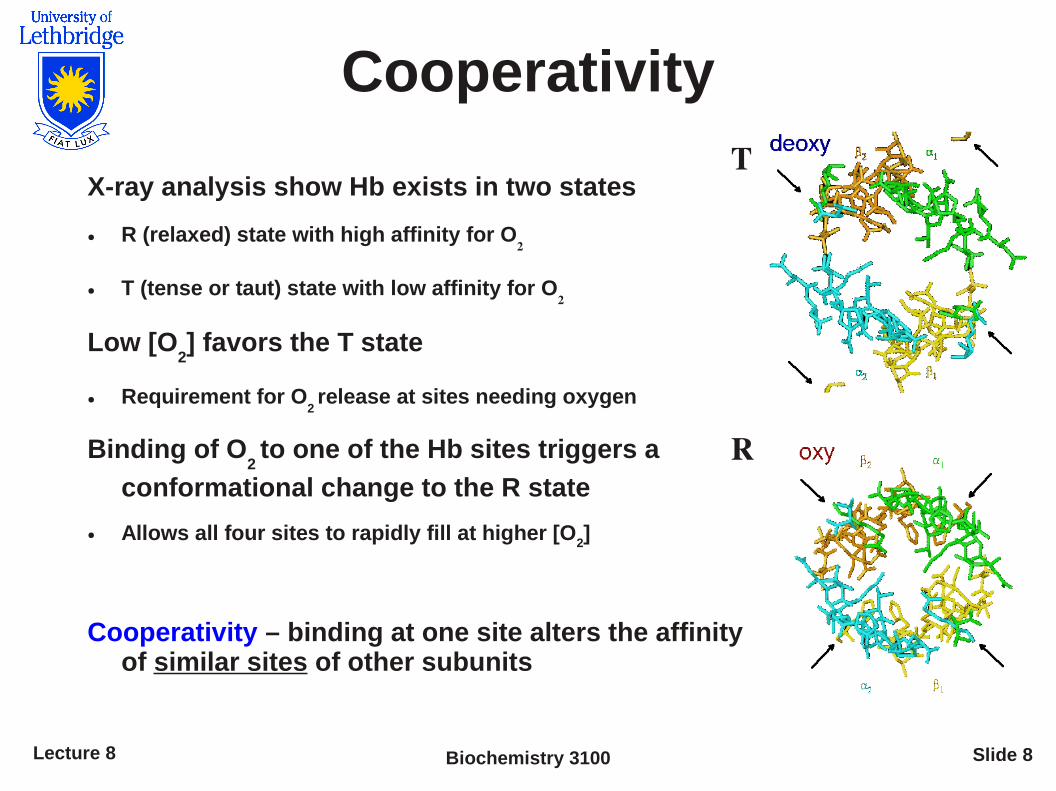
Cooperativity
X-ray analysis show Hb exists in two states
● R (relaxed) state with high affinity for O2
● T (tense or taut) state with low affinity for O2
Low [O2] favors the T state
● Requirement for O2 release at sites needing oxygen
Binding of O2 to one of the Hb sites triggers a
conformational change to the R state
● Allows all four sites to rapidly fill at higher [O2]
Cooperativity – binding at one site alters the affinity of similar sites of other subunits
T
R
Biochemistry 3100Lecture 8 Slide 9
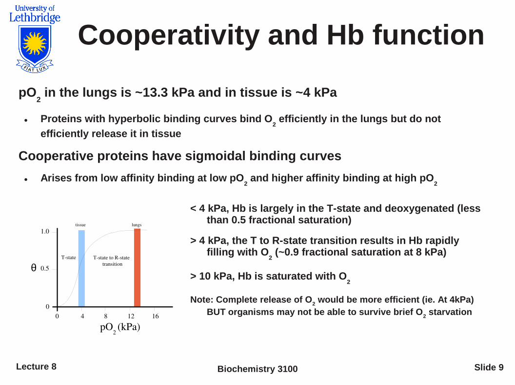
Cooperativity and Hb function
pO2 in the lungs is ~13.3 kPa and in tissue is ~4 kPa
● Proteins with hyperbolic binding curves bind O2 efficiently in the lungs but do not
efficiently release it in tissue
Cooperative proteins have sigmoidal binding curves
● Arises from low affinity binding at low pO2 and higher affinity binding at high pO
2
θ
0
1.0
0.5
0 4 8 12 16
pO2 (kPa)
tissue lungs
Tstate Tstate to Rstate transition
< 4 kPa, Hb is largely in the T-state and deoxygenated (less than 0.5 fractional saturation)
> 4 kPa, the T to R-state transition results in Hb rapidly filling with O
2 (~0.9 fractional saturation at 8 kPa)
> 10 kPa, Hb is saturated with O2
Note: Complete release of O2 would be more efficient (ie. At 4kPa)
BUT organisms may not be able to survive brief O2 starvation

Biochemistry 3100Lecture 8 Slide 10
Allosteric proteins
Allosteric proteins bind ligands at one site, undergo a conformational change and the binding properties of another site on the same protein are altered
– Cooperative proteins (eg Hb) are a special case of allosteric proteins
– All cooperative proteins are allosteric, but not all allosteric proteins are cooperative
Ligands that induce conformational changes in allosteric proteins are referred to as modulators
– Modulators may be inhibitors (induce less-active forms) or activators (induce more-active forms)
● Homotropic modulators are modulators that are identical to the ligand – occur in cooperative proteins (eg. O
2 is a homotropic activator of hemoglobin)
● Heterotropic modulator are different from the normal ligand (eg. 2,3-bisphosphoglycerate is a heterotropic inhibitor of hemoglobin)
Biochemistry 3100Lecture 8 Slide 11
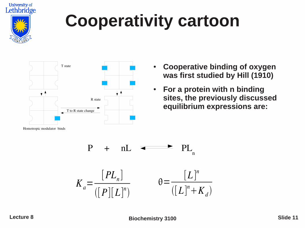
Cooperativity cartoon
T state
Homotropic modulator binds
R state
T to R state change
● Cooperative binding of oxygen was first studied by Hill (1910)
● For a protein with n binding sites, the previously discussed equilibrium expressions are:
P + nL PLn
K a=[PLn ]
[P ][L ]n=
[L ]n
[L ]nK d

Biochemistry 3100Lecture 8 Slide 12
Hill equation
The fractional occupancy equilibrium expression can be rearranged and converted to a linear form by taking the log of each side:
1−=
[L ]n
K d
log
1−=n log [L ]−log K d
Plotting log ( / (1 – )) vs log [L] is called the Hill plot
● The slope of the Hill plot n (Hill coefficient), reflect the degree of interaction between binding sites and is represented as n
H
(Note: nH only equals n if all ligands bind at the same instant)
nH values range from > 0 to the total number of binding sites, n.

Biochemistry 3100Lecture 8 Slide 13
Hill coefficient
nH < 1 negative cooperativity
(very rare)
nH = 1 no cooperativity
(typical)
nH > 1 positive cooperativity
(common)
nH = n complete cooperativity
(never)
log
(θ /
1 θ
)
2
0
0 1
Log pO2
Hb nH = 3
Hb Rstaten
H = 1
Hb Tstaten
H = 1
2 312
0
1
1
2
Note1: We must modify the equation for Hb by substituting pO
2 for [L] and P
50
n for Kd before plotting the data
Note2: Pn
50 occurs at y=0
log
1−=n log pO2−n log P50
n

Biochemistry 3100Lecture 8 Slide 14
Cooperativity models
Two models proposed to explain cooperative binding
(1) Concerted model – all subunits undergo the conformational change simultaneously
(2) Sequential model – subunit undergo the conformational change one at a time
Difficult to distinguish between the models as the concerted model is a special case of the sequential model (not mutually exclusive)
– The sequential model becomes the concerted model if the conformational change is sufficiently fast

Biochemistry 3100Lecture 8 Slide 15
Hb also transports CO2 and H+
Hb transports CO2 from tissues to the lungs and kidneys
Since CO2 is poorly soluble in water, some is hydrated to HCO
3
- by
carbonic anhydrase in a reaction that produces H+
Hb transports 40% of the H+ and ~20% of the CO2 formed in tissues
Binding* of H+ (Bohr effect) and CO2 are inversely related (inhibitory)
to the binding of O2
At low pH and high CO2, the binding of H+ and CO
2 aid in the release of O
2
Conversely in the capillaries of the lungs, CO2 is released and the pH
rises increasing Hb affinity for O2
The Bohr effect (modulation of Hb binding) is an important additional factor explaining why Hb (cooperative) is better suited as an oxygen carrier than Myoglobin
Biochemistry 3100Lecture 8 Slide 16
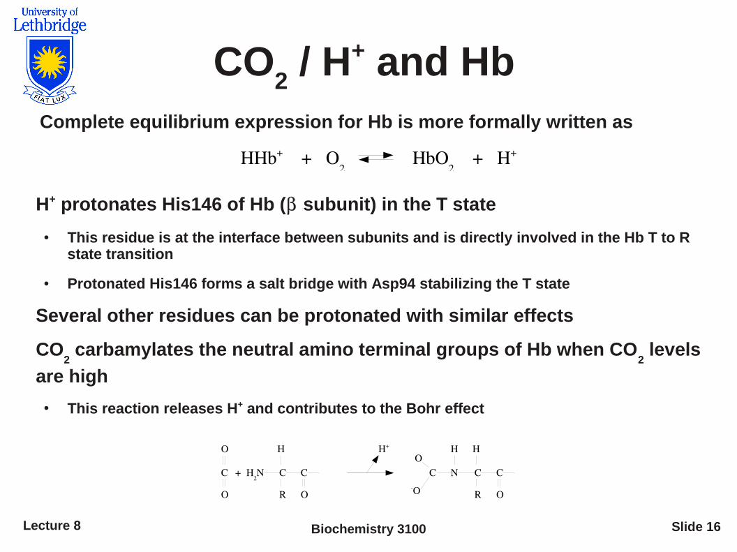
CO2 / H+ and Hb
Complete equilibrium expression for Hb is more formally written as
H+ protonates His146 of Hb ( subunit) in the T state
● This residue is at the interface between subunits and is directly involved in the Hb T to R state transition
● Protonated His146 forms a salt bridge with Asp94 stabilizing the T state
Several other residues can be protonated with similar effects
CO2 carbamylates the neutral amino terminal groups of Hb when CO
2 levels
are high
● This reaction releases H+ and contributes to the Bohr effect
HHb+ + O2
HbO2 + H+
H2N C C
R
H
O
C
O
O
+
H+
N C C
R
H
O
CO
OH

Biochemistry 3100Lecture 8 Slide 17
Heterotrophic modulators
2,3-bisphosphoglycerate (BPG) is a heterotrophic modulator of Hb
– BPG binds at a site distant from the O2 binding site and greatly reduces the
affinity of Hb for O2 under high CO
2 levels
– Leads to increased release of O2 compared to normal conditions
eg At high altitudes, pO2 and the fraction saturation of Hb is lower and less O
2
is released to tissues
– After several hours, BPG binding to Hb promotes release of O2 at low pO
2 and
restores normal levels of oxygen to tissues
Binding Curve showingeffect of low pressure
Binding Curve at low pressure in the presence of BPG
Biochemistry 3100Lecture 8 Slide 18
Summary
Hb reversibly binds O2, H+ and the heterotrophic regulator
BPG
Hb covalently binds CO2
Each of these ligands and CO2 affect Hb binding affinity for
O2 by stabilizing either the T or R states


