+ All Categories
Home > Documents > Chapter 10 The Knee Joint - Home | Kean University

Chapter 10 The Knee Joint - Home | Kean University

Date post: 18-Dec-2021
Category:
Upload: others
View: 2 times
Download: 0 times
Share this document with a friend
9
1 © 2007 McGraw-Hill Higher Education. All rights reserved. 10-1 Chapter 10 The Knee Joint Manual of Structural Kinesiology R.T. Floyd, EdD, ATC, CSCS © 2007 McGraw-Hill Higher Education. All rights reserved. 10-2 The Knee Joint Modified for Prentice WE: Arnheim’s principles of athletic training, ed 12, New York, 2006, McGraw-Hill; from Saladin, KS: Anatomy &physiology: the unity of forms and function, ed 2, New York, 2001, McGraw- Hill. © 2007 McGraw-Hill Higher Education. All rights reserved. 10-3 Bones © 2007 McGraw-Hill Higher Education. All rights reserved. 10-4 Bones Modified from Anthony CP, Kolthoff NJ: Textbook of anatomy and physiology, ed 9, St. Louis, 1975, Mosby. © 2007 McGraw-Hill Higher Education. All rights reserved. 10-5 Bones © 2007 McGraw-Hill Higher Education. All rights reserved. 10-6 Bones Modified from Anthony CP, Kolthoff NJ: Textbook of anatomy and physiology, ed 9, St. Louis, 1975, Mosby.
Transcript

1

© 2007 McGraw-Hill Higher Education. All rights reserved. 10-1

Chapter 10

The Knee Joint

Manual of Structural KinesiologyR.T. Floyd, EdD, ATC, CSCS

© 2007 McGraw-Hill Higher Education. All rights reserved. 10-2

The Knee Joint• Knee joint– largest joint in body– very complex– primarily a hinge jointModified for Prentice WE:

Arnheim’s principles of

athletic training, ed 12, New

York, 2006, McGraw-Hill;

from Saladin, KS: Anatomy

&physiology: the unity of

forms and function, ed 2,

New York, 2001, McGraw-

Hill.

© 2007 McGraw-Hill Higher Education. All rights reserved. 10-3

Bones• Enlarged femoral condyles articulate on enlarged tibial condyles• Medial & lateral tibial condyles (medial & lateral tibial plateaus) - receptacles for femoral condyles• Tibia – medial– bears most of weight© 2007 McGraw-Hill Higher Education. All rights reserved. 10-4

Bones• Fibula - lateral– serves as the attachment for knee joint structures– does not articulate with femur or patella– not part of knee jointModified from Anthony CP, Kolthoff NJ: Textbook of anatomy and

physiology, ed 9, St. Louis, 1975, Mosby.

© 2007 McGraw-Hill Higher Education. All rights reserved. 10-5

Bones• Patella– sesamoid (floating) bone– imbedded in quadriceps & patellar tendon– serves similar to a pulley in improving angle of pull, resulting in greater mechanical advantage in knee extension© 2007 McGraw-Hill Higher Education. All rights reserved. 10-6

Bones• Key bony landmarks– Superior & inferior patellar poles– Tibial tuberosity– Gerdy’s tubercle– Medial & lateral femoral condyles– Upper anterior medial tibialsurface– Head of fibulaModified from Anthony CP, Kolthoff NJ: Textbook of anatomy and

physiology, ed 9, St. Louis, 1975, Mosby.

2

© 2007 McGraw-Hill Higher Education. All rights reserved. 10-7

Bones• Three vasti muscles of quadriceps originate on proximal femur & insert on patellar superior pole– insertion is ultimately on tibial tuberosity via patella tendon• Iliotibial tract of tensor fasciae latae inserts on Gerdy’s tubercle• Sartorius, gracilis, & semitendinosus insert just below the medial condyle on upper anteromedial tibial surface© 2007 McGraw-Hill Higher Education. All rights reserved. 10-8

Bones• Semimembranosus inserts posteromedially on medial tibial condyle• Biceps femoris inserts primarily on fibula head• Popliteus originates on lateral aspect of lateral femoral condyle• Tibial collateral ligament originates on medial aspect of upper medial femoral condyle & inserts on medial tibial surface• Fibula collateral originates on lateral femoral condyle very close to popliteus origin & inserts on fibular head

© 2007 McGraw-Hill Higher Education. All rights reserved. 10-9

Joints• Knee joint proper (tibiofemoral joint)– classified as a ginglymus joint• Sometimes referred to as trochoginglymusjoint internal & external rotation occur during flexion• Some argue for condyloid classification• Patellofemoral joint– arthrodial classification – gliding nature of patella on femoral condyles© 2007 McGraw-Hill Higher Education. All rights reserved. 10-10

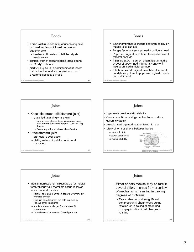
Joints• Ligaments provide static stability• Quadriceps & hamstrings contractions produce dynamic stability• Articular cartilage surfaces on femur & tibia• Menisci form cushions between bones– attached to tibia – deepen tibial fossa– enhance stabilityModified from Anthony CP, Kolthoff NJ: Textbook of anatomy and

physiology, ed 9, St. Louis, 1975, Mosby.

© 2007 McGraw-Hill Higher Education. All rights reserved. 10-11

Joints• Medial meniscus forms receptacle for medial femoral condyle, Lateral meniscus receives lateral femoral condyle– Thicker on outside border & taper down very thin to inside border– Can slip about slightly, but held in place by various small ligaments– Medial meniscus - larger & more open Cappearance– Lateral meniscus - closed C configuration© 2007 McGraw-Hill Higher Education. All rights reserved. 10-12

Joints– Either or both menisci may be torn in several different areas from a variety of mechanisms, resulting in varying degrees of problems• Tears often occur due significant compression & shear forces during rotation while flexing or extending during quick directional changes in running

3

© 2007 McGraw-Hill Higher Education. All rights reserved. 10-13

Joints• Anterior & posterior cruciate ligaments– cross within knee between tibia & femur– vital in respectively maintaining anterior & posterior stability, as well as rotatory stability• Anterior cruciate ligament (ACL) injuries– one of most common serious injuries to knee– mechanism often involves noncontact rotary forces associated with planting & cutting, hyperextension, or by violent quadriceps contraction which pulls tibia forward on femur © 2007 McGraw-Hill Higher Education. All rights reserved. 10-14

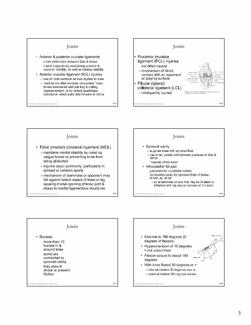
Joints• Posterior cruciateligament (PCL) injuries– not often injured– mechanism of direct contact with an opponent or playing surface• Fibular (lateral) collateral ligament (LCL)– infrequently injured Modified from Anthony CP, Kolthoff NJ: Textbook of anatomy and

physiology, ed 9, St. Louis, 1975, Mosby.

© 2007 McGraw-Hill Higher Education. All rights reserved. 10-15

Joints• Tibial (medial) collateral ligament (MCL)– maintains medial stability by resisting valgus forces or preventing knee from being abducted– injuries occur commonly, particularly in contact or collision sports – mechanism of teammate or opponent may fall against lateral aspect of knee or leg causing medial opening of knee joint & stress to medial ligamentous structures© 2007 McGraw-Hill Higher Education. All rights reserved. 10-16

Joints• Synovial cavity– supplies knee with synovial fluid – lies under patella and between surfaces of tibia & femur– "capsule of the knee”• Infrapatellar fat pad– just posterior to patellar tendon– an insertion point for synovial folds of tissue known as “plica”• an anatomical variant that may be irritated or inflamed with injuries or overuse of the knee

© 2007 McGraw-Hill Higher Education. All rights reserved. 10-17

Joints• Bursae– more than 10 bursae in & around knee– some are connected to synovial cavity– they absorb shock or prevent friction© 2007 McGraw-Hill Higher Education. All rights reserved. 10-18

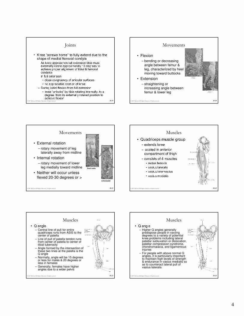
Joints• Extends to 180 degrees (0 degrees of flexion)• Hyperextension of 10 degrees or > not uncommon• Flexion occurs to about 140 degrees• With knee flexed 30 degrees or >– internal rotation 30 degrees occurs– external rotation 45 degrees occurs

4

© 2007 McGraw-Hill Higher Education. All rights reserved. 10-19

Joints• Knee “screws home” to fully extend due to the shape of medial femoral condyle– As knee approaches full extension tibia must externally rotate approximately 10 degrees to achieve proper alignment of tibial & femoral condyles– In full extension• close congruency of articular surfaces• no appreciable rotation of knee– During initial flexion from full extension• knee “unlocks” by tibia rotating internally, to a degree, from its externally rotated position to achieve flexion © 2007 McGraw-Hill Higher Education. All rights reserved. 10-20

Movements

• Flexion

– bending or decreasing angle between femur &

leg, characterized by heel moving toward buttocks

• Extension

– straightening or increasing angle between femur & lower leg

© 2007 McGraw-Hill Higher Education. All rights reserved. 10-21

Movements

• External rotation

– rotary movement of leg laterally away from midline

• Internal rotation

– rotary movement of lower leg medially toward midline

• Neither will occur unless flexed 20-30 degrees or >

© 2007 McGraw-Hill Higher Education. All rights reserved. 10-22

Muscles• Quadriceps muscle group– extends knee– located in anterior compartment of thigh– consists of 4 muscles• rectus femoris• vastus lateralis• vastus intermedius• vastus medialis

© 2007 McGraw-Hill Higher Education. All rights reserved. 10-23

Muscles• Q angle– Central line of pull for entire

quadriceps runs from ASIS to the center of patella

– Line of pull of patella tendon runs from center of patella to center of tibial tuberosity

– Angle formed by the intersection of these two lines at the patella is the Q angle

– Normally, angle will be 15 degrees or less for males & 20 degrees or less in females

– Generally, females have higher angles due to a wider pelvis

© 2007 McGraw-Hill Higher Education. All rights reserved. 10-24

Muscles• Q angle– Higher Q angles generally

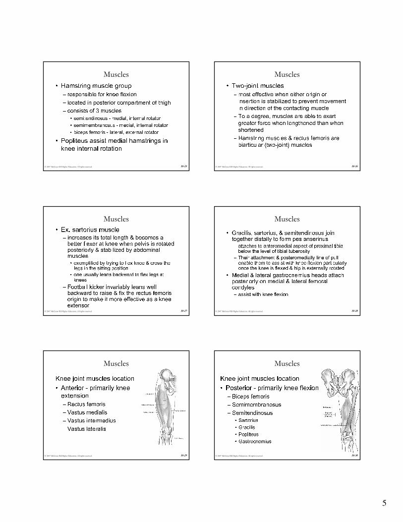
predispose people in varying degrees to a variety of potential knee problems including lateral patellar subluxation or dislocation, patellar compression syndrome, chondromalacia, and ligamentousinjuries

– For people with above normal Q angles, it is particularly important to maintain high levels of strength & endurance in vastus medialis so as to counteract lateral pull of vastus lateralis

5

© 2007 McGraw-Hill Higher Education. All rights reserved. 10-25

Muscles• Hamstring muscle group– responsible for knee flexion– located in posterior compartment of thigh– consists of 3 muscles• semitendinosus - medial, internal rotator• semimembranosus - medial, internal rotator• biceps femoris - lateral, external rotator• Popliteus assist medial hamstrings in knee internal rotation© 2007 McGraw-Hill Higher Education. All rights reserved. 10-26

Muscles• Two-joint muscles– most effective when either origin or insertion is stabilized to prevent movement in direction of the contacting muscle– To a degree, muscles are able to exert greater force when lengthened than when shortened– Hamstring muscles & rectus femoris are biarticular (two-joint) muscles

© 2007 McGraw-Hill Higher Education. All rights reserved. 10-27

Muscles• Ex. sartorius muscle– increases its total length & becomes a better flexor at knee when pelvis is rotated posteriorly & stabilized by abdominal muscles• exemplified by trying to flex knee & cross the legs in the sitting position• one usually leans backward to flex legs at knees– Football kicker invariably leans well backward to raise & fix the rectus femorisorigin to make it more effective as a knee extensor© 2007 McGraw-Hill Higher Education. All rights reserved. 10-28

Muscles• Gracilis, sartorius, & semitendinosus join together distally to form pes anserinus– attaches to anteromedial aspect of proximal tibia below the level of tibial tuberosity– Their attachment & posteromedially line of pull enable them to assist with knee flexion particularly once the knee is flexed & hip is externally rotated• Medial & lateral gastrocnemius heads attach posteriorly on medial & lateral femoral condyles– assist with knee flexion

© 2007 McGraw-Hill Higher Education. All rights reserved. 10-29

MusclesKnee joint muscles location• Anterior - primarily knee extension– Rectus femoris– Vastus medialis– Vastus intermedius– Vastus lateralis© 2007 McGraw-Hill Higher Education. All rights reserved. 10-30

MusclesKnee joint muscles location• Posterior - primarily knee flexion– Biceps femoris– Semimembranosus– Semitendinosus• Sartorius• Gracilis• Popliteus• Gastrocnemius

6

© 2007 McGraw-Hill Higher Education. All rights reserved. 10-31

Nerves• Femoral nerves innervates the knee extensors (quadriceps)– rectus femoris– vastus medialis– vastus intermedius– vastus lateralis© 2007 McGraw-Hill Higher Education. All rights reserved. 10-32

Nerves• Sciatic nerve– tibial division• semitendinosus, semimembranosus, biceps femoris (long head) – common peroneal(fibular) division• biceps femoris (short head)

© 2007 McGraw-Hill Higher Education. All rights reserved. 10-33

Quadriceps Muscles• Quadriceps muscles - vital in jumping– functions as a decelerator• when decreasing speed to change direction• when coming down from a jump– eccentric contraction during decelerating actions– controls slowing of movements initiated in previous phases of the sports skill© 2007 McGraw-Hill Higher Education. All rights reserved. 10-34

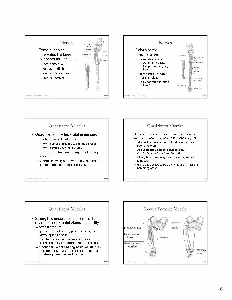
Quadriceps Muscles• Rectus femoris (two-joint), vastus medialis, vastus intermedius, vastus lateralis (largest)• All attach to patella then to tibial tuberosity via patellar tendon• All superficial & palpable except vastusintermedius (under rectus femoris)• Strength or power may be indicated by vertical jump test• Generally desired to be 25% to 33% stronger than hamstring group

© 2007 McGraw-Hill Higher Education. All rights reserved. 10-35

Quadriceps Muscles• Strength & endurance is essential for maintenance of patellofemoral stability– often a problem– quads are particularly prone to atrophy when injuries occur– may be developed by resisted knee extension activities from a seated position– functional weight bearing activities such as step-ups or squats are particularly useful for strengthening & endurance© 2007 McGraw-Hill Higher Education. All rights reserved. 10-36

Rectus Femoris Muscle

Flexion of hip

Extension of knee

Anterior pelvic rotation

7

© 2007 McGraw-Hill Higher Education. All rights reserved. 10-37

Vastus Lateralis Muscle

Extension of knee

© 2007 McGraw-Hill Higher Education. All rights reserved. 10-38

Vastus Intermedius Muscle

Extension of knee

© 2007 McGraw-Hill Higher Education. All rights reserved. 10-39

Vastus Medialis Muscle

Extension of knee

© 2007 McGraw-Hill Higher Education. All rights reserved. 10-40

Hamstring Muscles

• Hamstring muscle group

– Semitendinosus

– Biceps femoris

– Semimembranosus

© 2007 McGraw-Hill Higher Education. All rights reserved. 10-41

Hamstring Muscles

• Hamstring muscle strains very common

• “Running muscles” function in acceleration

• Antagonists to quadriceps muscles at knee

• Named for cordlike attachments at knee

• All originate on ischial tuberosity of pelvis

• Semitendinosus inserts on anteromedial tibia

• Semimembranosus inserts on posteromedialtibia

• Biceps femoris inserts on lateral tibial condyle& head of fibula

© 2007 McGraw-Hill Higher Education. All rights reserved. 10-42

Semitendinosus Muscle

Flexion of knee

Extension of hip

Internal rotation of hip

Internal rotation of flexed knee

Posterior pelvic rotation

8

© 2007 McGraw-Hill Higher Education. All rights reserved. 10-43

Semimembranosus Muscle

Flexion of knee

Extension of hip

Internal rotation of hip

Internal rotation of flexed knee

Posterior pelvic rotation

© 2007 McGraw-Hill Higher Education. All rights reserved. 10-44

Biceps Femoris Muscle

Flexion of knee

Extension of hip

External rotation of hip

External rotation of flexed knee

Posterior pelvic rotation

© 2007 McGraw-Hill Higher Education. All rights reserved. 10-45

Popliteus Muscle

Flexion of knee

Internal rotation of flexed knee

© 2007 McGraw-Hill Higher Education. All rights reserved. 10-46

Knee Extension

• Agonists

– Rectus Femoris

– Vastus Lateralis

– Vastus Intermedius

– Vastus Medialis

© 2007 McGraw-Hill Higher Education. All rights reserved. 10-47

Knee Flexion

• Agonists

– Biceps Femoris(Long & Short

Head)

– Semitendinosus

– Semimembranosus

© 2007 McGraw-Hill Higher Education. All rights reserved. 10-48

Knee Internal Rotation

• Agonists– Semitendinosus

– Semimembranosus

– Popliteus

9

© 2007 McGraw-Hill Higher Education. All rights reserved. 10-49

Knee External Rotation

• Agonists

– Biceps Femoris

© 2007 McGraw-Hill Higher Education. All rights reserved. 10-50

Web SitesRadiologic Anatomy Browser

http://radlinux1.usuf1.usuhs.mil/rad/iong

– This site has numerous radiological views of the musculoskeletal system.

University of Arkansas Medical School Gross Anatomy for Medical Students

http://anatomy.uams.edu/anatomyhtml/gross.html

– Dissections, anatomy tables, atlas images, links, etc.Loyola University Medical Center: Structure of the Human Body

www.meddean.luc.edu/lumen/meded/grossanatomy/index.htm– An excellent site with many slides, dissections, tutorials, etc.

for the study of human anatomyWheeless’ Textbook of Orthopaedics

www.wheelessonline.com/

– This site has an extensive index of links to the fractures, joints, muscles, nerves, trauma, medications, medical topics, lab tests, and links to orthopedic journals and other orthopedicand medical news.

© 2007 McGraw-Hill Higher Education. All rights reserved. 10-51

Web SitesPremiere Medical Search Engine

www.medsite.com

– This site allows the reader to enter any medical condition and it will search the net to find relevant articles.

Arthroscopy.com

www.arthroscopy.com/sports.htm– Patient information on various musculoskeletal problems of

the lower extremityVirtual Hospital

www.vh.org– Numerous slides, patient information, etc.

Human Anatomy Online

www.innerbody.com/image/musc08.html– Interactive musculoskeletal anatomy

The Hip and Knee Institute

www.hipsandknees.com/knee/index.html– Arthritis of the Knee Joint

© 2007 McGraw-Hill Higher Education. All rights reserved. 10-52

Web SitesAdam Healthcare Center

http://adam.about.com/surgery/100088.htm#

– Knee joint replacementAmerican Academy of Orthopaedic Surgeons

http://orthoinfo.aaos.org/category.cfm?topcategory=Knee

– Patient education library on the kneeEdheads Activities

www.edheads.org/activities/knee/

– Allows you to perform virtual knee surgeryGross Anatomy: The Functional Anatomy of the Knee Joint

www.upstate.edu/cdb/grossanat/limbs8.shtml– Functional Anatomy of the Knee

Knee Ligament Anatomy and Injury

www.orthoassociates.com/knee_lig.htm– Anatomy and injuries of the Knee and its ligaments

© 2007 McGraw-Hill Higher Education. All rights reserved. 10-53

Web SitesDuke Orthopaedics

www.wheelessonline.com/ortho/anatomy_and_kinematics_of_the_knee_joint

– Anatomy and Kinematics of the knee joint

Knee Injury: Meniscus

www.patient.co.uk/showdoc/27000672/– Understanding the knee joint and purpose of meniscus

Smart Play: The Kneewww.smartplay.net/ouch/bodybits/b_bitsknee.html

– Anatomy, functions, injuries, etc. of the knee

Patellofemoral Instabilitywww.massgeneral.org/ortho/PatellofemoralInstability.htm

– Patella Femoral AlignmentChiroweb.com

www.chiroweb.com/archives/21/24/03.html

– Abnormal Q Angle and Orthotic Support

© 2007 McGraw-Hill Higher Education. All rights reserved. 10-54

Web SitesThe Physician and Sportsmedicine

www.physsportsmed.com/issues/1997/05may/bach.htm

– Acute Knee Injuries: When to ReferThe Physician and Sportsmedicine

www.physsportsmed.com/issues/1999/10_01_99/laprade.htm

– Acute Knee Injuries: On-the-Field and Sideline Evaluation


Recommended