70
Chapter 11 Nervous System II
| Date post: | 02-Jan-2016 |
| Category: |
Documents |
| Upload: | adrienne-conway |
| View: | 11 times |
| Download: | 0 times |
Chapter 11
Nervous System II
PROTECTION OF THE CNS
The brain and spinal cord are protected (surrounded) by bones,
membranes, and fluid.
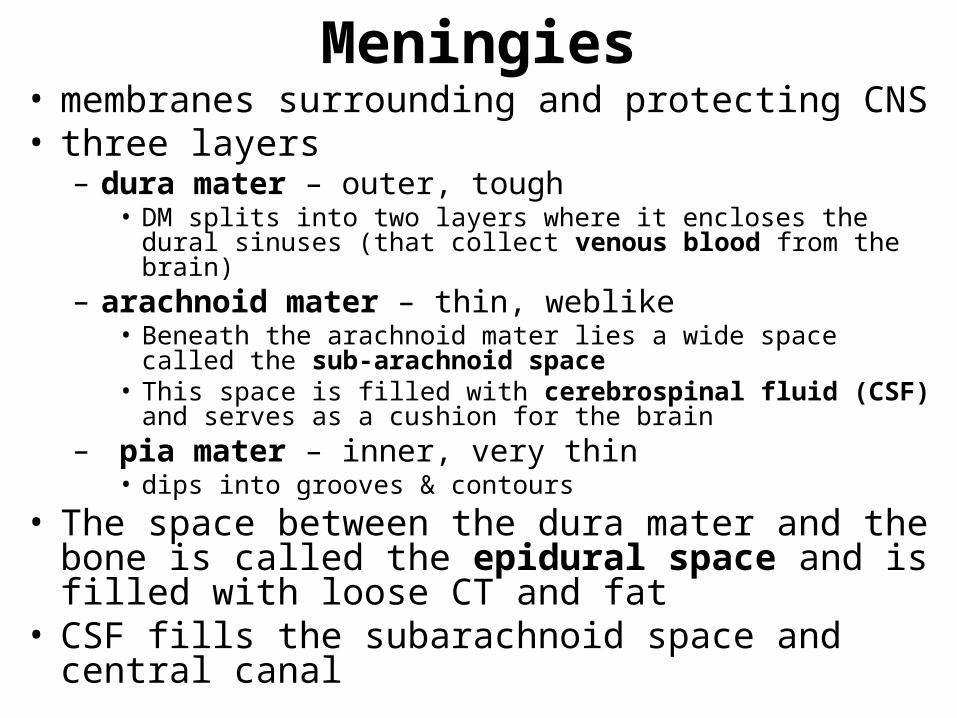
Meningies• membranes surrounding and protecting CNS• three layers
– dura mater – outer, tough• DM splits into two layers where it encloses the dural sinuses (that
collect venous blood from the brain)– arachnoid mater – thin, weblike
• Beneath the arachnoid mater lies a wide space called the sub-arachnoid space
• This space is filled with cerebrospinal fluid (CSF) and serves as a cushion for the brain
– pia mater – inner, very thin• dips into grooves & contours
• The space between the dura mater and the bone is called the epidural space and is filled with loose CT and fat
• CSF fills the subarachnoid space and central canal

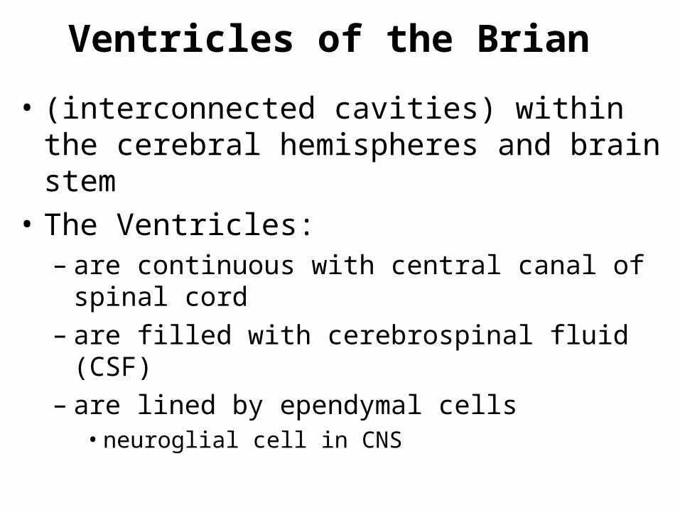
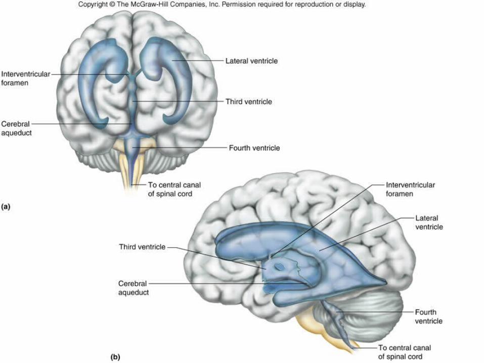
Ventricles of the Brian
• (interconnected cavities) within the cerebral hemispheres and brain stem
• The Ventricles:– are continuous with central canal of spinal cord– are filled with cerebrospinal fluid (CSF)– are lined by ependymal cells
• neuroglial cell in CNS

Cerebrospinal Fluid
• secreted by choroid plexus• circulates in ventricles, central canal of
spinal cord, and subarachnoid space• completely surrounds brain and spinal cord• clear liquid• provides nutrition and protection• helps maintain stable ion concentrations in
CNS

THE SPINAL CORD
The spinal cord is a nerve column that passes downward from brain into the vertebral canal.
Recall that it is part of the CNS
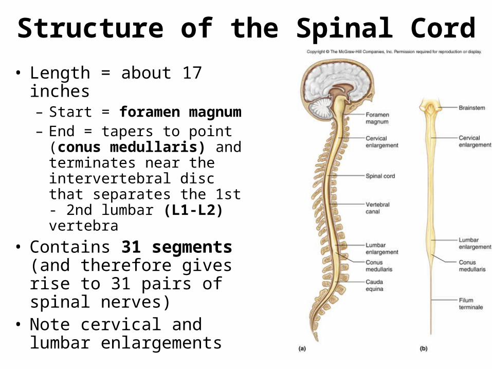
Structure of the Spinal Cord
• Length = about 17 inches– Start = foramen magnum– End = tapers to point
(conus medullaris) and terminates near the intervertebral disc that separates the 1st - 2nd lumbar (L1-L2) vertebra
• Contains 31 segments (and therefore gives rise to 31 pairs of spinal nerves)
• Note cervical and lumbar enlargements
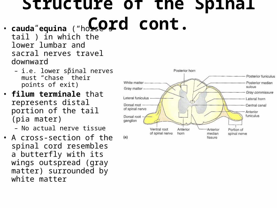
Structure of the Spinal Cord cont.• cauda equina (“horse’s
tail”) in which the lower lumbar and sacral nerves travel downward – i.e. lower spinal nerves
must “chase” their points of exit)
• filum terminale that represents distal portion of the tail (pia mater)– No actual nerve tissue
• A cross-section of the spinal cord resembles a butterfly with its wings outspread (gray matter) surrounded by white matter

Functions of the Spinal Cord
• Nerve Pathway = the route traveled by a nerve impulse through the nervous system
• Reflex arc = the simplest demonstration of a nerve pathway (spinal reflexes)– involves 2-3 neurons– involuntary response– often does not involve the brain
• Examples include:– knee-jerk or patellar reflex

Spinal Nerves• Spinal nerves extend to/from the spinal
cord and are part of the PNS • Ganglion = a bundle of cell bodies outside
the CNS• Dorsal Root Ganglion contains the cell
bodies of sensory (afferent) neurons bringing impulses to the CNS
• The fusion of the dorsal and ventral roots designates the beginning of the spinal nerve which then passes through its intervertebral foramen

Ascending and Descending Tracts
• The white matter of the spinal cord represents the location of our major nerve pathways called "nerve tracts"
• provide a 2-way system of communication:
• ascending tracts conduct sensory (afferent) impulses from body parts to brain
• descending tracts conduct motor (efferent) impulses from brain to effectors
• All pathways are paired (right and left)

BRAIN
The brain is the largest and most complex portion of the nervous system.
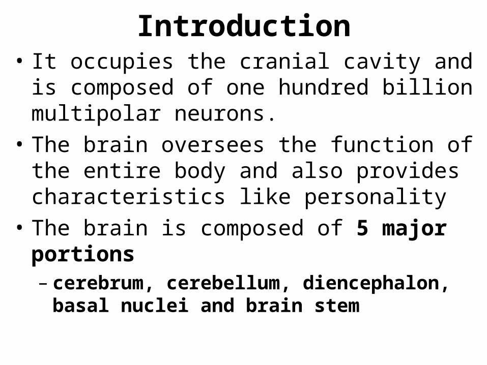
Introduction• It occupies the cranial cavity and is composed
of one hundred billion multipolar neurons.
• The brain oversees the function of the entire body and also provides characteristics like personality
• The brain is composed of 5 major portions– cerebrum, cerebellum, diencephalon, basal
nuclei and brain stem

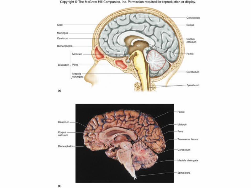
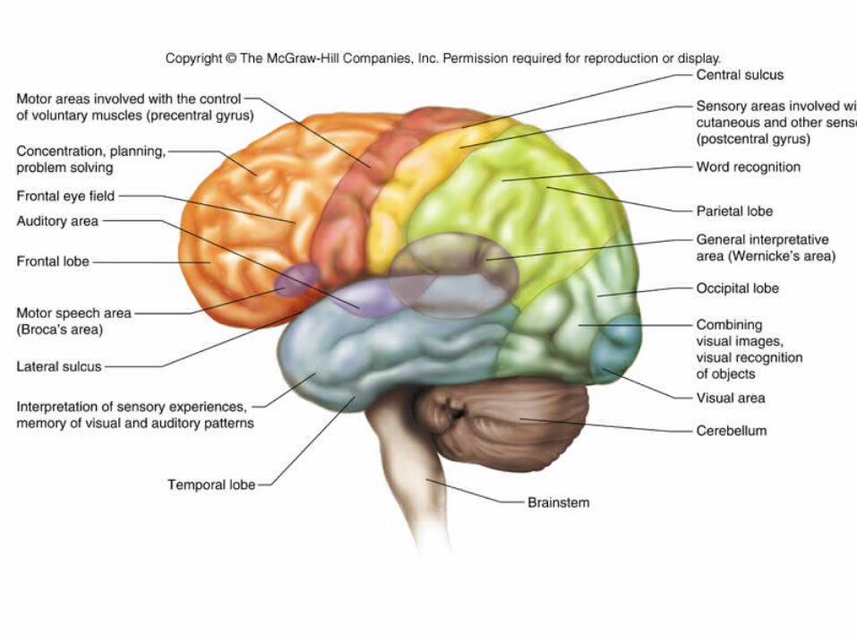
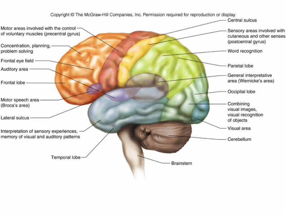
Structure of the Cerebrum
• Cerebrum = the largest portion of the brain, which is divided into two cerebral hemispheres. – Hemispheres are connected by a deep bridge of
nerve fibers called the corpus callosum– Surface ridges are called convolutions (gyri)– Convolutions are separated by two types of
grooves. (Sulci and Fissures)– Each hemisphere is divided into lobes, which are
named for the bones that cover them including frontal, parietal, temporal, and occipital lobes

Structure/Function of the Cerebrum• Composition:
– Bulk of cerebrum is white matter.– bundles of myelinated nerve fibers (by
oligodendrocyte)
• Cerebral cortex or the outer portion of cerebrum is composed of gray matter – bundles of neuron cell bodies– contains 75% of all neurons in nervous system
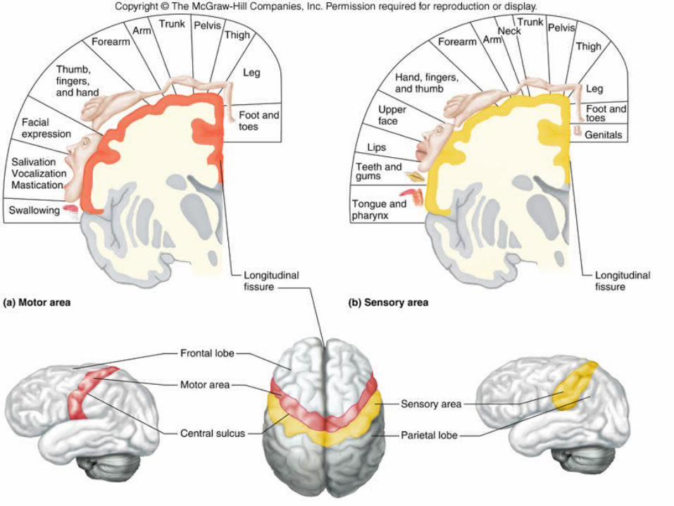
• Functional Regions of the Cerebral cortex– Responsible for all conscious behavior by
containing three kinds of functional areas, which include motor, sensory and association areas
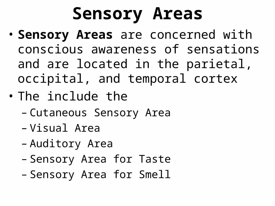
Sensory Areas• Sensory Areas are concerned with conscious
awareness of sensations and are located in the parietal, occipital, and temporal cortex
• The include the– Cutaneous Sensory Area– Visual Area– Auditory Area– Sensory Area for Taste– Sensory Area for Smell
Association Areas• regions that are not primary motor or
primary sensory areas
• widespread throughout the cerebral cortex
• Association traits include:– analyzing & interpreting sensory experiences– help provide memory, reasoning, verbalizing,
judgment and emotions

Hemisphere Dominance
• The left hemisphere is dominant is most individuals
• Dominant hemisphere controls
• speech• writing• reading• verbal skills• analytical skills• computational skills
• Nondominant hemisphere controls
• nonverbal tasks• motor tasks• understanding and interpreting musical and visual patterns• provides emotional and intuitive thought processes

Memory• Memory is the consequence of learning. Whereas
learning is the acquisition of new knowledge, memory is the persistence of that learning, with the ability to access it at a later time
• Two types of memory:– Short Term: working memory– Long Term: changes structure or function of neurons

Motor AreasMotor Areas are located in the frontal cortex:
• Primary motor cortex – initiates all voluntary muscle movements
• Broca's area– motor speech area
• Frontal Eye Field– controls voluntary movements of eyes and
eyelids

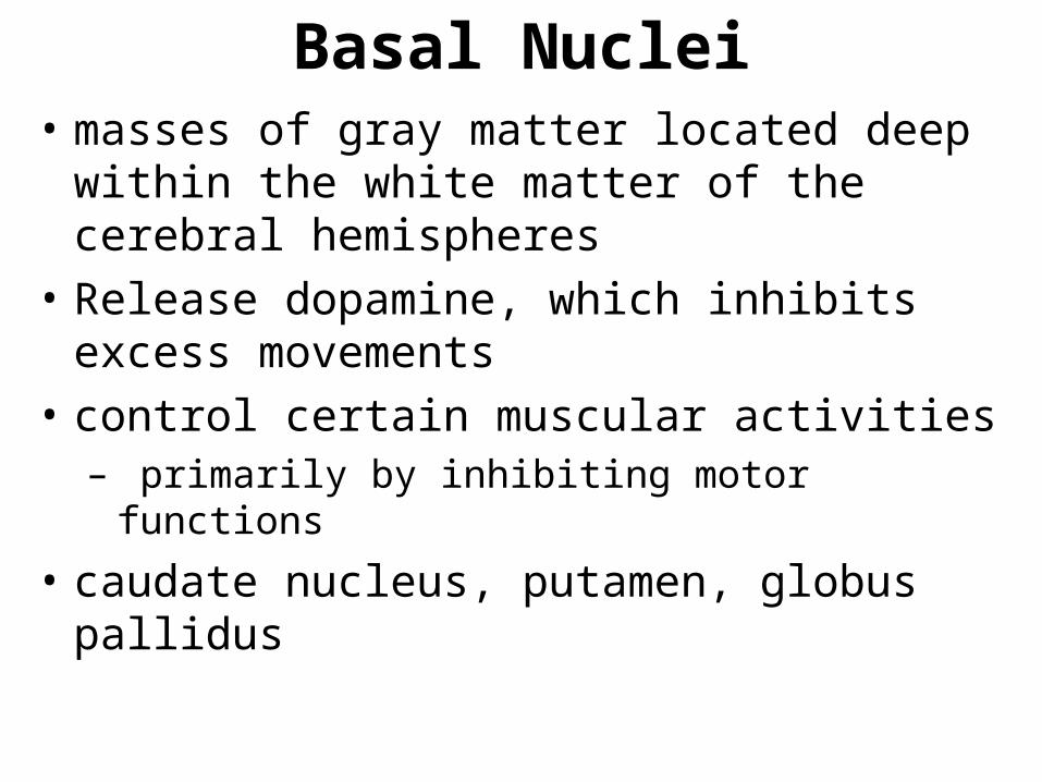
Basal Nuclei• masses of gray matter located deep within
the white matter of the cerebral hemispheres
• Release dopamine, which inhibits excess movements
• control certain muscular activities– primarily by inhibiting motor functions
• caudate nucleus, putamen, globus pallidus

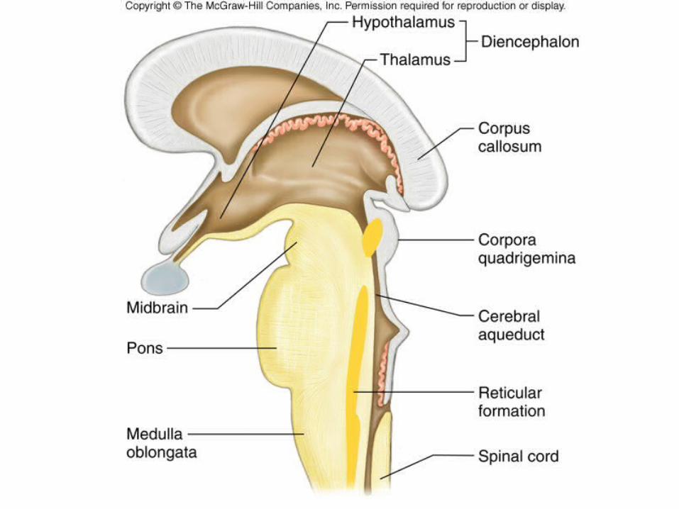
Diencephalon• includes two important areas of gray matter:• Thalamus
– gateway for sensory impulses heading to cerebral cortex
– receives all sensory impulses (except smell)– channels impulses to appropriate part of cerebral
cortex for interpretation
• Hypothalamus– maintains homeostasis by regulating visceral activities – links nervous and endocrine systems
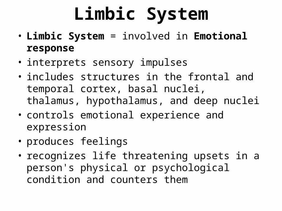
Limbic System• Limbic System = involved in Emotional
response • interprets sensory impulses• includes structures in the frontal and temporal
cortex, basal nuclei, thalamus, hypothalamus, and deep nuclei
• controls emotional experience and expression• produces feelings • recognizes life threatening upsets in a person's
physical or psychological condition and counters them

Brain Stem• The brain stem is composed of three
major parts – midbrain, pons, and medulla oblongata
• The brain stem serves as a pathway for fiber tracts running to (sensory impulses) and from (motor impulses) the cerebrum and is the sight where many cranial nerves (PNS) arise
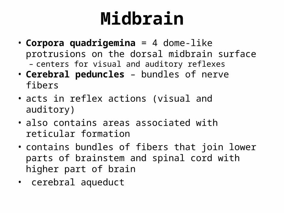
Midbrain• Corpora quadrigemina = 4 dome-like
protrusions on the dorsal midbrain surface– centers for visual and auditory reflexes
• Cerebral peduncles – bundles of nerve fibers• acts in reflex actions (visual and auditory)• also contains areas associated with reticular
formation• contains bundles of fibers that join lower parts of
brainstem and spinal cord with higher part of brain
• cerebral aqueduct

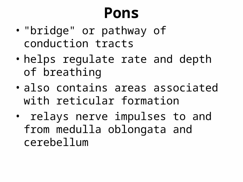
Pons• "bridge" or pathway of conduction tracts
• helps regulate rate and depth of breathing
• also contains areas associated with reticular formation
• relays nerve impulses to and from medulla oblongata and cerebellum
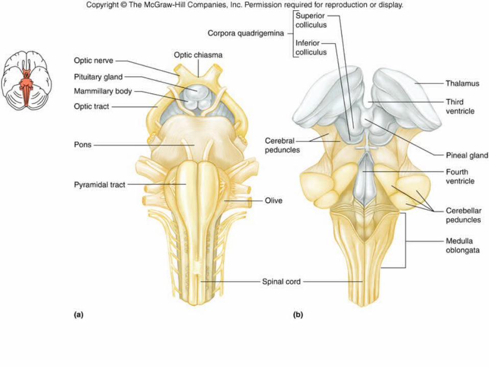

Medulla Oblongata• conducts ascending and descending
impulses between brain and spinal cord• contains an autonomic reflex center
involved in maintaining homeostasis of important visceral organs– contains cardiac, vasomotor, and respiratory
control centers – contains various nonvital reflex control
centers (coughing, sneezing, swallowing, vomiting)

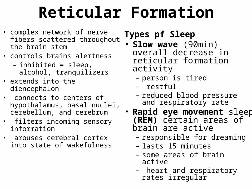
Reticular Formation• complex network of nerve fibers
scattered throughout the brain stem
• controls brains alertness– inhibited = sleep, alcohol,
tranquilizers • extends into the diencephalon• connects to centers of
hypothalamus, basal nuclei, cerebellum, and cerebrum
• filters incoming sensory information
• arouses cerebral cortex into state of wakefulness
Types pf Sleep• Slow wave (90min) overall
decrease in reticular formation activity– person is tired– restful– reduced blood pressure and
respiratory rate• Rapid eye movement
sleep (REM) certain areas of brain are active– responsible for dreaming– lasts 15 minutes– some areas of brain active– heart and respiratory rates
irregular

Cerebellum• note pattern of white matter (within gray matter)
= "arbor vitae" • integrates sensory information concerning
position of body parts• coordinates skeletal muscle activity• maintains posture
PERIFERAL NERVOUS SYSTEM

PSN Introduction• The peripheral nervous system (PNS) consists of
nerves that extend to and from the CNS organs • Cranial nerves arising from the brain
– Somatic fibers connecting to the skin and skeletal muscles– Autonomic fibers connecting to viscera
• Spinal nerves arising from the spinal cord– Somatic fibers connecting to the skin and skeletal muscles– Autonomic fibers connecting to viscera
• The PNS is divided into a sensory and motor branch• The motor branch of the PNS is further subdivided into
a somatic nervous system (from CNS to skin and skeletal muscles) and autonomic nervous system (from CNS to smooth muscle, cardiac muscle and endocrine glands)

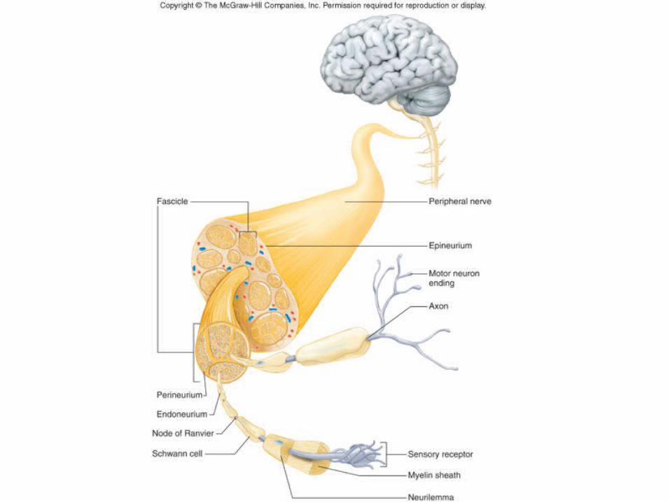
Structure Of Peripheral Nerves
• A nerve is a cord-like bundle of axons wrapped in CT
• Structure of a Nerve:
• Three types of CT wrappings (similar to muscle):– endoneurium around each axon (and myelin)– perineurium around each fascicle (bundle) of
axons– epineurium around each nerve

Nerve Fiber Classification• Mixed Nerves
– Nerves that carry impulses both to and from the CNS– contain both sensory and motor axons– most common; 2-way communication
• Sensory (afferent) Nerves – Nerves that only carry sensory impulses toward the
CNS– rare (only three pairs of cranial nerves)
• Motor (efferent) Nerves – Nerves that only carry motor impulses away from CNS– rare (only five pairs of cranial nerves)

Nerve Fiber Classification cont.• General somatic efferent
fibers– carry motor impulses from CNS
to skeletal muscles
• General somatic afferent fibers– carry sensory impulses to CNS
from skin and skeletal muscles
• General visceral efferent fibers– carry motor impulses away from
CNS to smooth muscles and glands
• General visceral afferent fibers– carry sensory impulses to CNS
from blood vessels and internal organs
• Special somatic efferent fibers– carry motor impulses from
brain to muscles used in chewing, swallowing, speaking, and forming facial expressions
• Special visceral afferent fibers– carry sensory impulses to
brain from olfactory and taste receptors
• Special somatic afferent fibers– carry sensory impulses to
brain from receptors of sight, hearing, and equilibrium
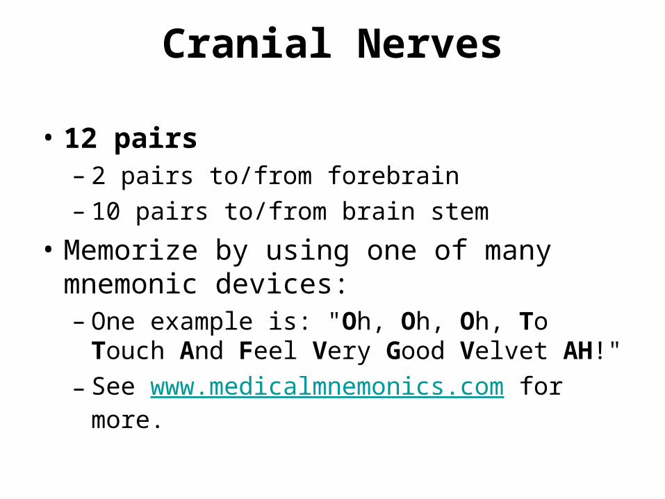
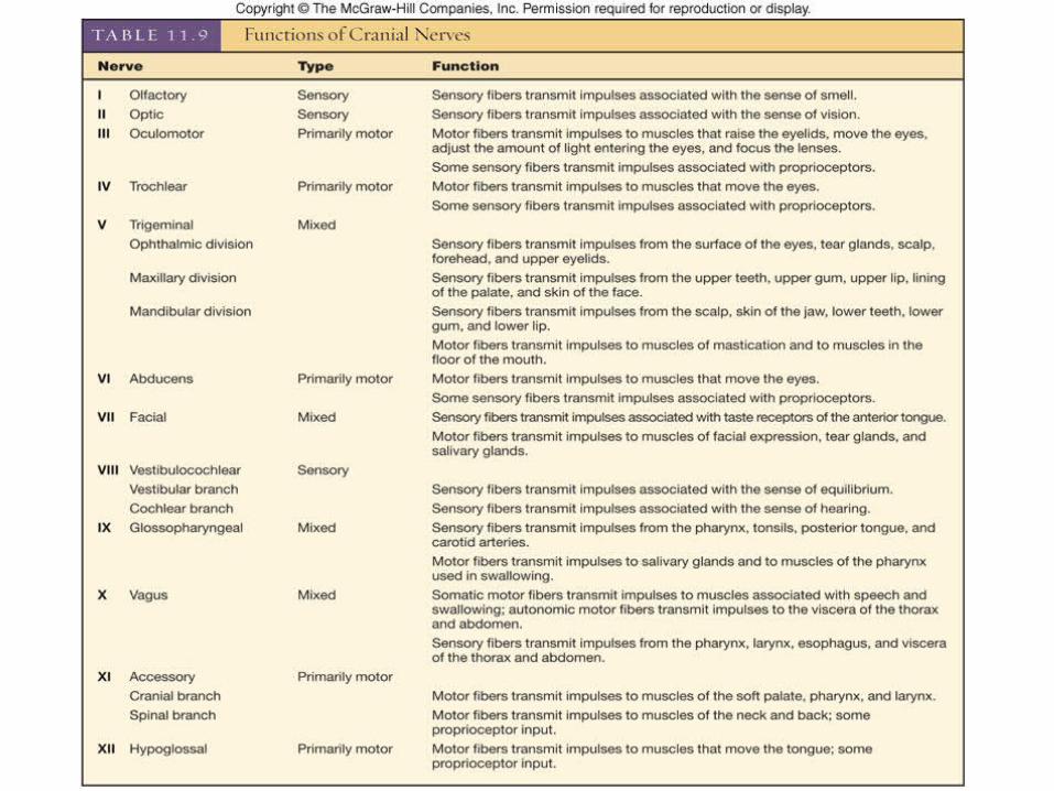
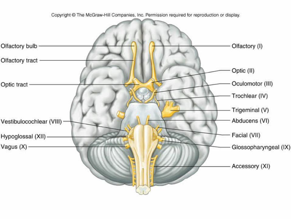
Cranial Nerves
• 12 pairs – 2 pairs to/from forebrain– 10 pairs to/from brain stem
• Memorize by using one of many mnemonic devices:– One example is: "Oh, Oh, Oh, To Touch And
Feel Very Good Velvet AH!"– See www.medicalmnemonics.com for more.

Spinal Nerves
• Introduction– Recall that a spinal nerve is formed from the
fusion of a dorsal and ventral root– Then the spinal nerve passes through its
intervertebral foramen– Spinal nerves are associated with the spinal
cord and are named for the region of the spinal cord from which they arise

Spinal Nerves cont.• General
Characteristics:
• 31 pairs: – 8 cervical nerves, 12
thoracic nerves, 5 lumbar nerves, 5 sacral nerves, 1 coccygeal nerve
• Composition = all mixed nerves

Spinal Nerve cont.
• Dorsal root (posterior or sensory root)– axons of sensory neurons in the dorsal root
ganglion
• Ventral root (anterior or motor root) – axons of motor neurons whose cell bodies
are in spinal cord
• Spinal nerve– union of ventral root and dorsal root


Dermatome
• an area of skin that the sensory nerve fibers of a particular spinal nerve innervate
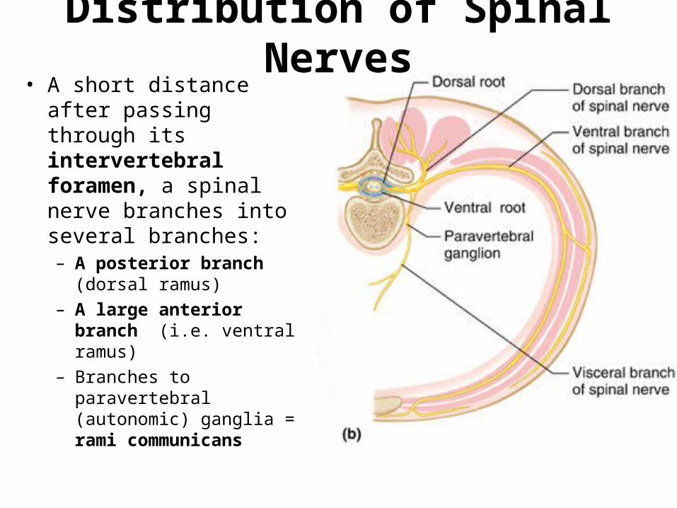
Distribution of Spinal Nerves• A short distance after
passing through its intervertebral foramen, a spinal nerve branches into several branches:– A posterior branch
(dorsal ramus)– A large anterior branch
(i.e. ventral ramus)– Branches to paravertebral
(autonomic) ganglia = rami communicans

Nerve plexus• Definition = a branching network (of the
anterior branches) of spinal nerves– The nerves do not extend directly to the body
part they innervate, instead they form networks.
• present in all spinal nerves except T2 - T12:– cervical plexus; neck muscles and diaphragm
(breathing)– brachial plexus; upper limb– lumbar plexus; anterior and medial thigh– sacral plexus; posterior lower limb, leg
Nerve plexus cont.• Each resulting branch of the plexus
contains the fibers from several spinal nerves
• Fibers from each spinal nerve are carried to the body periphery via several different routes or branches.
• Therefore, damage to one spinal segment cannot completely paralyze any limb muscle

Intercostal Nerves
• Nerves T2-T11 run in intercostal spaces
• Supply skin (sensory) and muscles (motor) in the surrounding area

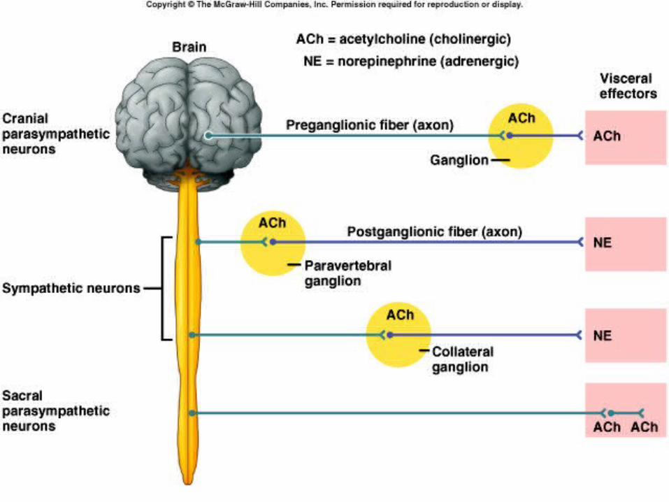
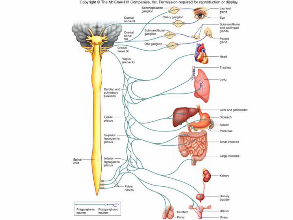
AUTONOMIC NERVOUS SYSTEM (ANS)

Autonomic Nervous System•functions without conscious effort• controls visceral activities• regulates smooth muscle, cardiac muscle, and glands• efferent fibers typically lead to ganglia outside CNS
•Controlled largely by CNS•Two Divisions
•sympathetic – prepares body for fight or flight situations•parasympathetic – prepares body for resting and digesting activities

Sympathetic Division• thoracolumbar divison – location of preganglionic neurons•preganglionic fibers leave spinal nerves through white rami and enter paravertebral ganglia•paraverterbral ganglia and fibers that connect them make up the sympathetic trunk

Sympathetic Division cont.• postganglionic fibers extend from sympathetic ganglia to visceral organs• postganglionic fibers usually pass through gray rami and return to a spinal nerve before proceeding to an effector

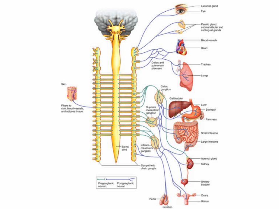

Parasympathetic Division• craniosacral division – location of preganglionic neurons•ganglia are near or within various organs
terminal ganglia•short postganglionic fibers
•continue to specific muscles or glands•preganglionic fibers of the head are included in nerves III, VII, and IX•preganglionic fibers of thorax and abdomen are parts of nerve X

Autonomic Neurotransmitters•Cholinergic Fibers
• release acetylcholine• preganglionic sympathetic and parasympathetic fibers• postganglionic parasympathetic fibers
•Adrenergic Fibers•release norepinephrine•most postganglionic sympathetic fibers