43
Chapter 15: The Autonomic Nervous System Copyright 2009, John Wiley & Sons, Inc.
| Date post: | 25-Dec-2015 |
| Category: |
Documents |
| Upload: | baldric-goodwin |
| View: | 215 times |
| Download: | 1 times |

Chapter 15: The Autonomic Nervous System
Copyright 2009, John Wiley & Sons, Inc.

General Properties of the ANS (Visceral Motor System)
Motor nervous system that controls glands, cardiac & smooth muscle Regulates unconscious processes that maintain homeostasis
blood pressure, body temperature, respiratory airflow Involved with visceral reflexes ANS carries out its actions without our intent (automatically)
biofeedback techniques- training that teaches some people to control hypertension, stress and migraine headaches.
Components: Autonomic sensory neurons (interoceptors) Integrating centers in CNS Autonomic motor to effectors

Visceral Reflex to High Blood Pressure
High BP detected by arterial stretch receptors, signal transmitted to CNS, efferent signals travel to the heart, heart slows reducing BP
Separate reflex arc for low BP exists

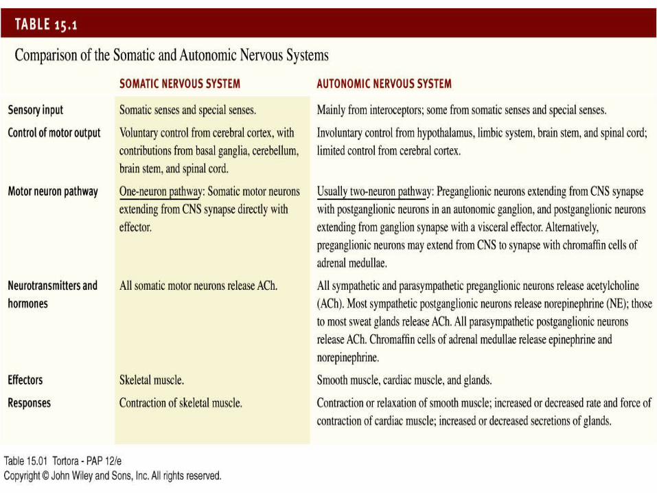
Comparison of Somatic and Autonomic Nervous Systems
Copyright 2009, John Wiley & Sons, Inc.
________ ___________
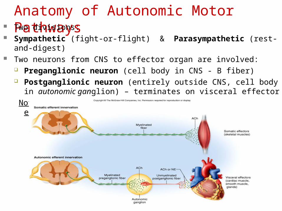
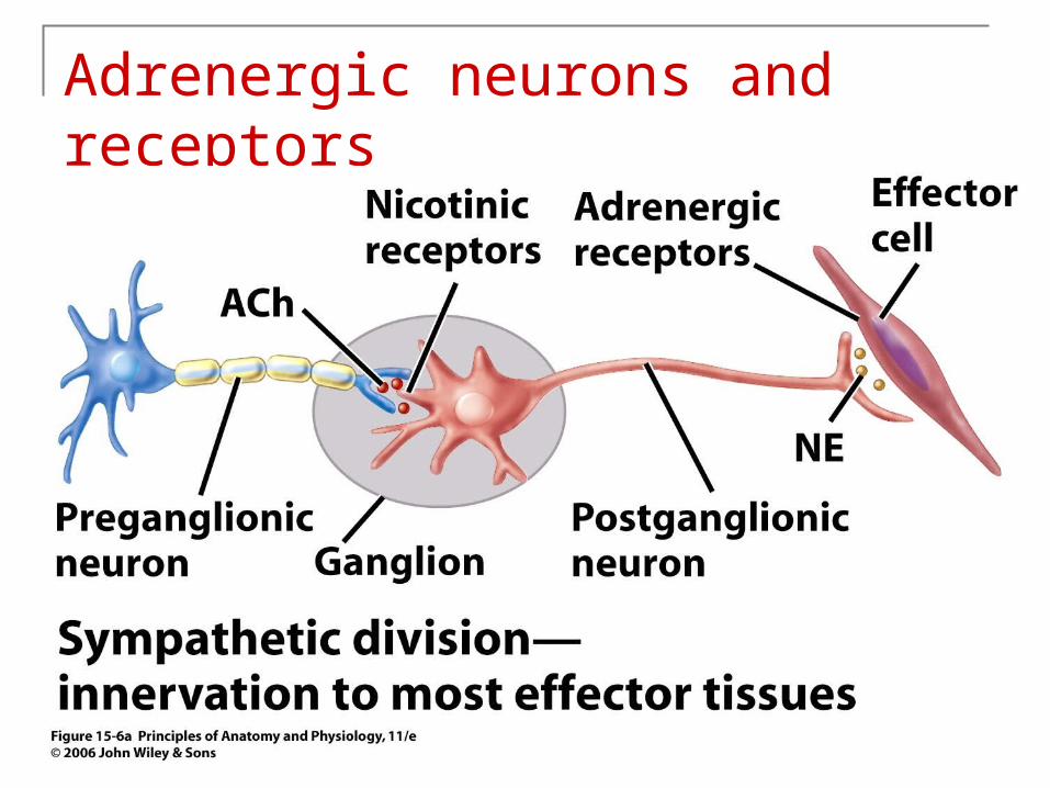
Anatomy of Autonomic Motor Pathways Two divisions:
Sympathetic (fight-or-flight) & Parasympathetic (rest-and-digest) Two neurons from CNS to effector organ are involved:
Preganglionic neuron (cell body in CNS - B fiber) Postganglionic neuron (entirely outside CNS, cell body in autonomic
ganglion) – terminates on visceral effector
Note : the Somatic motor division lacks ganglia entirely
Copyright 2009, John Wiley & Sons, Inc.
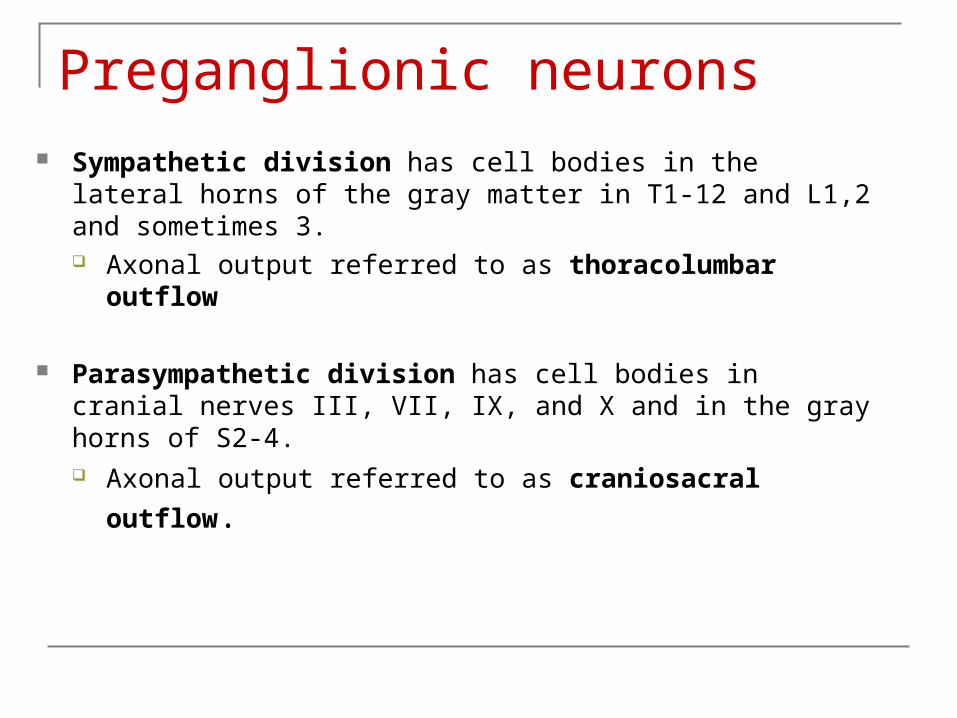
Preganglionic neurons Sympathetic division has cell bodies in the lateral horns of the
gray matter in T1-12 and L1,2 and sometimes 3. Axonal output referred to as thoracolumbar outflow
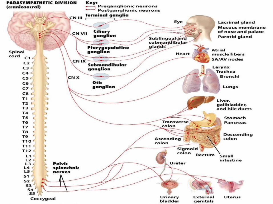
Parasympathetic division has cell bodies in cranial nerves III, VII, IX, and X and in the gray horns of S2-4.
Axonal output referred to as craniosacral outflow.
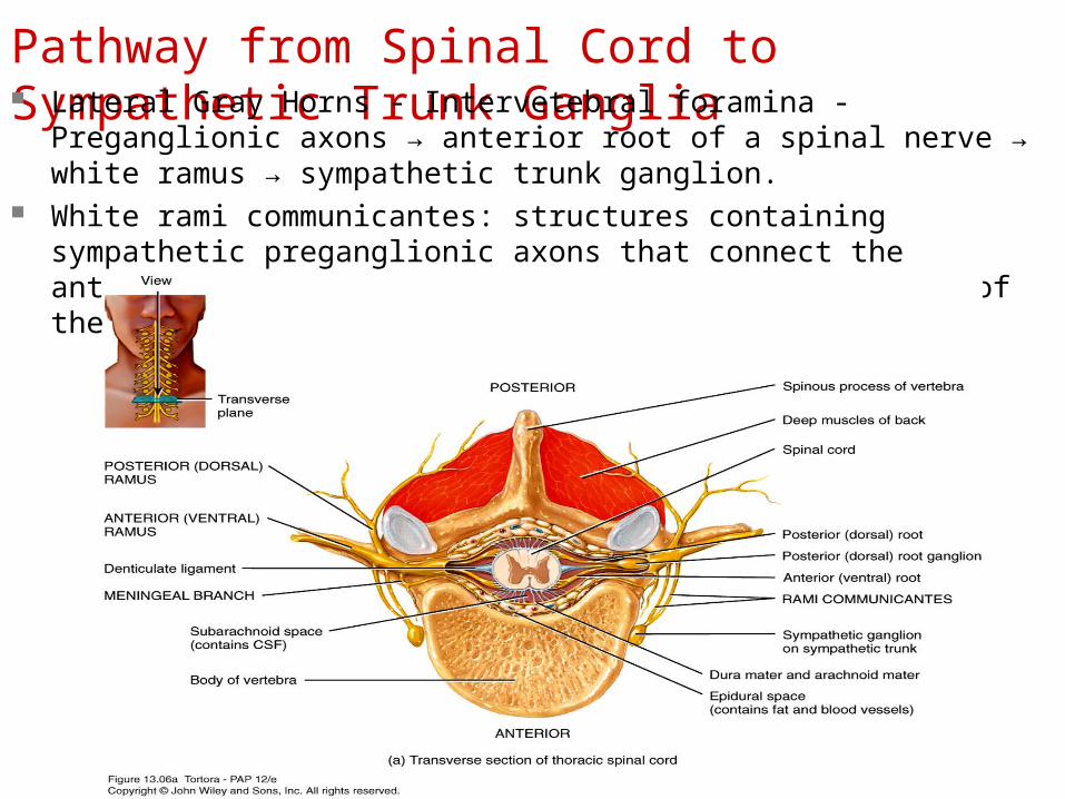
Pathway from Spinal Cord to Sympathetic Trunk Ganglia Lateral Gray Horns - Intervetebral foramina - Preganglionic axons →
anterior root of a spinal nerve → white ramus → sympathetic trunk ganglion. White rami communicantes: structures containing sympathetic preganglionic
axons that connect the anterior ramus of the spinal nerve with the ganglia of the sympathetic trunk.
Copyright 2009, John Wiley & Sons, Inc.
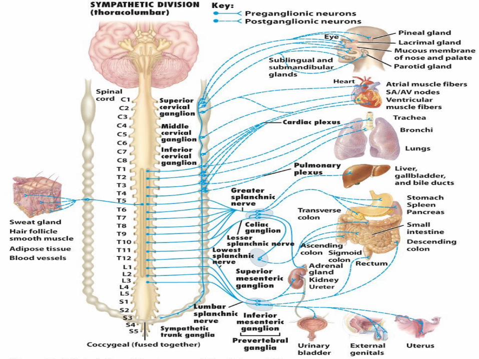

Sympathetic ganglia Sympathetic trunk/paravertebral/vertebral chain ganglia lie in a
vertical row close to both sides of the vertebral column from the base of skull to coccyx Superior, middle and inferior cervical ganglia Postganglionic neurons innervate organs above diaphragm.
Prevertebral/collateral ganglia lie anterior to vertebral column and close to abdominal arteries. Celiac, superior and inferior mesenteric ganglia Postganglionic neurons innervate organs below the diaphragm.

Parasympathetic ganglia Terminal ganglia are located close to area within wall of the visceral
organ. Ciliary ganglion Pterygopalatine ganglion Submandibular ganglion Otic ganglion
Axons are longer than those found in the sympathetic division.
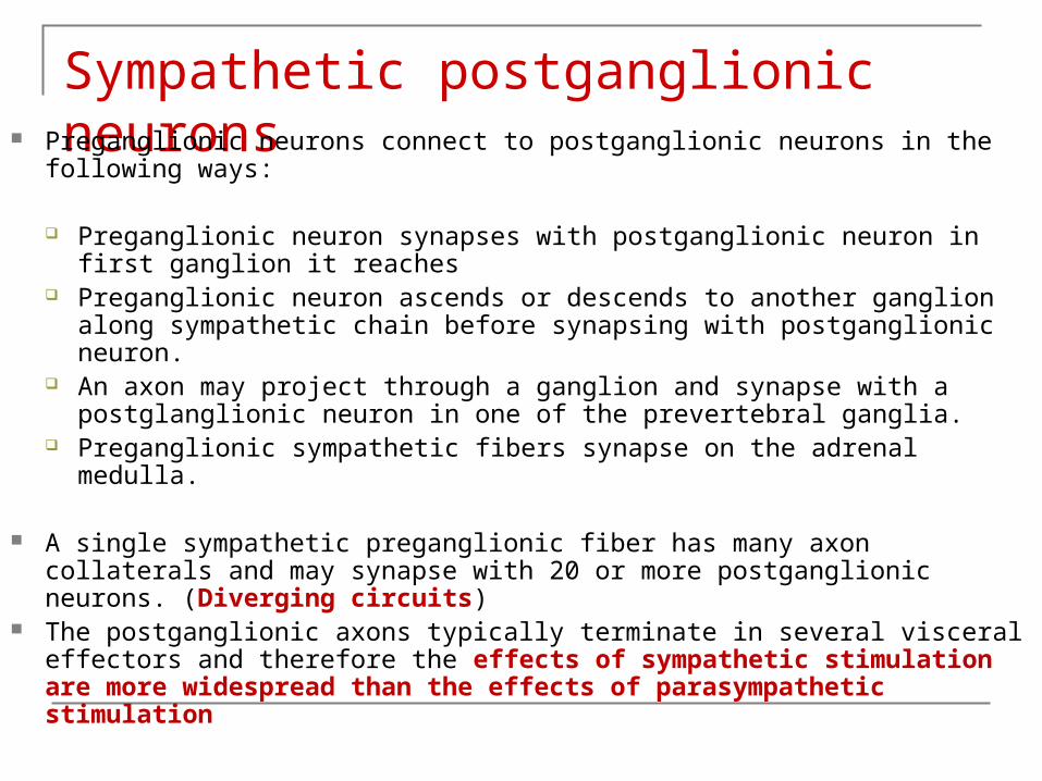
Sympathetic postganglionic neurons Preganglionic neurons connect to postganglionic neurons in the following
ways:
Preganglionic neuron synapses with postganglionic neuron in first ganglion it reaches
Preganglionic neuron ascends or descends to another ganglion along sympathetic chain before synapsing with postganglionic neuron.
An axon may project through a ganglion and synapse with a postglanglionic neuron in one of the prevertebral ganglia.
Preganglionic sympathetic fibers synapse on the adrenal medulla.
A single sympathetic preganglionic fiber has many axon collaterals and may synapse with 20 or more postganglionic neurons. (Diverging circuits)
The postganglionic axons typically terminate in several visceral effectors and therefore the effects of sympathetic stimulation are more widespread than the effects of parasympathetic stimulation

Copyright 2009, John Wiley & Sons, Inc.
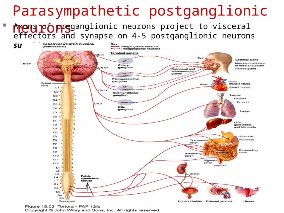
Parasympathetic postganglionic neurons Axons of preganglionic neurons project to visceral effectors and synapse on 4-5 postganglionic neurons supplying a single visceral organ.
Autonomic plexuses Made up a tangled web of ganglia and axons (sympathetic &
parasympathetic) found close to major arteries.
Cardiac plexus—heart Pulmonary plexus—pulmonary branches Celiac plexus—celiac and mesenteric arteries, liver, gall bladder,
pancreas, stomach, spleen, and kidneys & adrenal medulla Superior & Inferior Mesenteric plexus—large and small intestines Hypogastric plexus—pelvic viscera (urinary bladder & genital organs Renal plexus—kidneys and ureters
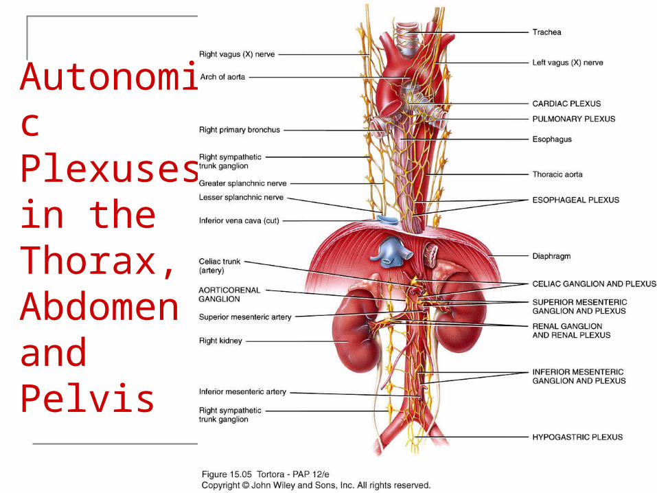
Autonomic Plexuses in the Thorax, Abdomen and Pelvis
Copyright 2009, John Wiley & Sons, Inc.

Pathways from Sympathetic Trunk Ganglia to Visceral Effectors Axons leave the sympathetic trunk in 4
possible ways:
1. - spinal nerves
2. - cephalic periarterial nerves
3. - sympathetic nerves
4. - splanchnic nerves
Copyright 2009, John Wiley & Sons, Inc.
Spinal nerves Gray ramus: Axons of some postganglionic
neurons leave the sympathetic trunk by entering a short pathway called a gray ramus and merge with the anterior ramus of a spinal nerve.
Gray rami communicantes: structures containing sympathetic postganglionic axons that connect the ganglia of the sympathetic trunk to spinal nerves.
Serve skin in neck, trunk, limbs, sweat glands, and arrector pili
Copyright 2009, John Wiley & Sons, Inc.

Postganglionic neurons in the Sympathetic Division
Copyright 2009, John Wiley & Sons, Inc.
Cephalic Periarterial Nerves
Some sympathetic preganglionic neurons that enter the sympathetic trunk ascend to the superior cervical ganglion where they synapse with postganglionic neurons. Some of these leave the sympathetic trunk by forming cephalic periarterial nerves.
Serve visceral effectors in the skin of the face and head.
Copyright 2009, John Wiley & Sons, Inc.

Sympathetic Nerves
Some axons of the postganglionic neurons leave the trunk by forming sympathetic nerves.
Innervate the heart and lungs.
Copyright 2009, John Wiley & Sons, Inc.
Splanchnic Nerves to the Adrenal Medulla Some sympathetic preganglionic axons pass,
without synapsing, through the sympathetic trunk, greater splanchnic nerves and celiac ganglion into the adrenal medulla (modified sympathetic ganglia).
Release hormones into blood- 80% epinephrine, 20% norepinephrine, w/ some dopamine.
Copyright 2009, John Wiley & Sons, Inc.
Splanchnic Nerves continued.. Some sympathetic preganglionic axons pass
through the sympathetic trunk without terminating in it. Beyond the trunk they form nerves called splanchnic nerves which extend to prevertebral ganglia.
T5-T9 or T10- Greater splanchnic nerve. (serves: stomach, spleen, liver, kidneys, and small intestines)
T10-T11- Lesser splanchnic nerve. (serves: blood vessels of small intestine and proximal colon)
L1-L4- Lumbar splanchnic nerve. Terminate in the inferior mesenteric ganglion
Copyright 2009, John Wiley & Sons, Inc.
Cranial Parasympathetic Outflow The cranial outflow has four pairs of ganglia
and are associated with the vagus nerve.1. Ciliary ganglia-muscle fibers in eye ball
2. Pterygopalatine ganglia-membranes and glands of head
3. Submandibular ganglia-sublingual salivary glands
4. Otic ganglia-parotid salivary glands Vagus nerve carries nearly 80% of the total
craniosacral flow.
Copyright 2009, John Wiley & Sons, Inc.
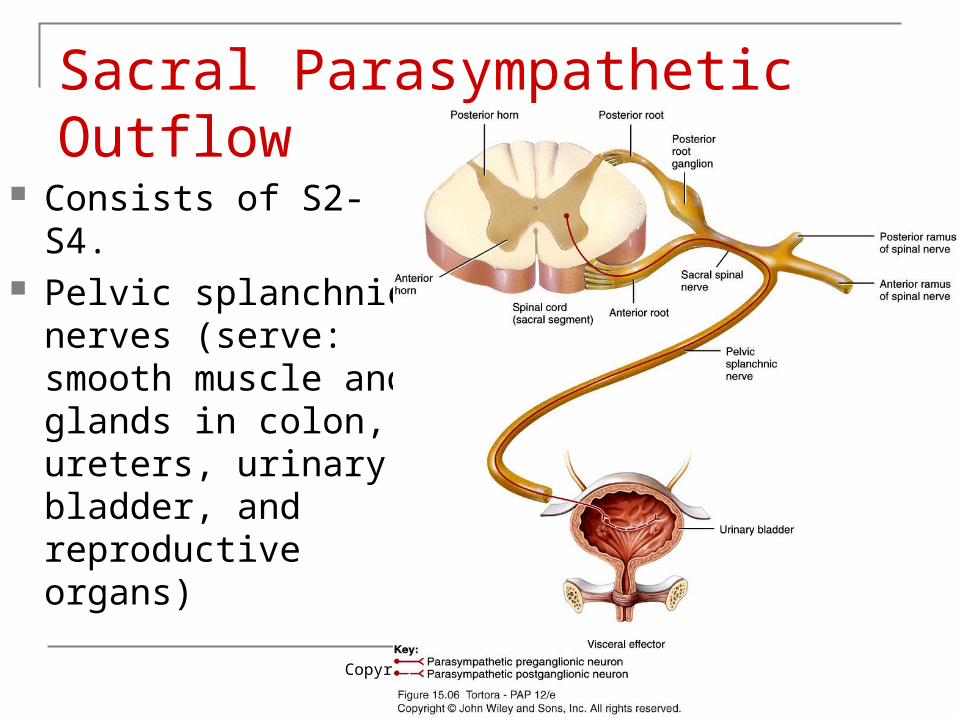
Sacral Parasympathetic Outflow
Consists of S2-S4. Pelvic splanchnic
nerves (serve: smooth muscle and glands in colon, ureters, urinary bladder, and reproductive organs)
Copyright 2009, John Wiley & Sons, Inc.
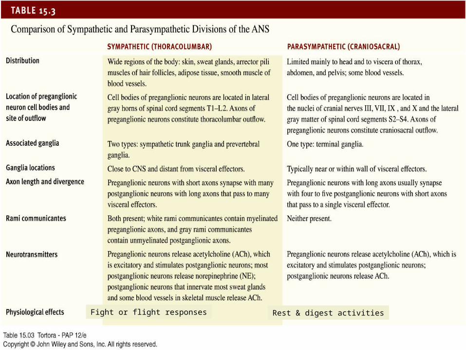
Fight or flight responses Rest & digest activities
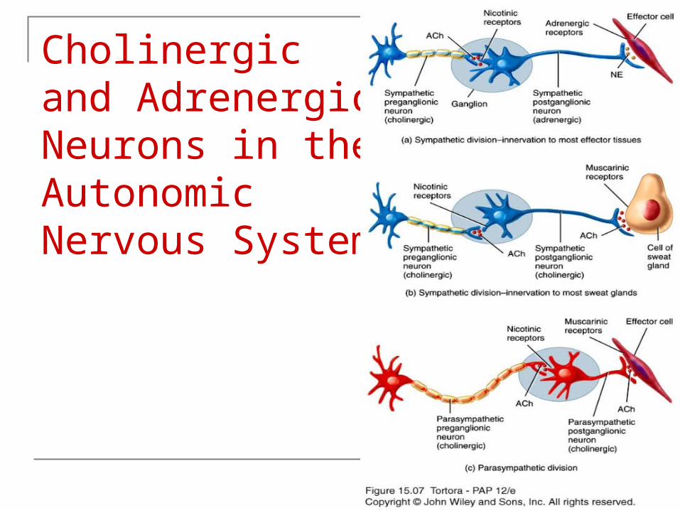
Cholinergic and Adrenergic Neurons in the Autonomic Nervous System

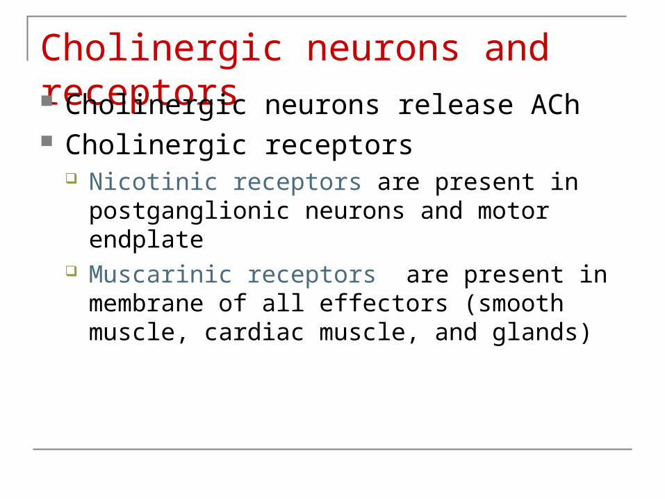
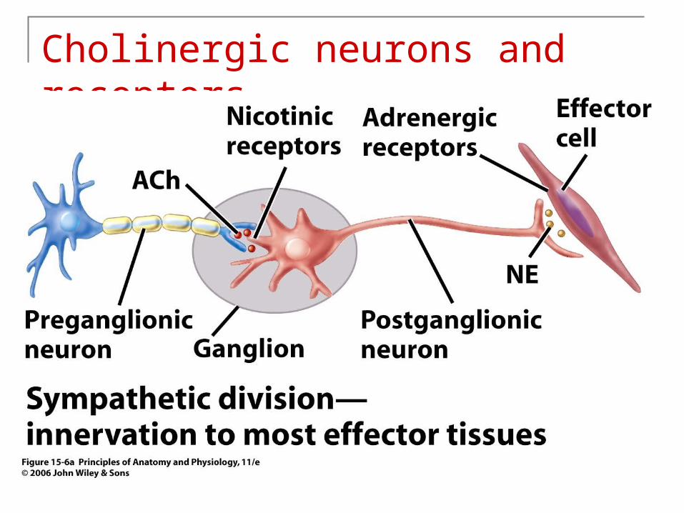
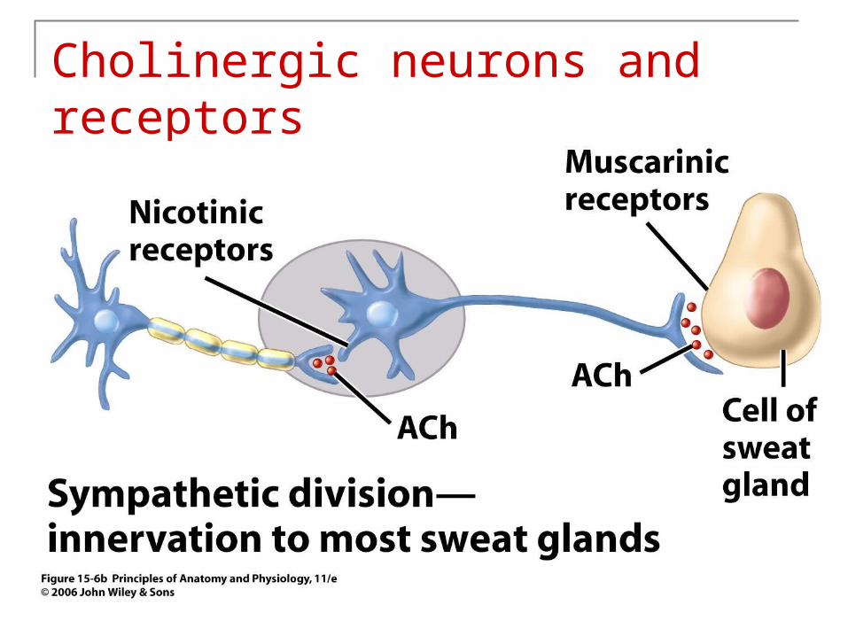
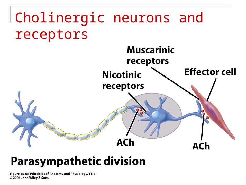
Cholinergic Neurons & Cholinergic Receptors:Cholinergic neurons → release acetylcholine (ACh).
Cholinergic neurons include-
1.All sympathetic and parasympathetic preganglionic neurons.
2.Sympathetic postganglionic neurons that innervate most sweat glands.
3.All parasympathetic postganglionic neurons.
Cholinergic Receptors:
1.Nicotinic receptors – present in postganglion neurons for Para. and Sym. and motor end plate
2.Muscarinic receptors – present in membrane of all effectors(smooth
muscle, cardiac muscle and glands)
Copyright 2009, John Wiley & Sons, Inc.
Cholinergic neurons and receptors Cholinergic neurons release ACh Cholinergic receptors
Nicotinic receptors are present in postganglionic neurons and motor endplate
Muscarinic receptors are present in membrane of all effectors (smooth muscle, cardiac muscle, and glands)
Cholinergic neurons and receptors
Cholinergic neurons and receptors
Cholinergic neurons and receptors

Adrenergic neurons and receptors Adrenergic neurons release norepinephrine
(NE) Adrenergic receptors
Alpha and beta receptors are on visceral effectors 1 and 1 are excitatory 1 and 2 are inhibitory 3 is on brown adipose tissue and is involved in
thermogenesis
Adrenergic neurons and receptors
Physiology of the ANS
Autonomic tone- a balance between the sympathetic and parasympathetic activity.
Regulated by the hypothalamus.
Copyright 2009, John Wiley & Sons, Inc.
Sympathetic Responses Stress ↑ sympathetic system ↑ fight-or-flight response. ↑ production of ATP. Dilation of the pupils. ↑ heart rate and blood pressure. Dilation of the airways.
Constriction of blood vessels that supply the kidneys and gastrointestinal tract. ↑ blood supply to the skeletal muscles, cardiac muscle, liver and adipose
tissue ↑ glycogenolysis ↑ blood glucose.
↑ lipolysis.
Copyright 2009, John Wiley & Sons, Inc.
Parasympathetic Responses Rest-and-digest response. Conserve and restore body energy. ↑ digestive and urinary function. ↓ body functions that support physical activity. SLUDD—salivation, lacrimation, urination, digestion, defecation Decreased heart rate Decreased diameter of airways Decreased diameter of the pupils
Copyright 2009, John Wiley & Sons, Inc.
Autonomic reflexes Regulate controlled conditions in the body
Blood pressure Digestion Defecation Urination
The reflex arc organizes the response.
Receptor—distal end of the sensory neuron Sensory neuron--projects to CNS Integration center—hypothalamus and brain stem and spinal cord Motor neurons project from CNS through one or two synapses Effector—effects on smooth muscle, cardiac muscle, and glands

Visceral Reflex Arc
Copyright © 2005 Pearson Education, Inc., publishing as Benjamin Cummings
Autonomic reflexes Receptor—distal end of the sensory neuron Sensory neuron--projects to CNS Integration center—hypothalamus and brain stem and spinal cord Motor neurons project from CNS through one or two synapses
Effector—effects on smooth muscle, cardiac muscle, and glands.
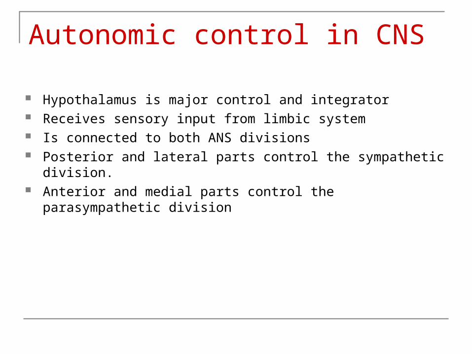
Autonomic control in CNS
Hypothalamus is major control and integrator Receives sensory input from limbic system Is connected to both ANS divisions Posterior and lateral parts control the sympathetic division. Anterior and medial parts control the parasympathetic division


