56
Chapter 19 Regulation of Metabolism
| Date post: | 25-Dec-2015 |
| Category: |
Documents |
| Upload: | kelly-ross |
| View: | 216 times |
| Download: | 3 times |
Chapter 19
Regulation of Metabolism
Copyright © The McGraw-Hill Companies, Inc. Permission required for reproduction or display.
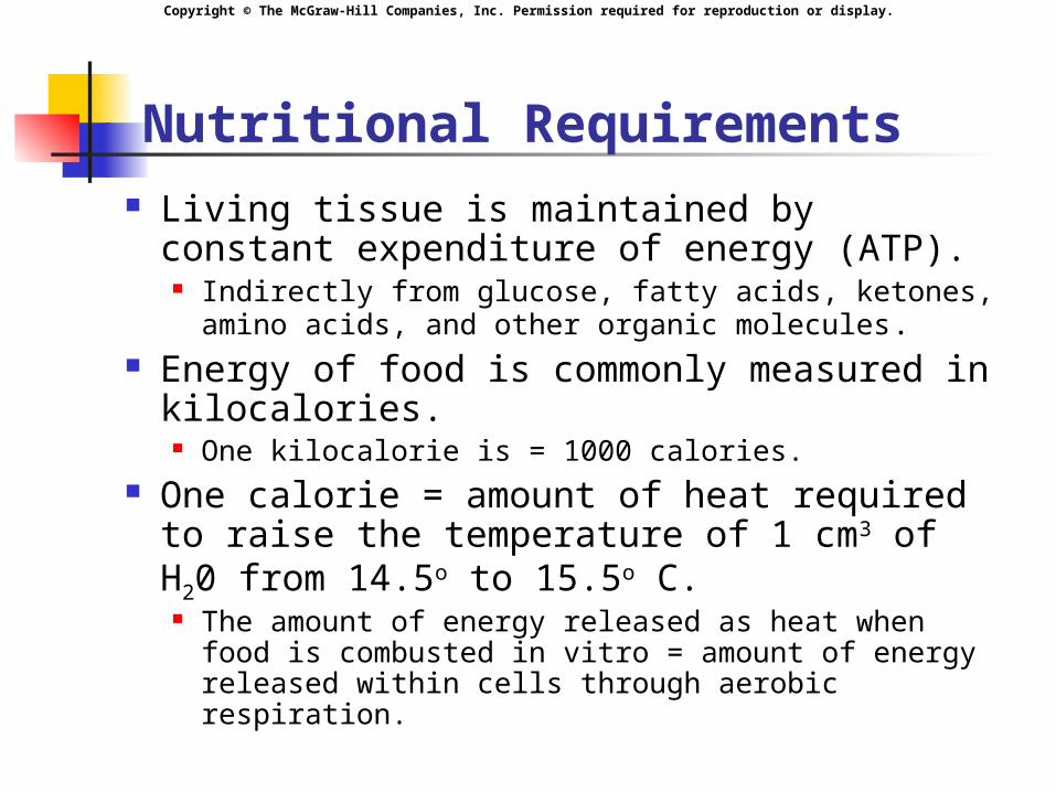
Nutritional Requirements Living tissue is maintained by constant
expenditure of energy (ATP). Indirectly from glucose, fatty acids, ketones,
amino acids, and other organic molecules. Energy of food is commonly measured in
kilocalories. One kilocalorie is = 1000 calories.
One calorie = amount of heat required to raise the temperature of 1 cm3 of H20 from 14.5o to 15.5o C.
The amount of energy released as heat when food is combusted in vitro = amount of energy released within cells through aerobic respiration.
Copyright © The McGraw-Hill Companies, Inc. Permission required for reproduction or display.
Metabolic Rate and Caloric Requirements
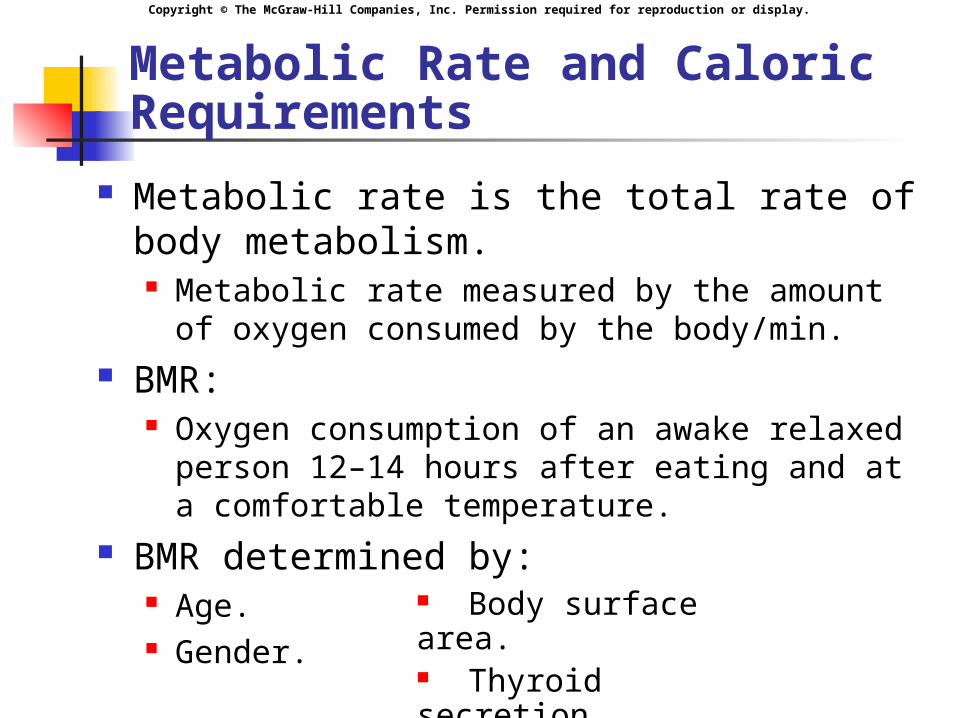
Metabolic rate is the total rate of body metabolism. Metabolic rate measured by the amount of
oxygen consumed by the body/min. BMR:
Oxygen consumption of an awake relaxed person 12–14 hours after eating and at a comfortable temperature.
BMR determined by: Age. Gender.
Body surface area. Thyroid secretion.
Copyright © The McGraw-Hill Companies, Inc. Permission required for reproduction or display.
Anabolic Requirements
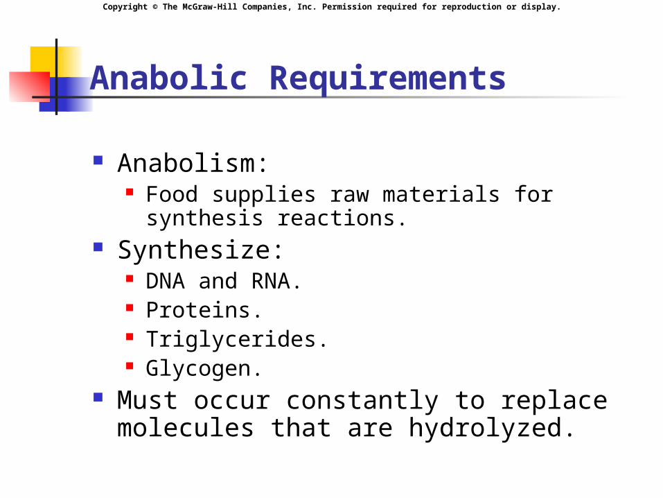
Anabolism: Food supplies raw materials for synthesis
reactions. Synthesize:
DNA and RNA. Proteins. Triglycerides. Glycogen.
Must occur constantly to replace molecules that are hydrolyzed.
Copyright © The McGraw-Hill Companies, Inc. Permission required for reproduction or display.
Aerobic Requirements (continued)
Catabolism: Hydrolysis (break down monomers
down to C02 and H20.): Hydrolysis reactions and cellular
respiration. Gluconeogenesis. Glycogenolysis. Lipolysis.
Copyright © The McGraw-Hill Companies, Inc. Permission required for reproduction or display.
Turnover Rate
Rate at which a molecule is broken down and resynthesized.
Average daily turnover for carbohydrates is 250 g/day.
Some glucose is reused to form glycogen. Only need about 150 g/day.
Average daily turnover for protein is 150 g/day. Some protein may be reused for protein synthesis.
Only need 35 g/day. 9 essential amino acids.
Average daily turnover for fats is 100 g/day. Little is actually required in the diet.
Fat can be produced from excess carbohydrates. Essential fatty acids:
Linoleic and linolenic acids.
Copyright © The McGraw-Hill Companies, Inc. Permission required for reproduction or display.
Vitamins and Minerals
Vitamins: Small organic molecules that serve as coenzymes in
metabolic reactions or have highly specific functions. Must be obtained from the diet because the
body does not produce them, or does so in insufficient amounts.
2 classes of vitamins: Fat-soluble:
A,D, E, and K. Water-soluble:
B1, B2, B3, B6, B12, pantothenic acid, biotin, folic acid, and vitamin C.
Copyright © The McGraw-Hill Companies, Inc. Permission required for reproduction or display.
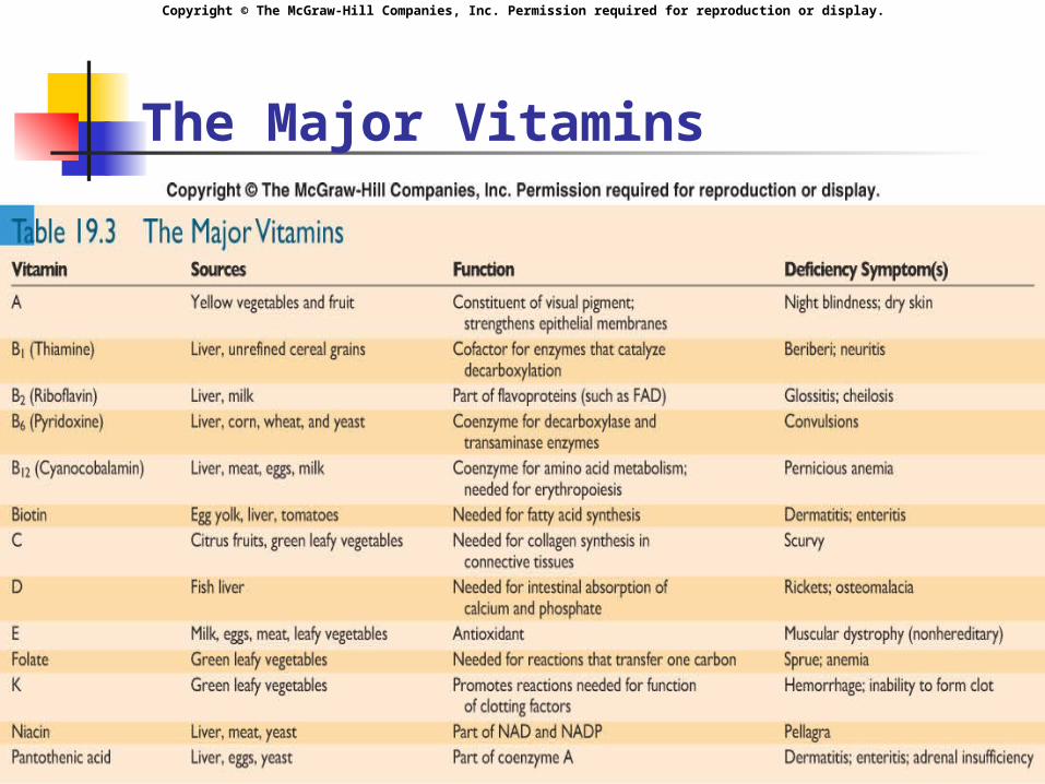
Vitamins
Water-soluble vitamins: Serve as coenzymes in the metabolism of
carbohydrates, lipids, and proteins. May serve as antioxidants.
Fat-soluble vitamins: Bind to nuclear receptors. Serve as antioxidants. Assist in regulation of fetal development. Regulate Ca2+ balance.
Copyright © The McGraw-Hill Companies, Inc. Permission required for reproduction or display.
The Major Vitamins
Copyright © The McGraw-Hill Companies, Inc. Permission required for reproduction or display.
Minerals
Needed as cofactors for specific enzymes and other critical functions.
Trace elements: Required in small amounts from 50 g
to 18 mg/day.
Copyright © The McGraw-Hill Companies, Inc. Permission required for reproduction or display.
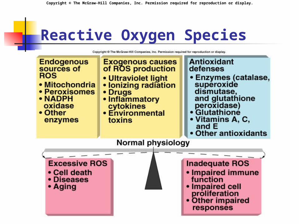
Free Radicals and Antioxidants
Electrons are located in orbitals. Each orbital contains a maximum of 2
electrons. Free radical:
When an orbital has an unpaired electron. Highly reactive in the body. Oxidize or reduce other atoms.
Major free radicals called: Reactive oxygen or nitrogen species:
Oxygen or nitrogen as unpaired electron.
Copyright © The McGraw-Hill Companies, Inc. Permission required for reproduction or display.
Free Radicals and Antioxidants (continued)
Functions of free radicals: Help to destroy bacteria. Produce vasodilation.
NO radical, superoxide radical, and hydroxy radical. Exert oxidative stress contributing to disease
states. Excess production of free radicals can damage lipids,
proteins, and DNA. Promotes apoptosis, contributes to aging, inflammatory
disease, heart disease, CVA, HTN, and degenerative disease.
Promotes malignant growth. Protective mechanism against oxidative stress.
Can react with free radicals by picking up unpaired electrons.
Glutathione, vitamin C, and vitamin E.
Copyright © The McGraw-Hill Companies, Inc. Permission required for reproduction or display.
Reactive Oxygen Species
Insert fig. 19.1
Copyright © The McGraw-Hill Companies, Inc. Permission required for reproduction or display.
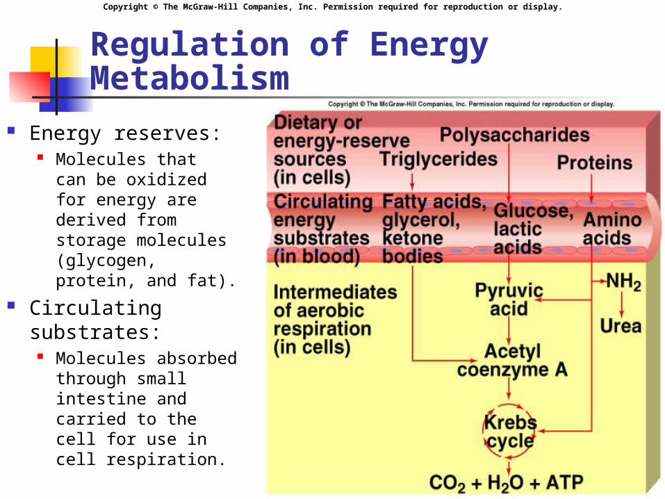
Regulation of Energy Metabolism
Energy reserves: Molecules that
can be oxidized for energy are derived from storage molecules (glycogen, protein, and fat).
Circulating substrates:
Molecules absorbed through small intestine and carried to the cell for use in cell respiration.
Insert fig. 19.2
Copyright © The McGraw-Hill Companies, Inc. Permission required for reproduction or display.
Eating
Eating behaviors partially controlled by hypothalamus.
Lesions in vetromedial area produce hyperphagia (obesity).
Lesions in lateral hypothalamus produces hypophagia (weight loss).
Endorphins, NE, serotonin, and CCK affect hunger and satiety.
Copyright © The McGraw-Hill Companies, Inc. Permission required for reproduction or display.
Regulatory Functions of Adipose Tissue
Adipostat regulatory system (negative feedback loops) to defend amount of adipose tissue. Differentiation of adipocytes require nuclear
receptor protein (PPAR which is activated when bound to 15-D PGJ2:
Stimulates adipogenesis by promoting development of preadipocytes into mature adipocytes.
Number of adipocytes increase after birth. Differentiation promoted by high [fatty acids].
Adipocytes store fat within large vacuoles. May secrete hormones involved in regulation
of metabolism.
Copyright © The McGraw-Hill Companies, Inc. Permission required for reproduction or display.
Regulatory Functions of Adipose Tissue (continued)
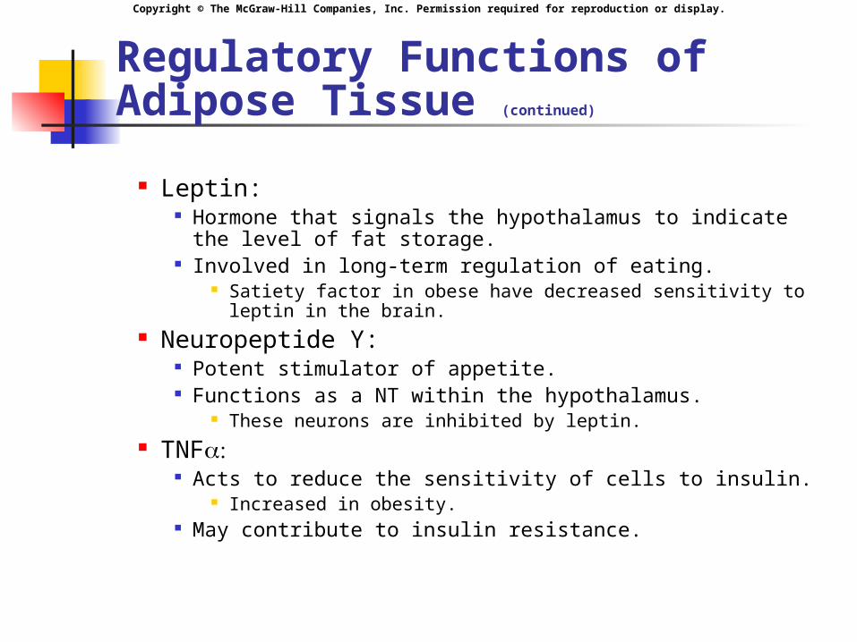
Leptin: Hormone that signals the hypothalamus to indicate the
level of fat storage. Involved in long-term regulation of eating.
Satiety factor in obese have decreased sensitivity to leptin in the brain.
Neuropeptide Y: Potent stimulator of appetite. Functions as a NT within the hypothalamus.
These neurons are inhibited by leptin. TNF
Acts to reduce the sensitivity of cells to insulin. Increased in obesity.
May contribute to insulin resistance.
Copyright © The McGraw-Hill Companies, Inc. Permission required for reproduction or display.
Regulation of Hunger
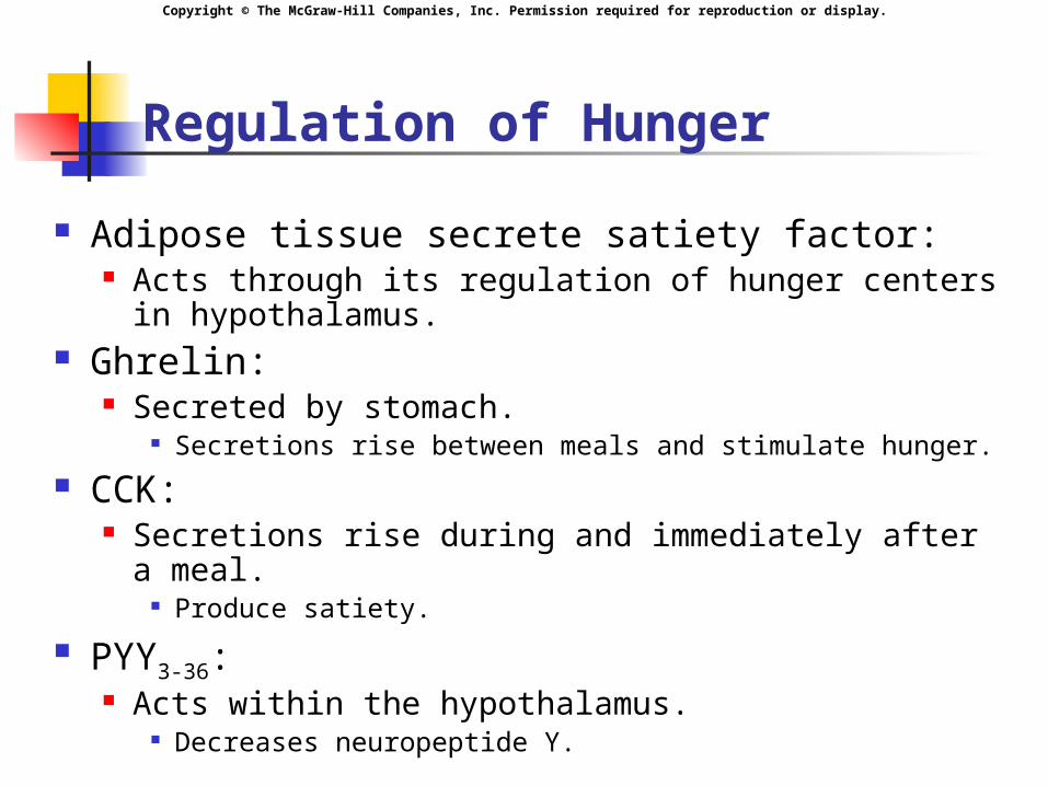
Adipose tissue secrete satiety factor: Acts through its regulation of hunger centers in
hypothalamus. Ghrelin:
Secreted by stomach. Secretions rise between meals and stimulate hunger.
CCK: Secretions rise during and immediately after a
meal. Produce satiety.
PYY3-36: Acts within the hypothalamus.
Decreases neuropeptide Y.
Copyright © The McGraw-Hill Companies, Inc. Permission required for reproduction or display.
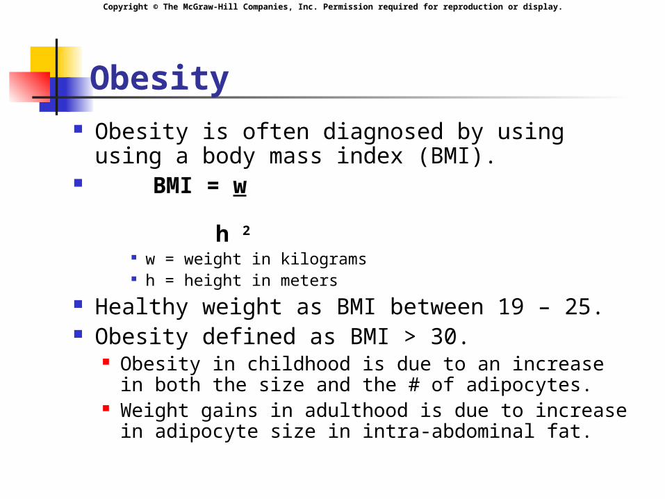
Obesity Obesity is often diagnosed by using using
a body mass index (BMI). BMI = w
h 2
w = weight in kilograms h = height in meters
Healthy weight as BMI between 19 – 25. Obesity defined as BMI > 30.
Obesity in childhood is due to an increase in both the size and the # of adipocytes.
Weight gains in adulthood is due to increase in adipocyte size in intra-abdominal fat.
Copyright © The McGraw-Hill Companies, Inc. Permission required for reproduction or display.
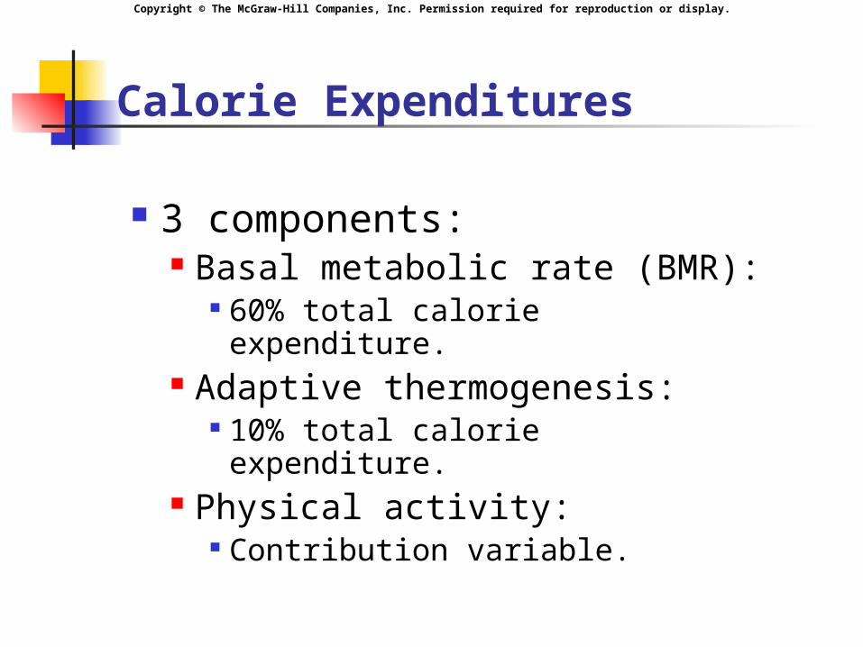
Calorie Expenditures
3 components: Basal metabolic rate (BMR):
60% total calorie expenditure. Adaptive thermogenesis:
10% total calorie expenditure. Physical activity:
Contribution variable.
Copyright © The McGraw-Hill Companies, Inc. Permission required for reproduction or display.
Hormonal Regulation of Metabolism
Absorptive state: Absorption of energy. 4 hour period after eating. Increase in insulin secretion.
Postabsorptive state: Fasting state. At least 4 hours after the meal. Increase in glucagon secretion.
Copyright © The McGraw-Hill Companies, Inc. Permission required for reproduction or display.
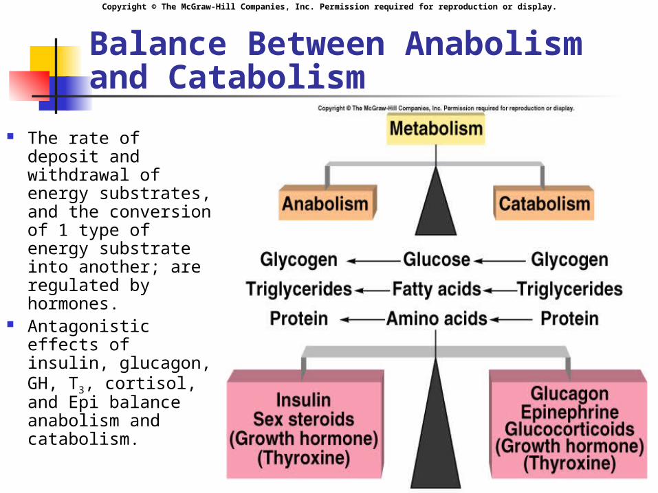
Balance Between Anabolism and Catabolism
The rate of deposit and withdrawal of energy substrates, and the conversion of 1 type of energy substrate into another; are regulated by hormones.
Antagonistic effects of insulin, glucagon, GH, T3, cortisol, and Epi balance anabolism and catabolism.
Insert fig. 19.4
Copyright © The McGraw-Hill Companies, Inc. Permission required for reproduction or display.
Energy Regulation of Pancreas
Islets of Langerhans contain 3 distinct cell types: cells
Secreteglucagon. cells
Secreteinsulin. cells
Secrete somatostatin.
Copyright © The McGraw-Hill Companies, Inc. Permission required for reproduction or display.
Regulation of Insulin and Glucagon
Mainly regulated by blood [glucose].
Lesser effect: blood [amino acid]. Regulated by negative feedback.
Glucose enters the brain by facilitated diffusion.
Normal fasting [glucose] is 65–105 mg/dl.
Copyright © The McGraw-Hill Companies, Inc. Permission required for reproduction or display.
Regulation of Insulin and Glucagon (continued)
When blood [glucose] increases: Glucose binds to GLUT2 receptor
protein in cells, stimulating the production and release of insulin.
Insulin: Stimulates skeletal muscle cells and
adipocytes to incorporate GLUT4 (glucose facilitated diffusion carrier) into plasma membranes.
Promotes anabolism.
Copyright © The McGraw-Hill Companies, Inc. Permission required for reproduction or display.
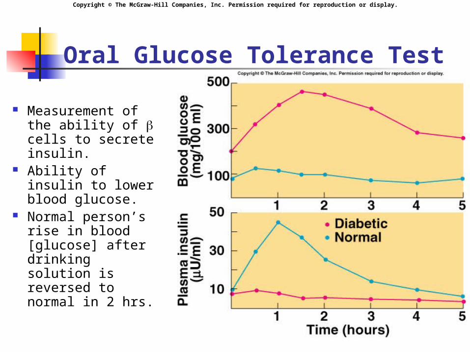
Oral Glucose Tolerance Test
Measurement of the ability of cells to secrete insulin.
Ability of insulin to lower blood glucose.
Normal person’s rise in blood [glucose] after drinking solution is reversed to normal in 2 hrs.
Insert fig. 19.8
Copyright © The McGraw-Hill Companies, Inc. Permission required for reproduction or display.
Regulation of Insulin and Glucagon
Parasympathetic nervous system: Stimulates insulin secretion.
Sympathetic nervous system: Stimulates glucagon secretion.
GIP: Stimulates insulin secretion.
GLP-1: Stimulates insulin secretion.
CCK: Stimulates insulin secretion.
Copyright © The McGraw-Hill Companies, Inc. Permission required for reproduction or display.
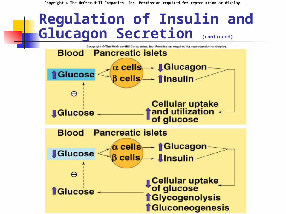
Regulation of Insulin and Glucagon Secretion (continued)
Copyright © The McGraw-Hill Companies, Inc. Permission required for reproduction or display.
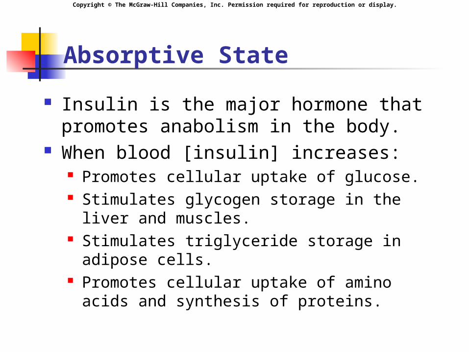
Absorptive State
Insulin is the major hormone that promotes anabolism in the body.
When blood [insulin] increases: Promotes cellular uptake of glucose. Stimulates glycogen storage in the liver and
muscles. Stimulates triglyceride storage in adipose
cells. Promotes cellular uptake of amino acids and
synthesis of proteins.
Copyright © The McGraw-Hill Companies, Inc. Permission required for reproduction or display.
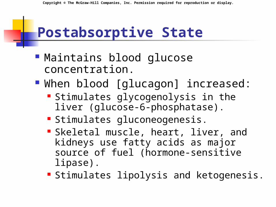
Postabsorptive State
Maintains blood glucose concentration.
When blood [glucagon] increased: Stimulates glycogenolysis in the liver
(glucose-6-phosphatase). Stimulates gluconeogenesis. Skeletal muscle, heart, liver, and
kidneys use fatty acids as major source of fuel (hormone-sensitive lipase).
Stimulates lipolysis and ketogenesis.
Copyright © The McGraw-Hill Companies, Inc. Permission required for reproduction or display.
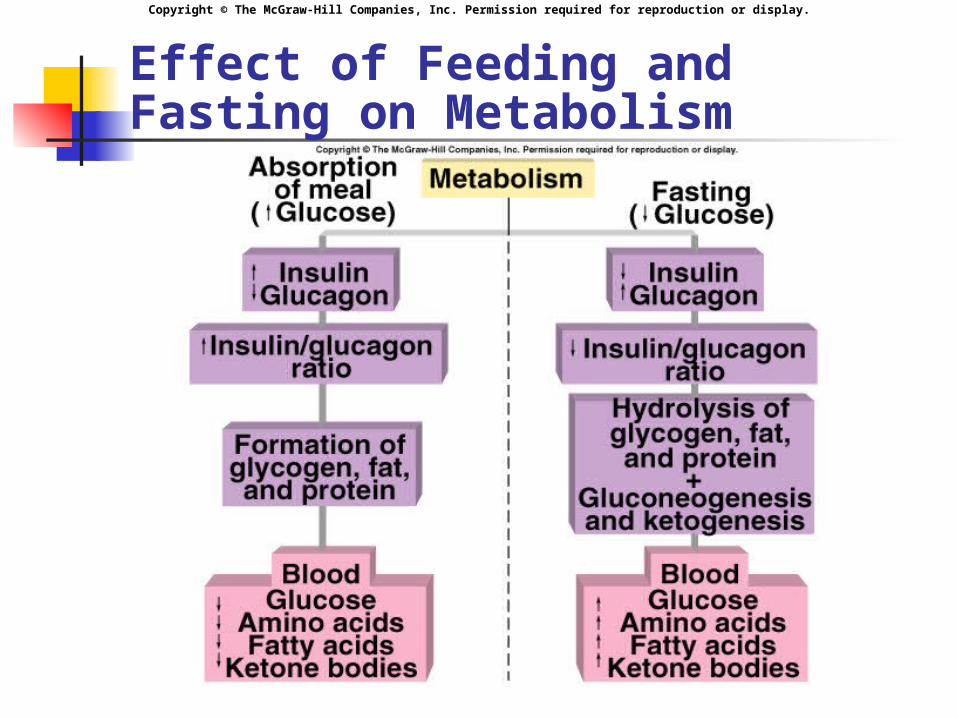
Insert fig. 19.10
Effect of Feeding and Fasting on Metabolism
Copyright © The McGraw-Hill Companies, Inc. Permission required for reproduction or display.
Diabetes Mellitus
Chronic high blood [glucose]. 2 forms of diabetes mellitus:
Type I: insulin dependent diabetes (IDDM).
Type II: non-insulin dependent diabetes (NIDDM).
Copyright © The McGraw-Hill Companies, Inc. Permission required for reproduction or display.
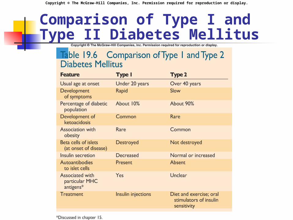
Comparison of Type I and Type II Diabetes Mellitus
Insert table 19.6
Copyright © The McGraw-Hill Companies, Inc. Permission required for reproduction or display.
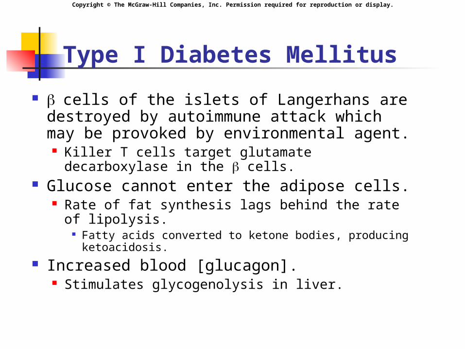
Type I Diabetes Mellitus
cells of the islets of Langerhans are destroyed by autoimmune attack which may be provoked by environmental agent. Killer T cells target glutamate decarboxylase in
the cells. Glucose cannot enter the adipose cells.
Rate of fat synthesis lags behind the rate of lipolysis.
Fatty acids converted to ketone bodies, producing ketoacidosis.
Increased blood [glucagon]. Stimulates glycogenolysis in liver.
Copyright © The McGraw-Hill Companies, Inc. Permission required for reproduction or display.
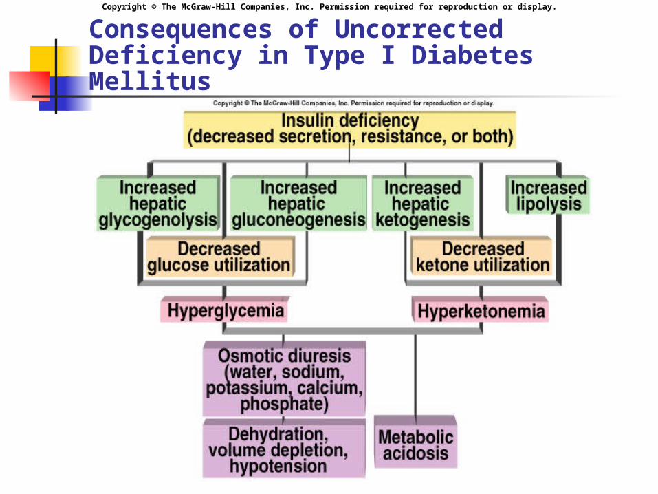
Consequences of Uncorrected Deficiency in Type I Diabetes Mellitus
Insert fig. 19.11
Copyright © The McGraw-Hill Companies, Inc. Permission required for reproduction or display.
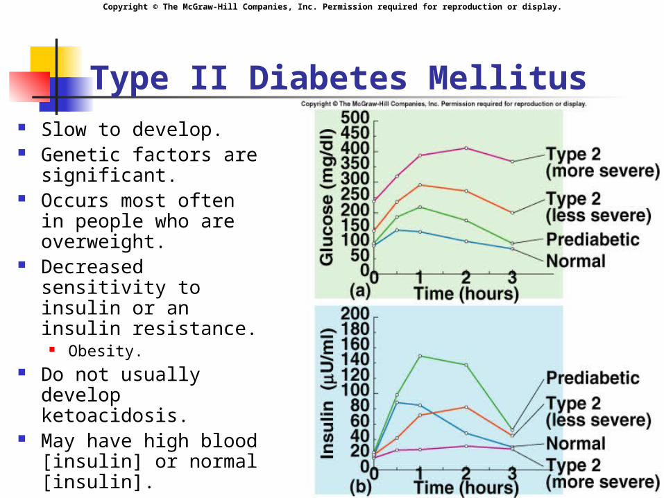
Type II Diabetes Mellitus Slow to develop. Genetic factors are
significant. Occurs most often in
people who are overweight.
Decreased sensitivity to insulin or an insulin resistance.
Obesity. Do not usually
develop ketoacidosis. May have high blood
[insulin] or normal [insulin].
Insert fig. 19.12
Copyright © The McGraw-Hill Companies, Inc. Permission required for reproduction or display.
Treatment in Diabetes
Change in lifestyle: Increase exercise:
Increases the amount of membrane GLUT-4 carriers in the skeletal muscle cells.
Weight reduction. Increased fiber in diet. Reduce saturated fat.
Copyright © The McGraw-Hill Companies, Inc. Permission required for reproduction or display.
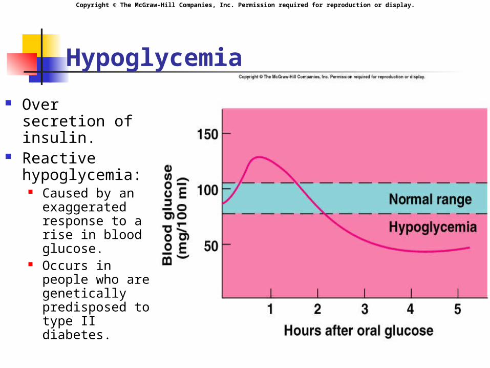
Hypoglycemia
Over secretion of insulin.
Reactive hypoglycemia:
Caused by an exaggerated response to a rise in blood glucose.
Occurs in people who are genetically predisposed to type II diabetes.
Insert fig. 19.13
Copyright © The McGraw-Hill Companies, Inc. Permission required for reproduction or display.
Metabolic Regulation
Anabolic effects of insulin are antagonized by the hormones of the adrenals, thyroid, and anterior pituitary. Insulin, T3, and GH can act
synergistically to stimulate protein synthesis.
Copyright © The McGraw-Hill Companies, Inc. Permission required for reproduction or display.
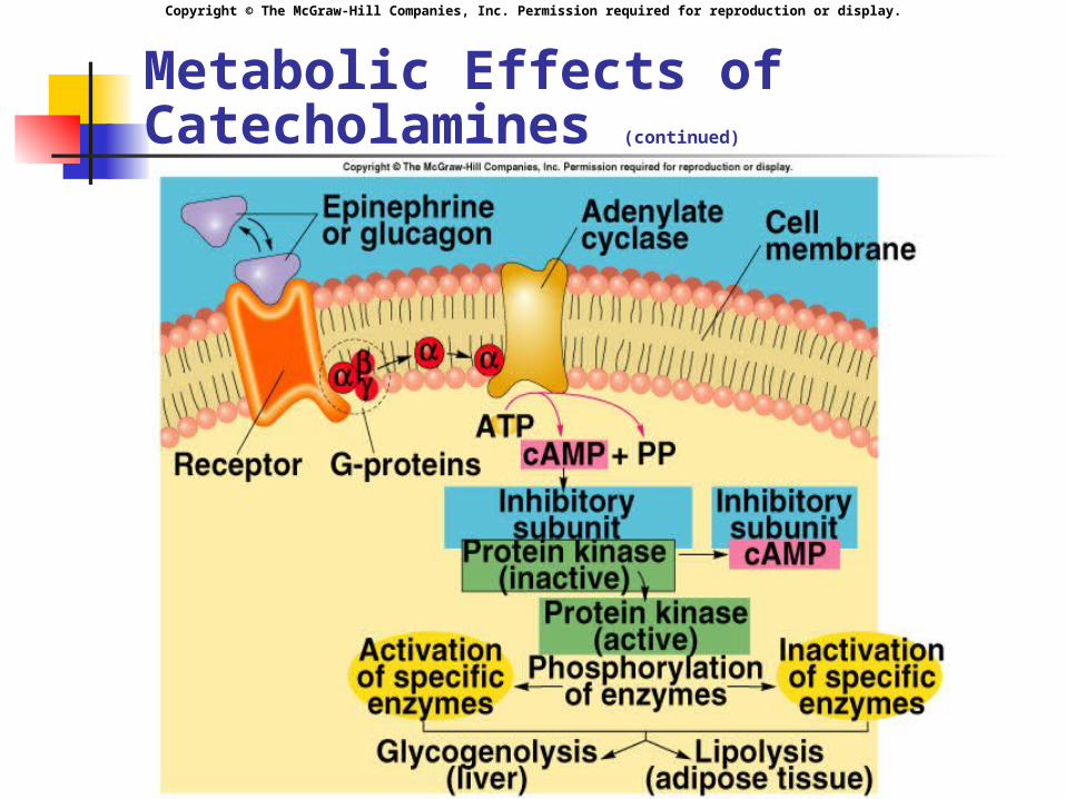
Metabolic Effects of Catecholamines
Metabolic effects similar to glucagon. Stimulate glycogenolysis.
Stimulate release of glucose from the liver. Stimulate lipolysis and release of fatty acids.
NE stimulates 3 receptors in brown fat. Contains uncoupling protein that dissociates
electron transport from ATP production.
Copyright © The McGraw-Hill Companies, Inc. Permission required for reproduction or display.
Metabolic Effects of Catecholamines (continued)
Copyright © The McGraw-Hill Companies, Inc. Permission required for reproduction or display.
Metabolic Effects of Glucocorticoids
Glucocorticoids secreted in response to release of ACTH.
Support the effects of increased glucagon.
Promote lipolysis and ketogenesis. Promote protein breakdown in the
muscles. Increases blood [amino acids].
Promote liver gluconeogenesis.
Copyright © The McGraw-Hill Companies, Inc. Permission required for reproduction or display.
Thyroxine
Active form is T3. Stimulates cellular respiration by:
Production of uncoupling proteins. Stimulation of active transport Na+/K+
pumps: Lowers cellular [ATP].
Increases metabolic heat. Increases metabolic rate. Contributes to proper growth and
development of CNS in children.
Copyright © The McGraw-Hill Companies, Inc. Permission required for reproduction or display.
Growth Hormone (Somatotropin)
Inhibited by somatostatin. Stimulates growth in children and
adolescents. Stimulated by:
GHRH. Increase in blood [amino acids]. Decrease in blood [glucose].
Pulsatile, increasing during sleep, decreasing during day.
Copyright © The McGraw-Hill Companies, Inc. Permission required for reproduction or display.
Growth Hormone (continued)
IGF-1: Liver produces and secretes IGF-1 in
response to GH. Stimulates cell division and growth of
cartilage. IGF-2:
Has more insulin-like actions. Promotes anabolism and catabolism.
Stimulates cellular uptake of amino acids and protein synthesis.
Decreases glucose utilization by the tissues. Raises blood [glucose].
Copyright © The McGraw-Hill Companies, Inc. Permission required for reproduction or display.
Effects of Growth Hormone on Body Growth
Gigantism: Excess GH secretion in children.
Maintain normal body proportions. Acromegaly:
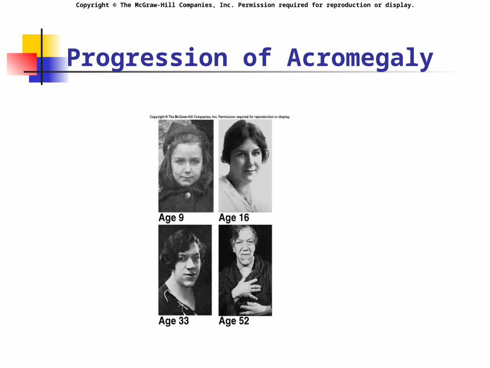
Excess GH secretion in adults after the epiphyseal discs are sealed.
No increase in height. Growth of soft tissue.
Elongation of jaw, deformities in hands, feet, and bones of face.
Dwarfism: Inadequate secretion of GH during childhood.
Copyright © The McGraw-Hill Companies, Inc. Permission required for reproduction or display.
Progression of Acromegaly
Copyright © The McGraw-Hill Companies, Inc. Permission required for reproduction or display.
Bone Deposition and Resorption
Ca2+ and phosphate concentrations are affected by: Bone formation and resorption. Intestinal absorption of Ca2+ and P04
3-. Urinary excretion.
Osteoblasts: Secrete an organic matrix of collagen proteins. Deposit hydroxyapatite crystals.
Osteoclasts: Secrete enzymes to dissolve hydroxyapatite.
Formation and resorption of bone occur constantly at rates determined by osteoblasts and osteoclasts.
Copyright © The McGraw-Hill Companies, Inc. Permission required for reproduction or display.
Bone Deposition and Resorption (continued)
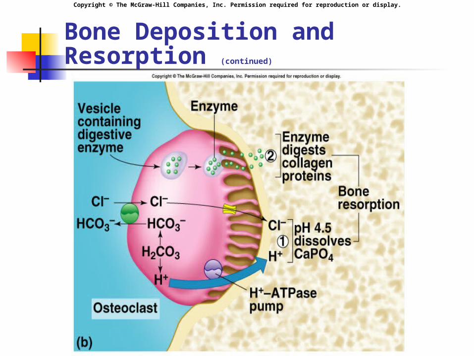
Bone resorption occurs when an osteoclast attaches to the bone matrix and forms ruffled membrane.
Osteoclast secretes products that dissolve both Ca2+ and P04
3- ; and digest the matrix. Transport of H+ by H+ ATPase pump in ruffled border. Cl- channel allows Cl- to flow to H+ to maintain electrical
neutrality. Protein matrix digested by cathepsin K.
Cytoplasm prevented from becoming to basic by a Cl-/HC03
- pump.
Copyright © The McGraw-Hill Companies, Inc. Permission required for reproduction or display.
Bone Deposition and Resorption (continued)
Copyright © The McGraw-Hill Companies, Inc. Permission required for reproduction or display.
Parathyroid Hormone (PTH)
Single most important hormone in the control of blood [Ca2+]. Stimulated by decreased blood [Ca2+].
Stimulates osteoclasts to reabsorb bone. Stimulates kidneys to reabsorb Ca2+ from
glomerular filtrate, and inhibit reabsorption of P04
3-. Promotes formation of 1,25 vitamin D3. Many cancers secrete PTH-related protein
that interacts with PTH receptors. Produce hypercalcemia.
Copyright © The McGraw-Hill Companies, Inc. Permission required for reproduction or display.
Calcitonin
Works with PTH and 1,25 vitamin D3 to regulate blood [Ca2+].
Stimulated by increased plasma [Ca2+]. Inhibits the activity of osteoclasts. Stimulates urinary excretion of Ca2+
and P043- by inhibiting reabsorption.
Physiological significance in adults is questionable.
Copyright © The McGraw-Hill Companies, Inc. Permission required for reproduction or display.
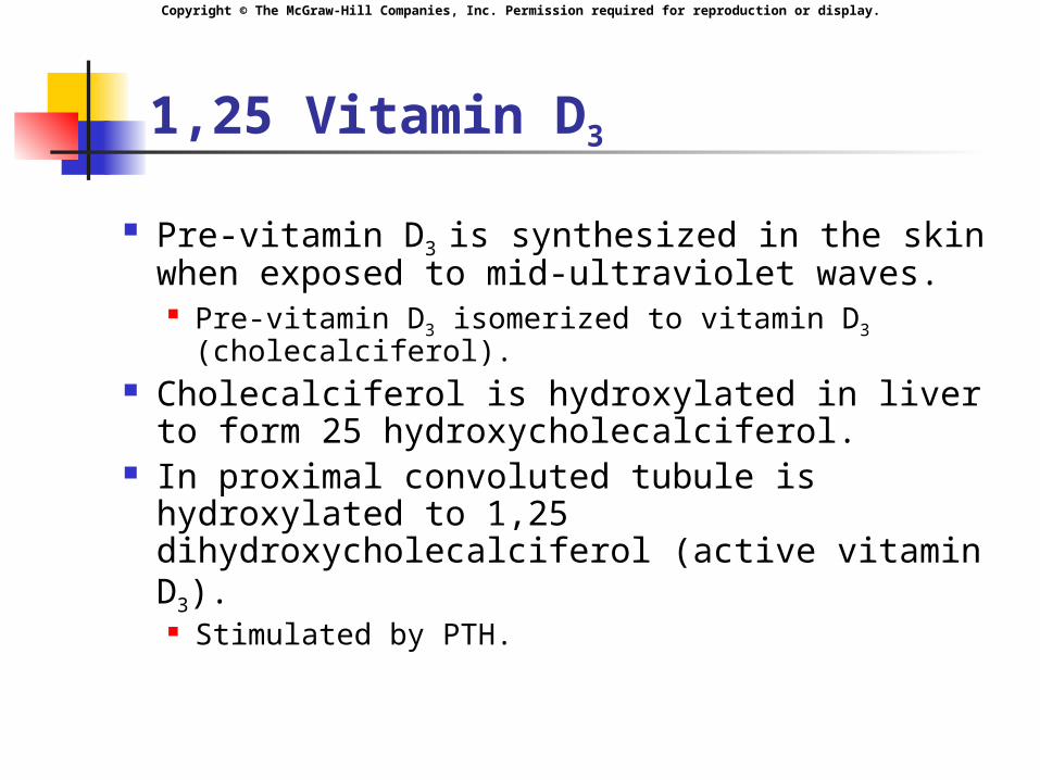
1,25 Vitamin D3
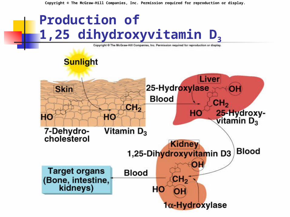
Pre-vitamin D3 is synthesized in the skin when exposed to mid-ultraviolet waves. Pre-vitamin D3 isomerized to vitamin D3
(cholecalciferol). Cholecalciferol is hydroxylated in liver
to form 25 hydroxycholecalciferol. In proximal convoluted tubule is
hydroxylated to 1,25 dihydroxycholecalciferol (active vitamin D3). Stimulated by PTH.
Copyright © The McGraw-Hill Companies, Inc. Permission required for reproduction or display.
Production of 1,25 dihydroxyvitamin D3
Insert fig. 19.20
Copyright © The McGraw-Hill Companies, Inc. Permission required for reproduction or display.
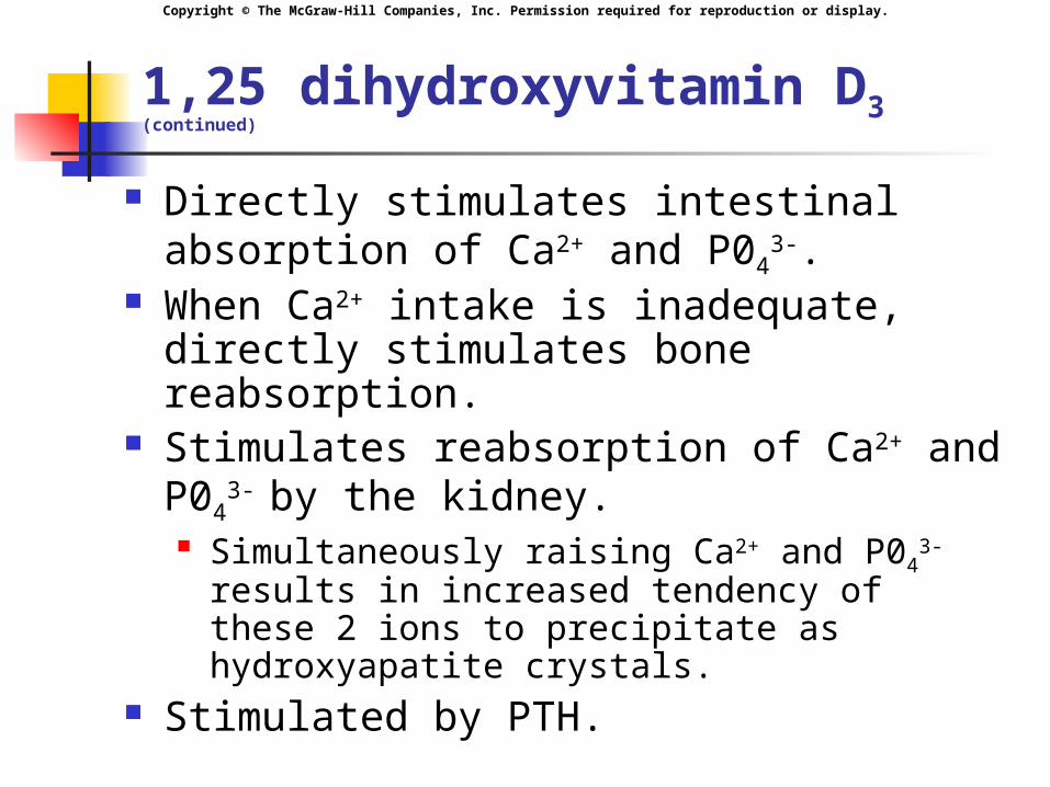
1,25 dihydroxyvitamin D3 (continued)
Directly stimulates intestinal absorption of Ca2+ and P04
3-. When Ca2+ intake is inadequate, directly
stimulates bone reabsorption. Stimulates reabsorption of Ca2+ and P04
3-
by the kidney. Simultaneously raising Ca2+ and P04
3- results in increased tendency of these 2 ions to precipitate as hydroxyapatite crystals.
Stimulated by PTH.
Copyright © The McGraw-Hill Companies, Inc. Permission required for reproduction or display.
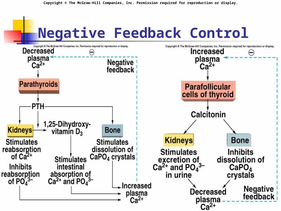
Negative Feedback Control
Insert fig. 19.23


