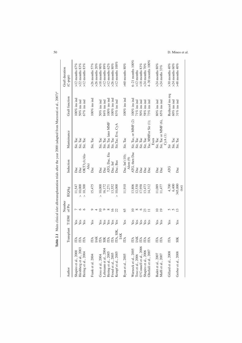
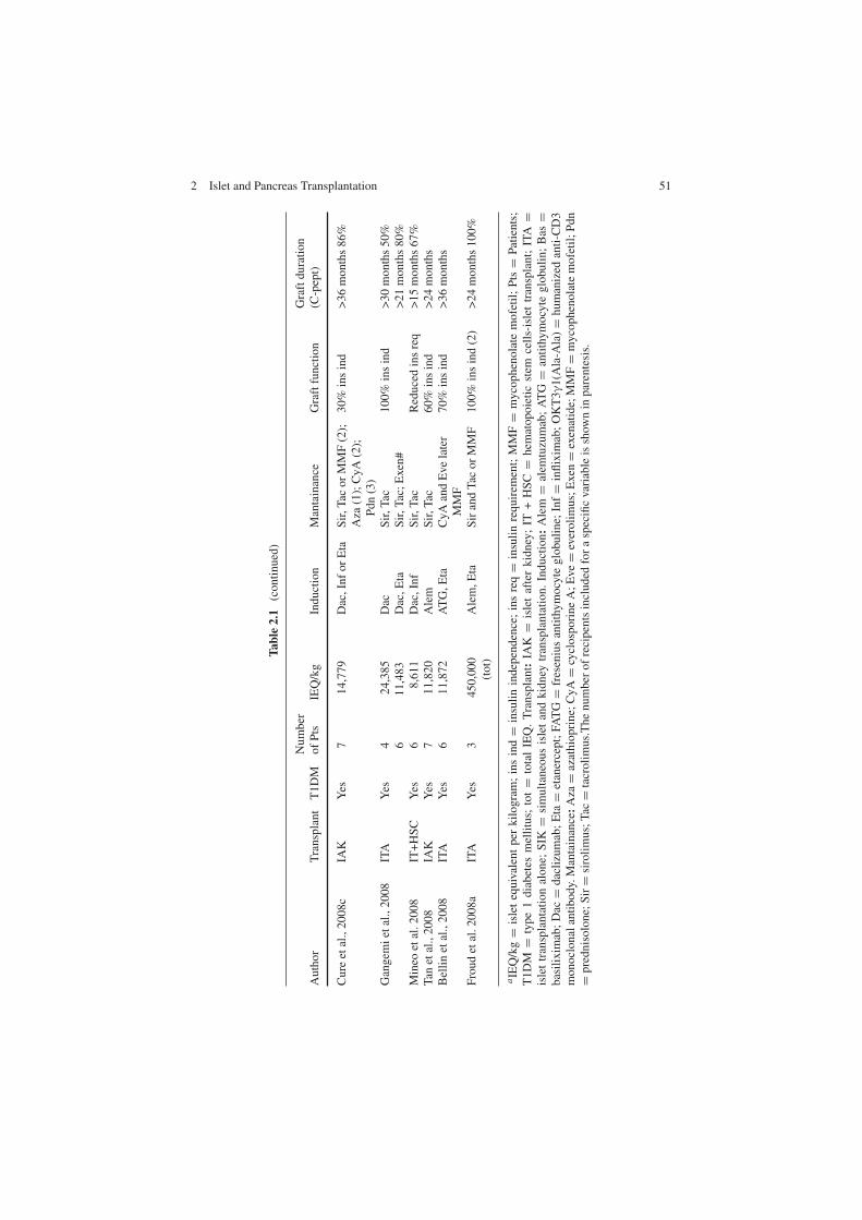
Chapter 2 Islet and Pancreas Transplantation Davide Mineo, Gaetano Ciancio, George W. Burke, Rodolfo Alejandro, and Camillo Ricordi Abstract Islet allotransplantation for patients with brittle type 1 diabetes melli- tus (T1DM) is a minimally invasive and relatively safe procedure that can induce sustained, normalized glucose control and restore C-peptide secretion, with reduc- tion of hypoglycemic episodes, stabilization or delay of chronic complications, and better quality of life. Current immunosuppressive protocols have significantly improved short-term outcomes, whereas long-term results are still inadequate (from 80% to 10% insulin-independence from 1 to 5 years post-transplant). Principal limitations include: imperfections in the islet isolation process, auto- and alloimmu- nity, allosensitization, immunosuppression-related toxicity, and unsuitability of the intrahepatic implantation site. More efficient isolation methods, safer and more effi- cient immunosuppressive agents in tolerogenic strategies, and alternative transplant site(s) may resolve these limitations in the near future. Simultaneous pancreas– kidney (SPK) transplantation is the optimal treatment for patients with T1DM with end-stage renal disease. Restoration of normoglycemia after pancreas trans- plant, as well as of renal function after kidney transplant, results in significant improvement of neuropathy, retinopathy, and nephropathy. Novel immunosuppres- sive therapies, improvements in surgical techniques, and better understanding of postoperative recipient care have improved results of SPK transplants consistently over the past decade. Future directions include optimization of immunosuppression, allowing freedom from insulin injection therapy while maintaining normoglycemia, and avoidance of chronic transplant glomerulopathy, with durable normalization of kidney function, thus improving quality of life as well as extending patient survival. 2.1 Type 1 Diabetes Mellitus Type 1 diabetes mellitus (T1DM) is a cell-specific autoimmune disease triggered by environmental factors (e.g., viral infections, toxins, diet nutrients or anti- gens) in genetically predisposed individuals [e.g., human leukocyte antigen (HLA) D. Mineo (B ) Diabetes Research Institute, University of Miami, Miami, FL, USA e-mail: [email protected]41 S. Efrat (ed.), Stem Cell Therapy for Diabetes, Stem Cell Biology and Regenerative Medicine, DOI 10.1007/978-1-60761-366-4_2, C Humana Press, a part of Springer Science+Business Media, LLC 2010
Transcript
Chapter 2
Islet and Pancreas Transplantation
Davide Mineo, Gaetano Ciancio, George W. Burke,
Rodolfo Alejandro, and Camillo Ricordi
Abstract Islet allotransplantation for patients with brittle type 1 diabetes melli-
tus (T1DM) is a minimally invasive and relatively safe procedure that can induce
sustained, normalized glucose control and restore C-peptide secretion, with reduc-
tion of hypoglycemic episodes, stabilization or delay of chronic complications,
and better quality of life. Current immunosuppressive protocols have significantly
improved short-term outcomes, whereas long-term results are still inadequate (from
80% to 10% insulin-independence from 1 to 5 years post-transplant). Principal
limitations include: imperfections in the islet isolation process, auto- and alloimmu-
nity, allosensitization, immunosuppression-related toxicity, and unsuitability of the
intrahepatic implantation site. More efficient isolation methods, safer and more effi-
cient immunosuppressive agents in tolerogenic strategies, and alternative transplant
site(s) may resolve these limitations in the near future. Simultaneous pancreas–
kidney (SPK) transplantation is the optimal treatment for patients with T1DM
with end-stage renal disease. Restoration of normoglycemia after pancreas trans-
plant, as well as of renal function after kidney transplant, results in significant
improvement of neuropathy, retinopathy, and nephropathy. Novel immunosuppres-
sive therapies, improvements in surgical techniques, and better understanding of
postoperative recipient care have improved results of SPK transplants consistently
over the past decade. Future directions include optimization of immunosuppression,
allowing freedom from insulin injection therapy while maintaining normoglycemia,
and avoidance of chronic transplant glomerulopathy, with durable normalization of
kidney function, thus improving quality of life as well as extending patient survival.
2.1 Type 1 Diabetes Mellitus
Type 1 diabetes mellitus (T1DM) is a cell-specific autoimmune disease triggered
by environmental factors (e.g., viral infections, toxins, diet nutrients or anti-
gens) in genetically predisposed individuals [e.g., human leukocyte antigen (HLA)
D. Mineo (B)Diabetes Research Institute, University of Miami, Miami, FL, USAe-mail: [email protected]
dation, delayed intimal media thickening), with fewer cardiovascular events and
better survival in IT recipients (90 vs. 50% at 7 years). Overall, together with
the improvement in glycemic control, IT seems to be protective for kidney graft
function and to increase its longevity. The prolonged C-peptide secretion may con-
tribute to such beneficial effects by reducing nerve dysfunction and increasing blood
flow in cardiac and renal districts, with myocardial and glomerular vasodilatation,
improving cardiovascular and kidney function, and slowing the progression of dia-
betic macro- and microangiopathy (Johansson et al., 2000; Wahren et al., 2000;
Hansen et al., 2002; Fiorina et al., 2003a, b; Fiorina et al., 2005a, b; Lee et al.,
2005; Venturini et al., 2006; Ryan et al., 2006; Del Carro et al., 2007; Fung et al.,
2007; Maffi et al., 2007; Senior et al., 2007; Thompson et al., 2008; Warnock et al.,
2008, Leitao et al., 2009).
At islet graft dysfunction, long- and short-acting insulin analogues (e.g., glargine
and lispro), and/or the incretin-mimetic exenatide, are gradually started. The lat-
ter seems to have direct effects on β cells (increased glucose-dependent insulin
secretion, restored first-phase secretion, better insulin processing, and higher amylin
54 D. Mineo et al.
Fig. 2.2 Intravenous glucose (IVGTT) (a) and mixed-meal (MMTT) (b and c) tolerance
tests, pre- and post-islet allotransplantation. Reproduced with permission from Faradji et al.,2008
2 Islet and Pancreas Transplantation 55
Fig. 2.3 Continuous glucose monitoring system (CGMS) profiles pre- (a) and post-islet (b)
allotransplantation, and at islet graft dysfunction (c). Different lines represent different days ofglucose monitoring. Reproduced with permission from Gorn et al., 2008
56 D. Mineo et al.
synthesis) and indirect effects on glucose metabolism (reduced glucagon secre-
tion, lower hepatic gluconeogenesis, reduced gastric empting, and delayed glucose
absorption). Whether reduction of apoptosis or regeneration of β cells can occur, as
observed in experimental models, is not yet clear. Exenatide may also aid in pro-
tecting β cells from immunosuppression-related toxicity. Several side effects (e.g.,
vomiting, nausea), the risk of pancreatitis, and the possible worsening of preexisting
diabetic gastroparesis may limit its use (D’Amico et al., 2005; Ranta et al., 2006;
Cure et al., 2008b; Ranganath, 2008).
2.2.3.2 Islet Graft Monitoring
The clinical management of islet transplant recipients relies on the combina-
tion of several immune responses and metabolic parameters together with blood
trough levels of immunosuppressants and recipient clinical status, including
immunosuppressive-related side effects and toxicity symptoms.
The immune alloresponse is monitored principally by mixed lymphocyte allore-
action (MLR) and panel reactive alloantibody (PRA) assays for cellular and
humoral reactivity, respectively. Evaluation of cytotoxic gene expression levels (e.g.,
granzyme B) or ATP production in in-vitro stimulated CD4+ T-lymphocytes may
represent helpful tools for confirming the clinical picture and the islet graft course,
together with cytokine measurement and characterization or other soluble markers.
Recurrent autoimmunity can be detected by reappearance of T1DM-specific autoan-
tibodies (e.g., anti-GAD65, anti-IA2, and anti-insulin) and seems to be associated
with lower insulin-independence rates and shorter islet graft survival. Histological
signs of selective destruction of β-cell allograft as well as autoreactive cytotoxic and
memory T cells against specific β-cell epitopes have been also described (Stegall
et al., 1996; Bosi et al., 2001; Han et al., 2004; Pinkse et al., 2005; Huurman et al.,
2008; Huurman et al., 2009; Mineo et al., 2008b; Monti et al., 2008, Saini et al.,
2008).
Monitoring islet graft function for detection or prediction of β-cell dysfunction
or failure is based on insulin requirements and blood HbA1c, glucose, C-peptide,
and insulin levels measured in the fasting state or after stimulation testing (e.g.,
intravenous arginine tolerance test, IVGTT, and MMTT). Several indices of islet
graft function are derived from these measurements (e.g., acute insulin or C-peptide
release, fasting C-peptide/glucose ratio, 90-min glucose). Composite indices are
also calculated based on insulin requirements, HbA1c, and the number of infused
IEQ, such as the beta score. The use of CGMS or of the MAGE index derived
from daily glucose measurements with finger-sticks can help detect early graft
dysfunction. Unfortunately, none of these indices is completely reliable or standard-
ized, resulting in detection of metabolic alterations when it is too late to intervene
with modifications of the immunosuppressive therapy for rescuing the islet graft
(Teuscher et al., 1998; Geiger et al., 2005; Rickels et al., 2005b; Faradji et al., 2007b;
Rickels et al., 2007b; Gorn et al., 2008; Baidal et al., 2009).
To date, limited imaging methods are clinically available for visualizing or
monitoring the islet graft in vivo. Luciferase-transduced bioluminescence optical
2 Islet and Pancreas Transplantation 57
imaging, despite high sensitivity, has limited depth penetration and is not applicable
to human studies. High-sensitivity (e.g., 3-tesla) magnetic resonance imaging of
islets labeled with different tracers (e.g., superparamagnetic iron nanoparticles)
is being tested in animal settings with promising results for clinical applica-
tion. Positron emission tomography with 18-fluorodeoxy-D-glucose has been used
recently in human setting to assess intrahepatic islet engraftment and survival in the
immediate postinfusion period. Percutaneous hepatic biopsy is not routinely used
owing to procedure-related risks (e.g., bleeding) and lack of certainty of retrieval of
islet graft tissue (Eich et al., 2007; Medarova and Moore, 2008).
2.2.4 Complications and Limitations
2.2.4.1 Recipient- and Graft-Related Complications
Acute complications during the islet infusion procedure are rare (<2–6%), and
include: intraabdominal bleeding, pleural or abdominal effusions, peripheral portal
vein branches thrombosis, and transient transaminitis. Novel radiological tech-
niques, intracatheter-tract coagulants, and recipient peritransplant antithrombotic
prophylaxis have reduced their incidence. Intrahepatic focal steatosis and amyloid
deposits may follow IT, but their effect on islet graft function and survival is still
unclear (Bhargava et al., 2004; Froud et al., 2004; Barshes et al., 2005; Hafiz et al.,
2005; Westermark et al., 2008).
The extended period of the islet allograft survival in recent protocols has involved
long-term immunosuppression-related side effects in virtually all recipients, primar-
ily common or opportunistic infections (mainly skin, respiratory, and urinary tracts),
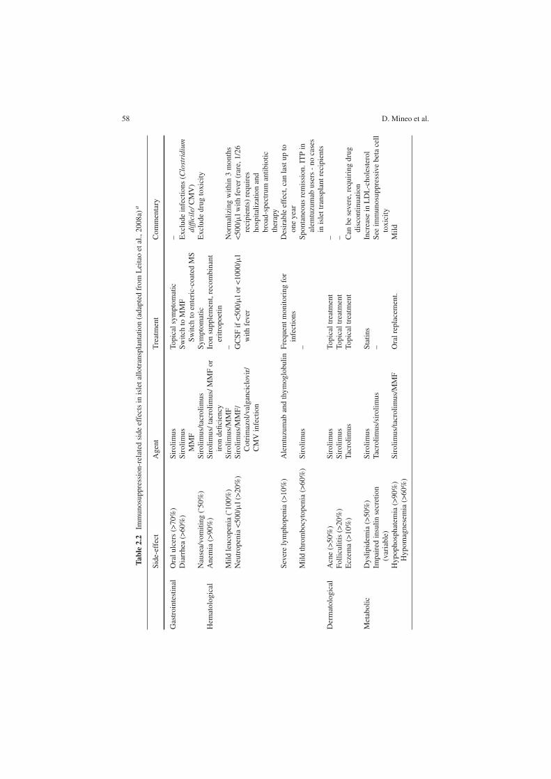
and direct immunosuppressive toxicity (Table 2.2). Several serious adverse events
have been observed that required hospitalization and specific therapy (e.g., profound
neutropenia, pneumonia, ovarian cysts), but only one death could be attributed to
immunosuppression (viral meningitis). Extremely rare are viral reactivations (e.g.,
EBV, CMV) or de novo malignancies, with only 13 neoplasms reported (two pap-
illary thyroid carcinomas, six squamous and two basal-cell skin carcinomas, one
ovarian and one breast cancer, one pulmonary nodule) in approximately 400 IT
recipients according to data of CITR (Cure et al., 2004; Hafiz et al., 2004; 2005;
Faradji et al., 2007a, Alejandro et al., 2008; Cure et al., 2008c).
Sirolimus has opposing effects on insulin secretion and action, which appear to
be cell- , species- , and dose-dependent, and act by inhibition of insulin-receptor
signal transduction and of the kinases regulating the β-cell cycle. Beta-cell dys-
function and reduction of insulin secretion seem to occur only at doses higher than
those used in the clinical setting, whereas increased basal and glucose-stimulated
insulin levels with reduced apoptosis have been seen at therapeutic concentrations.
In skeletal muscle and adipose cells, long-term exposure seems to reduce insulin-
dependent glucose uptake and insulin sensitivity, whereas in the short term opposite
effects have been observed. Reversible, dose-dependent dyslipidemia also occurs
(Subramanian and Trence, 2007; Vantyghem et al., 2007).
58 D. Mineo et al.
Tab
le2.2
Immunosuppression-related
sideeffectsin
isletallotransplantation(adaptedfrom
Leitaoetal.,2008a)
a
Side-effect
Agent
Treatment
Commentary
Gastrointestinal
Oralulcers(>70%)
Sirolimus
Topicalsymptomatic
–Diarrhea
(>60%)
Sirolimus
MMF
Switch
toMMF
Switch
toenteric-coated
MS
Excludeinfections(C
lost
rid
ium
dif
fici
le/CMV)
Nausea/vomiting(˜50%)
Sirolimus/tacrolimus
Symptomatic
Excludedrugtoxicity
Hem
atological
Anem
ia(>90%)
Sirolimus/tacrolimus/MMFor
irondeficiency
Ironsupplement,recombinant
eritropoetin
Mildleucopenia(˜100%)
Sirolimus/MMF
–Norm
alizingwithin
3months
Neutropenia<500/µl(>20%)
Sirolimus/MMF/
Cotrim
azol/valganciclovir/
CMVinfection
GCSFif<500/µlor<1000/µl
withfever
<500/µlwithfever
(rare,1/26
recipients)requires
hospitalizationand
broad-spectrum
antibiotic
therapy
Severelymphopenia(>10%)
Alemtuzumab
andthymoglobulin
Frequentmonitoringfor
infections
Desirableeffect,canlastupto
oneyear
Mildthrombocytopenia(>60%)
Sirolimus
–Spontaneousremission.ITPin
alem
tuzumab
users-nocases
inislettransplantrecipients
Dermatological
Acne(>50%)
Sirolimus
Topicaltreatm
ent
–Folliculitis(>20%)
Sirolimus
Topicaltreatm
ent
–Eczem
a(>10%)
Tacrolimus
Topicaltreatm
ent
Can
besevere,requiringdrug
discontinuation
Metabolic
Dyslipidem
ia(>50%)
Sirolimus
Statins
Increase
inLDL-cholesterol
Impairedinsulinsecretion
(variable)
Tacrolimus/sirolimus
–See
immunosuppressivebetacell
toxicity
Hypophosphatem
ia(>90%)
Hypomagnesem
ia(>60%)
Sirolimus/tacrolimus/MMF
Oralreplacement.
Mild
2 Islet and Pancreas Transplantation 59
Tab
le2.2
(continued)
Side-effect
Agent
Treatment
Commentary
Gonadal
Ovariancysts(>60%)
Altered
menses(>60%)
Sirolimus
Horm
onal
Surgeryin
selected
cases
Oligo-oram
enorrhea.
Gonadothropinsdysregulation.
Nopolycysticpattern
atultrasound
Cardiovascular
Transienthypotension(rare)
Alemtuzumab
Symptomatic
–Increase
inbloodpressure
(>30%)
Tacrolimus/sirolimus
Initiation/increase
inantihypertensivemedication
–
Peripheraledem
a(>50%)
Sirolimus
Diuretics
–Renal
Proteinuria/albuminuria,decrease
inglomerularfiltrationrate
(variable)
Tacrolimus/sirolimus
ACEi/ARB,statins
Stableifadequatetreatm
entof
diabeticnephropathyclassical
risk
factors(i.e.,
LDL-cholesterolandblood
pressure)isprovided
Neurological
Tremors,paresthesias,headache,
milddepression(>10%)
Tacrolimus
Switch
toMMF
Can
besevere:disablingpain
syndromeand
leukoencephalopathy
aACEi=
angiotensin-converting-enzymeinhibitors;ARB
=angiotensinogen
receptors
blockers;CMV
=cytomegalovirus;GCSF
=granulocyte
colony-
stim
ulatingfactor;ITP
=im
munethrombocytopenicpurpura;LDL
=low-density
lipoprotein;MS
=mycophenolatesodium.
60 D. Mineo et al.
MPA may also have a detrimental effect on β cells, by reducing insulin secretion
and inducing apoptosis, as well as on peripheral insulin sensitivity, with most of such
data coming from experimental settings, whereas lipid metabolism is not affected.
The enteric-coated formulation mycophenolate sodium has recently shown better
gastrointestinal tolerability and absorption than MMF and is increasingly used to
avoid toxicity from other immunosuppressive drugs (Havrdova et al., 2005; Gao
et al., 2007; Subramanian and Trence, 2007; Park et al., 2009).
All the immunosuppressive agents can interfere with islet engraftment and β-
cell self-renewal. Indeed, sirolimus has antiproliferative and antiangiogenic effects
on duct and islet cells that may impair β-cell engraftment and neovascularization as
well as viability and regeneration. Tacrolimus andMPA also have negative effects on
duct and islet cell proliferation and differentiation, preventing β-cell neogenesis or
replication. No negative effects of everolimus, a newly introduced mTOR inhibitor,
on glucose metabolism, have yet been reported, although it can induce dyslipidemia
(Bussiere et al., 2006; Cantaluppi et al., 2006a, b; Marcelli-Tourvieille et al., 2007;
Nir et al., 2007; Zahr et al., 2007).
Renal toxicity is still a major side effect of immunosuppressive therapy.
Tacrolimus may cause acute vasomotor vasculopathy with tubular necrosis and/or
chronic fibrotic vasculopathy with glomerulosclerosis and interstitial fibrosis.
Moreover, sirolimus may induce acute renal dysfunction and/or chronic proteinuria
by increasing glomerular permeability and injury or by suppressing the compen-
satory renal cell proliferation and repair capacity. Their combined use in IT can
have synergic negative effects on renal function per se or may cause the progression
of diabetic nephropathy, especially in the presence of pretransplant abnormalities
(e.g., microalbuminuria, reduced eGFR), whereas the alternative use of MPA-based
regimens could prevent renal injury (Rangan, 2006; Williams and Haragsim, 2006).
Supportive therapy is normally used to counteract systemic immunosuppressant-
related side effects, such as angiotensin converting enzyme inhibitors (ACEi)
or angiotensinogen receptor blockers (ARB), statins or ezetimibe, together with
bone marrow stimulants (e.g., granulocyte-colony stimulating factor, erythropoi-
etin), anti-infective prophylaxes, and dietary supplements (e.g., iron). In most
cases prompt treatment of complications minimizes recipient morbidity without any
sequela (Hafiz et al., 2005; Faradji et al. 2007).
2.2.4.2 Transplant-Related Limitations
Similarly to the pretransplantation period, many factors can contribute to a signif-
icant post-transplantation islet loss especially during the early postinfusion phase,
reducing the effective number of functioning islets available during the follow-up
period. Because of that often a second or third donor islet infusion is required to
achieve insulin independence and durable normalization of glucose control in the
recipients.
In particular, during islet infusion, an intravascular instant blood-mediated
inflammatory reaction (IBMIR) seems responsible for destroying 50–70% of the
infused β cells. An upregulation of tissue factor and other molecules on islet
2 Islet and Pancreas Transplantation 61
cell surface after the isolation process is capable of triggering innate immunity
via activation of coagulation, complement, inflammation, and natural antibod-
ies, destroying the islets. Peritransplant anticoagulant prophylaxis with heparin
can counteract this reaction (Moberg et al., 2002; Johansson et al., 2005; Eich
et al., 2007).
A progressive intrahepatic islet graft dysfunction and loss also occurs owing
to: poor revascularization, chronic hypoxia, absent reinnervations, proinflammatory
milieu, drug toxicity, glucolipotoxicity, fat and amyloid deposition, islet functional
overload, premature apoptosis, and lack of regeneration. Several ongoing experi-
ments are aimed at identifying alternative and less hostile implantation sites for islet
allograft, with the omental pouch, the thymus, or the bone marrow being the most
attractive. Recently, islet autotransplantation in the forearm muscle of a child with
genetically determined pancreatitis has shown a prolonged (over 2 years) restora-
tion of insulin secretion and normalized glucose control, while requiring minimal
exogenous insulin therapy (Desai et al., 2003; Bhargava et al., 2004; Pileggi et al.,
2006; Huang et al., 2008; Merani et al., 2008; Rafael et al., 2008; Westermark et al.,
2008; Lau and Carlsson, 2009).
Finally, a major concern of IT is the recipient-wide allosensitization from the
multiple HLA-mismatched donor infusions performed to achieve insulin indepen-
dence, hypothetically jeopardizing the chances of receiving future organ transplants
(e.g., kidney or pancreas). Pretransplant PRA levels higher than 15–20% and
donor-specific antibodies (DSA) seem associated with reduced islet graft survival.
Post-transplant positive PRA levels and de novo DSA may occur after drug dose
reduction for persistent or serious side effects (e.g., infections) but their impact on
islet graft loss is still unclear. Allosensitization seems absent or minimal under the
recommended trough levels of immunosuppression, which also seem able to con-
tain low PRA levels (<5–15%), but it occurs constantly when immunosuppression
is discontinued, such as after islet graft failure. High PRA levels (>50%) with DSA
and cross-reacting non-DSA may persist for a long time. A slower immunosup-
pressive tapering could minimize or prevent sudden and massive antigen exposition
from residual islet graft (Mohanakumar et al., 2006; Rickels et al., 2006b; Campbell
et al., 2007a, b; Cardani et al., 2007).
2.2.4.3 Current Challenges and Future Perspectives
Several technical and clinical limitations still persist in IT (Fig. 2.4). Various cyto-
protective strategies and agents are currently under investigation to improve organ
preservation and islet yield and survival through the isolation process, such as perflu-
orocarbons in a two-layer method, new lytic enzyme blends or purification methods,
and JNK or caspase inhibitors (Kin et al., 2006; Barbaro et al., 2007; Emamaullee
et al., 2007; Sabek et al., 2008; Varona-Santos et al., 2008).
New immunological and possibly tolerogenic strategies, including more selec-
tive lymphodepleting drugs, costimulatory blockade, and anti-inflammatory agents,
are being tested for increasing islet allograft longevity, preventing allorejection and
62 D. Mineo et al.
Drug Toxicity ImmunityIntra -
hepatic Site
Islet
Processing Pancreas
recovery Donor/Organ
1 2 3 4 5 6
Insulin Independence
12
Time
Beta
Islet Transplant
654
4
3
5 6
Beta-Cell Toxicity
Lipo-Toxicity
Anti-Proliferation
Anti-
Angiogenesis
IBMIR (acute)
Allo-Rejection
Auto-Immunity
Allo-Sensitization
Hypoxia
Gluco-
toxicity
Apoptosis
Enzyme
Purification
Culture
Cell-Viability
Cell-Potency
Recovery Technique
Warm/Cold Ischemia
Preservation Solution
Organ Shipment
Age/BMI
Organ fibrosis
Fat Infiltration
Brain Death
Intensive Care
1 2 4
Insulin Independence
12
Time
Beta
- C
ell
Ma
ss
Islet Transplant
654
4
3
5 6
Fig. 2.4 Main challenges in clinical islet allotransplantation. Reproduced with permission fromMineo et al. 2008c
recurrent autoimmunity, and reducing recipient side effects and islet graft toxic-
ity (Vincenti and Kirk, 2008). The Clinical Islet Transplant Consortium, including
centers in North America and Europe, is starting different phase II–III trials using
agent lisofylline, and IBMIR-blocker low-molecular-weight dextran) to improve
outcomes. Standardized procedures are also used, with the goal of obtaining
approval for IT as a standard health-care procedure, thus allowing for insurance
reimbursement. Indeed, costs of IT are very high, approximately $250,000 in the
first 2 years post-transplantation, and only a few countries (e.g., Canada) have
included this procedure as an optional treatment for selected patients with T1DM.
2.2.5 Conclusions
IT as treatment for brittle T1DM has recently achieved successful graft function,
with long-term metabolic improvements and minimal procedure-related complica-
tions. Unfortunately, islet recovery from isolation and post-transplant graft durabil-
ity with the current methods and protocols are still unsatisfactory. Several limitations
remain, including auto- and alloimmunity, allosensitization, immunosuppressive-
related toxicity, and implantation-site unsuitability. In the near future, improvements
in both the isolation process and islet cytoprotection, as well as new, less toxic
immunological agents together with tolerogenic protocols, and alternative implanta-
tion sites, may overcome such challenges (Ricordi, 2003; Ricordi and Strom, 2004;
Shapiro, 2008).
2 Islet and Pancreas Transplantation 63
2.3 Simultaneous Pancreas–Kidney Transplantation
Simultaneous pancreas–kidney transplantation (SPK) is considered the best treat-
ment option for patients with T1DM and end-stage renal disease (ESRD). The pan-
creas transplant can restore euglycemia, providing long-term insulin independence;
increase patient survival; stabilize or improve diabetic retinopathy and neuropathy;
and, in combination with the kidney transplant, eliminate the need for long-term
dialysis (Gruessner and Sutherland, 2005; Leichtman et al., 2008).
More potent immunosuppression agents, improvements in surgical techniques,
and better understanding of postoperative complications have led to consistent
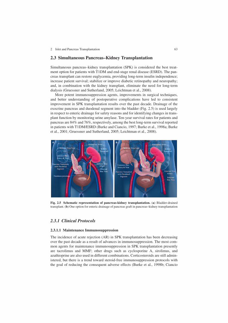
improvement in SPK transplantation results over the past decade. Drainage of the
exocrine pancreas and duodenal segment into the bladder (Fig. 2.5) is used largely
in respect to enteric drainage for safety reasons and for identifying changes in trans-
plant function by monitoring urine amylase. Ten-year survival rates for patients and
pancreas are 84% and 76%, respectively, among the best long-term survival reported
in patients with T1DM/ESRD (Burke and Ciancio, 1997; Burke et al., 1998a; Burke
et al., 2001; Gruessner and Sutherland, 2005; Leichtman et al., 2008).
Fig. 2.5 Schematic representation of pancreas-kidney transplantation. (a) Bladder-drainedtransplant. (b) One option for enteric drainage of pancreas graft in pancreas–kidney transplantation
2.3.1 Clinical Protocols
2.3.1.1 Maintenance Immunosuppression
The incidence of acute rejection (AR) in SPK transplantation has been decreasing
over the past decade as a result of advances in immunosuppression. The most com-
mon agents for maintenance immunosuppression in SPK transplantation presently
are tacrolimus and MMF; other drugs such as cyclosporine A, sirolimus, and
azathioprine are also used in different combinations. Corticosteroids are still admin-
istered, but there is a trend toward steroid-free immunosuppression protocols with
the goal of reducing the consequent adverse effects (Burke et al., 1998b; Ciancio
64 D. Mineo et al.
et al., 2000a; Burke et al., 2004b; Gruessner and Sutherland, 2005; Cantarovich and
Vistoli, 2009; Mineo et al., 2008c; Singh and Stratta, 2008).
2.3.1.2 Induction Therapy
The recent therapeutic protocols in kidney and kidney–pancreas transplantation
attempt to reduce the incidence and severity of AR as well as prevent long-term
chronic (vascular) allograft dysfunction (CAD). The methodologies include reduc-
tion of CNIs and of their short- and long-term nephrotoxicity, reduction or avoidance
of corticosteroids, use of adjunctive maintenance antiproliferative agents (e.g.,
mTOR inhibitors), and utilization of new agents, such as nonlymphodepleting mon-
oclonal antibodies (daclizumab or basiliximab), or lymphodepleting monoclonal
(alemtuzumab) and polyclonal (e.g., rATG) antibodies. The percentage of patients
treated with induction therapy has been increasing and was more than 75% in the
most recently reported data from the International Pancreas Transplant Registry
(IPTR) 2004 (Gruessner and Sutherland, 2005).
Daclizumab
A series of studies has been published analyzing the safety and efficacy of
daclizumab as induction therapy in SPK transplant recipients (Bruce et al. 2000;
Burke et al., 2001; Lo et al., 2001a, b; Stratta et al., 2001; Burke et al., 2002a,
b; Stratta et al., 2002). The results of a multicenter survey using daclizumab as
induction therapy showed a low incidence of AR when used in combination with
tacrolimus, MMF, and corticosteroids in SPK transplant recipients (Bruce et al.,
2001). The survey reported experience with 71 SPK transplant recipients receiving
4–5 daclizumab doses (n = 45) or 1–3 doses (n = 26). There were no differences
in patient and kidney graft survival rates, 98 vs. 96% and 92 vs. 92%, respec-
tively. However, there was a trend toward improved pancreas graft survival rates
in the group receiving 4–5 doses, compared with 1–3 doses (96 vs. 85%, p =
0.07). Although more patients receiving 1–3 doses had rejection (54%) than patients
receiving 4–5 doses (24%), there was no dose–response relationship between the
total number of doses or the adjusted total milligram/kilogram dose and time to
rejection. All patients with functioning grafts had good renal and pancreatic allo-
graft function at 6 and 12 months. The overall incidence of major infection was
27%, and there were no differences in the incidence of infection between the two
groups. No major adverse events were attributed to daclizumab use. In conclusion,
excellent short-term outcomes were noted in this retrospective, multicenter survey of
initial experience with daclizumab induction in combination with tacrolimus, MMF,
and corticosteroids in SPK transplant recipients.
The safety and efficacy of two dosing regimens of daclizumab as an adjunctive
immunosuppressive agent versus no antibody induction in SPK transplant recipients
receiving tacrolimus and MMF as primary immunosuppression were investigated
in a multicenter, open label, comparative trial (Stratta et al., 2002). SPK trans-
plant recipients were randomized to one of three groups: daclizumab 1 mg/kg every
2 Islet and Pancreas Transplantation 65
14 days for five doses (Group I), daclizumab 2 mg/kg every 14 days for two doses
(Group II), and no antibody induction (Group III). A total of 166 patients were ran-
domized into the three groups [Group I (n = 70), Group II (n = 74), Group III
(n = 22)]. At a minimum follow-up of 3 months, patient, kidney and pancreas graft
survival rates were similar among the three groups. However, the rates of acute renal
allograft rejection were 18% for Group I, 8% for Group II, and 36% for Group III
(p < 0.005). The probabilities of either kidney or pancreas allograft rejection were
22% for Group I, 8% for Group II, and 38% for Group III. At 3 months, the actuarial
event-free survival (no AR, allograft loss, or death) rates were 67%, 81%, and 50%
in Group I, II, and III, respectively. Although the follow-up was short, this study
emphasized the important role of induction antibodies in reducing AR.
Daclizumab in Combination with rATG
The use of new immunosuppressive agents continues to be associated with reduced
rates of AR episodes in SPK transplant recipients (Burke et al., 2002a, b). Forty-
two SPK transplant recipients were included in a prospective, randomized trial in
which they received rATG and daclizumab, tacrolimus, and corticosteroids as base-
line immunosuppression. They were then randomized to receive either MMF or
sirolimus in addition to baseline immunosuppression. Twenty-two patients received
MMF and 20 received sirolimus. There were three episodes of AR (7.1%). These
were in the MMF group, all in patients who were off either MMF (wound infection,
pneumonia) or corticosteroids. Each of these episodes was corticosteroid-resistant,
but responsive to antibody therapy (OKT3 or rATG). Actuarial patient, kidney, and
pancreas allograft survivals were 100%, 100%, and 95% in the sirolimus group and
100%, 100%, and 100% in the MMF group (Burke et al., 2002b).
A similar study (Gallon et al., 2007) reported the effect of two tacrolimus-based
maintenance regimens on long-term renal allograft function in SPK transplant recip-
are required because there is no reason to monitor urinary activity. However, rejec-
tion episodes may progress undiagnosed before treatment is started, and this delay
increases the possibility of allograft loss.
2.3.4 Conclusions
The optimal treatment for T1DM in the context of ESRD, where the primary goal
is to restore normal glucose metabolism and then kidney function, is achieved by
whole pancreas and kidney allograft transplantation. The administration of lym-
phodepleting agents continues to increase, whereas use of IL2 receptor antagonists
is declining. The main goal of induction therapy is to provide a strong and long-
term immunosuppressive effect for protocols that include steroid avoidance, CNIs
minimization, or even monotherapy maintenance. The current trend is to reduce or
minimize the number of immunosuppressive drugs in order to prevent or avoid side
effects and adverse events. The challenge is to find the balance between benefit
(protection from AR and long-term graft function) and risk (side effects, infection,
cancers).
Acknowledgments We thank Mr. John Wilkes for assistance in reviewing and editing themanuscript.
For further information including transplant data and annual reportsU.S. Department of Health and Human Services (http://www.hhs.gov); Organ Procurement
and Transplantation Network (http://www.optn.org); Scientific Registry of TransplantRecipients (http://www.ustransplant.org); Health Resources and Services Administration(http://www.hrsa.gov); Collaborative Islet Transplant Registry (http://www.citregistry.org); andClinical Islet Transplant consortium (www.citisletstudy.org).
References
Alejandro R, Barton FB, Hering BJ, et al. (2008) Collaborative Islet Transplant RegistryInvestigators. 2008 Update from the Collaborative Islet Transplant Registry (CITR).Transplantation. 86:1783–1788.
Alejandro R, Lehmann R, Ricordi C, et al. (1997) Long-term function (6 years) of islet allograftsin type 1 diabetes. Diabetes. 46:1983–1989.
74 D. Mineo et al.
Badet L, Benhamou PY, Wojtusciszyn A, et al. (2007) Expectations and strategies regarding islet
transplantation: metabolic data from the GRAGIL 2 trial. Transplantation. 84:89–96.
Baidal DA, Faradji RN, Messinger S, et al. (2009) Early metabolic markers of islet allograft
dysfunction. Transplantation. 87:689–697.
Baidal DA, Froud T, Ferreira JV, et al. (2003) The bag method for islet cell infusion. Cell
Transplant. 12:809–813.
Barbaro B, Salehi P, Wang Y, et al. (2007) Improved human pancreatic islet purification with the
refined UIC-UB density gradient. Transplantation. 84:1200–1203.
Barshes NR, Lee TC, Goodpastor SE, et al. (2005) Transaminitis after pancreatic islet translanta-
tion. J Am Coll Surg. 200:353–361.
Bellin MD, Kandaswamy R, Parkey J, et al. (2008) Prolonged insulin independence after islet
allotransplants in recipients with type 1 diabetes. Am J Transplant. 8:2463–2470.
Bhargava R, Senior PA, Ackerman TE, et al. (2004) Prevalence of hepatic steatosis after islet
transplantation and its relation to graft function. Diabetes. 53:1311–1317.
Bosi E, Braghi S, Maffi P, et al. (2001) Autoantibody response to islet transplantation in type 1
diabetes. Diabetes. 50:2464–2471.
Bretzel RG, Brandhorst D, Brandhorst H, et al. (1999) Improved survival of intraportal pan-
creatic islet cell allografts in patients with type-1 diabetes mellitus by refined peritransplant
management. J Mol Med. 77:140–143.
Bruce DS, Sollinger HW, Humar A, et al. (2001) Multicenter survey of daclizumab induction in
Stratta RJ, Alloway RR, Hodge E, et al. (2002) A multicenter, open-label, comparative trial
of two daclizumab dosing strategies versus no antibody induction in combination with
tacrolimus, mycophenolate mofetil, and steroids for the prevention of acute rejection in simul-
taneous kidney-pancreas transplant recipients: 6-month interim analysis. Transplant Proc.
34:1903–1905.
Stratta RJ, Alloway RR, Lo A, et al. (2001) A multicenter trial of two daclizumab dosing strategies
versus no antibody induction in simultaneous kidney-pancreas transplantation: interim analysis.
Transplant Proc. 33:1692–1693.
Stratta RJ, Gaber AO, Shokouh-Amiri MH, et al. (2000) A prospective comparison of systemic-
bladder versus portal-enteric drainage in vascularized pancreas transplantation. Surgery.
127:217–226.
Subramanian S, Trence DL. (2007) Immunosuppressive agents: effects on glucose and lipid
metabolism. Endocrinol Metab Clin North Am. 36:891–905.
Sureshkumar KK, Patel BM, Markatos A, et al. (2005) Quality of life after organ transplantation
in type 1 diabetics with end-stage renal disease. Clin Transplant. 20:19–25.
Tan J, Yang S, Cai J, et al. (2008) Simultaneous islet and kidney transplantation in seven patients
with type 1 diabetes and end-stage renal disease using a glucocorticoid-free immunosuppres-
sive regimen with alemtuzumab induction. Diabetes. 57:2666–2671.
Teuscher AU, Kendall DM, Smets YF, et al. (1998) Successful islet autotransplantation in humans:
functional insulin secretory reserve as an estimate of surviving islet cell mass. Diabetes.
47:324–330.
Tharavanij T, Betancourt A, Messinger S, et al. (2008) Improved long-term health-related quality
of life after islet transplantation. Transplantation. 86:1161–1167.
The Diabetes Control and Complications Trial Research Group. (1993) The effect of intensive
treatment of diabetes on the development and progression of long-term complications in
insulin-dependent diabetes mellitus. N Engl J Med. 329:977–986.
2 Islet and Pancreas Transplantation 83
The Diabetes Control and Complications Trial Research Group. (1997) Hypoglycemia in thediabetes control and complications trial. Diabetes. 46:271–286.
Thompson DM, Begg IS, Harris C, et al. (2008) Reduced progression of diabetic retinopathyafter islet cell transplantation compared with intensive medical therapy. Transplantation. 85:1400–1405.
Toso C, Baertschiger R, Morel P, et al. (2006) Sequential kidney/islet transplantation: efficacyand safety assessment of a steroid-free immunosuppression protocol. Am J Transplant. 6:1049–1058.
Troppmann C, Gruessner AC, Benedetti E, et al. (1996) Vascular graft thrombosis after pancreatictransplantation: univariate and multivariate operative and operative risk factor analysis. J AmColl Surg. 182:285–316.
Tzakis AG, Ricordi C, Alejandro R, et al. (1990) Pancreatic islet transplantation after upperabdominal exenteration and liver replacement. Lancet. 336:402–405.
Vantyghem MC, Marcelli-Tourvielle S, Pattou F, et al. (2007) Effects of non-steroid immunosup-pressive drugs on insulin secretion in transplantation. Ann Endocrinol. 68:21–27.
Varona-Santos JL, Pileggi A, Molano RD, et al. (2008) c-Jun N-terminal kinase 1 is deleterious tothe function and survival of murine pancreatic islets. Diabetologia. 51:2271–2280.
Venturini M, Fiorina P, Maffi P, et al. (2006) Early increase of retinal arterial and venous bloodflow velocities at color Doppler imaging in brittle type 1 diabetes after islet transplant alone.Transplantation. 81:1274–1277.
Vincenti F, Kirk AD. (2008) What’s next in the pipeline. Am J Transplant. 8:1972–1981.Wahren J, Ekberg K, Johansson J, et al. (2000) Role of C-peptide in human physiology. Am J
Physiol Endocrinol Metab. 278:759–768.Warnock GL, Meloche RM, Thompson D, et al. (2005) Improved human pancreatic islet isolation
for a prospective cohort study of islet transplantation vs best medical therapy in type 1 diabetesmellitus. Arch Surg. 140:735–744.
Warnock GL, Thompson DM, Meloche RM, et al. (2008) A multi-year analysis of islet trans-plantation compared with intensive medical therapy on progression of complications in type 1diabetes. Transplantation. 86:1762–1766.
Westermark GT, Westermark P, Berne C, et al. (2008) Nordic Network for Clinical IsletTransplantation. Widespread amyloid deposition in transplanted human pancreatic islets. NEngl J Med. 359:977–979.
Wilkin TJ. (2008) Diabetes 1 and 2, or one and the same? Progress with the accelerator hypothesis.Pediatr Diabetes. 9:23–32.
Williams D, Haragsim L. (2006) Calcineurin nephrotoxicity. Adv Chronic Kidney Dis. 13:47–55.Yoon JW, Jun HS. (2005) Autoimmune destruction of pancreatic β cells. Am J Ther. 12:580–591.Zahr E, Molano RD, Pileggi A, et al. (2007) Rapamycin impairs in vivo proliferation of islet
β-cells. Transplantation. 84:1576–1583.Zimmet P, Alberti KG, Shaw J. (2001) Global and societal implications of the diabetes epidemic.