KEY KNOWLEDGE This chapter is designed to enable students to: • investigate the defining characteristics of prokaryotic and eukaryotic cells • identify cell structure and organisation • identify cell organelles and understand their functions • investigate the different modes of transport of materials across plasma membranes • understand and apply the principle of the surface-area-to-volume ratio. CHAPTER 2 STRUCTURE AND FUNCTION OF CELLS FIGURE 2.1 Here we see a specialised white blood cell attacking many small rod-shaped bacterial cells at the site of an infection. These white blood cells that are part of the body’s immune system are produced in the bone marrow and travel via the bloodstream to sites of infection. While various cells have different functions, they share common structural features. In this chapter, we will explore the structure of cells and the passage of materials into cells.
Transcript
Key KnowledgeThis chapter is designed to enable students to:• investigatethedefiningcharacteristicsofprokaryoticandeukaryotic
Chapter 2StruCture and funCtion of CellSfigure 2.1 Here we see a specialisedwhitebloodcellattackingmanysmallrod-shapedbacterialcellsatthesiteofaninfection.Thesewhitebloodcellsthatarepartofthebody’simmunesystemareproducedinthebonemarrowand travelviathebloodstreamtositesofinfection.Whilevariouscellshavedifferentfunctions,theysharecommonstructuralfeatures.Inthischapter,wewillexplorethestructureofcellsandthepassageofmaterialsintocells.
26 Nature of biology book 1
Clues from a pondIn July 1991, in Connecticut, USA, two young boys were fishing at a local pond. They were attacked by three older boys and severely beaten until almost unconscious. The older boys then threw the younger boys into the pond where they were in danger of drowning. Fortunately one of the young boys was suf-ficiently conscious to save himself and his friend.
Three suspects were apprehended, but what material evidence was available to place them at the crime scene?
Diatoms are single-celled golden-brown algae that are common in both sea water and fresh water. Whatever the shape of a diatom cell (figure 2.2), its plasma membrane is surrounded by a wall made of silica (glass) and pectin. The wall is in two parts that fit together like a lid on a box, with the cell inside the box. Contact between a diatom cell and its environment takes place through thousands of tiny holes in its ‘glass’ coat.
figure 2.2 (a)Eachspeciesofdiatomhasadistinctiveshape. (b)Aclose-upofthesilicashellofadiatom(×580).Thesilicaandpectinwallsofdiatomssurvivelongafterthecellsthatmadethemhavedied.(c)Anevencloserviewofthesurfaceofthesilicashell(× 2340) shows the detailed patterns and perforations.
(a) (b) (c)
There are innumerable different species of diatoms commonly found, but research has shown that the ratio of numbers of each of the species is different from location to location. The ratio at a particular site is characteristic of that site, even if it changes from season to season.
Let’s return to the attacks at the pond and the need for material evidence. Samples were collected from the pond and compared with diatoms found in the mud on the shoes of both the victims and the suspects. The same 25 different freshwater species were isolated from each of the samples. Statistical testing indicated that there was no difference in the population ratios in each of the samples. So the police had material evidence that the suspects were at the scene of the crime. They were, in fact, guilty of the attack.
Diatoms are used extensively in forensic cases where it is important to estab-lish whether death occurred in water and, if so, what kind of water and at which location. Each pond and stream has its own populations — and diatoms that live in still water do not generally populate running waters. It is the presence of a rigid wall that survives after the death of the diatom cell that leaves a trace, and this can be followed long after the death of a person.
The popular face of forensic science, as it is portrayed in many television programs, tends to rely heavily on evidence gained from the DNA and other analyses of animal tissues, such as blood, skin, hair and semen. However, for-ensic botany also has an important role. In addition to diatom studies, the type of plant material found at a crime scene and knowledge about where it grows may be an important clue. Did a person die where the body was found or was it transported from elsewhere?
Analysis of food in the stomach can indicate the time of death after a meal. A plant cell has a wall of cellulose surrounding its plasma membrane.
odd faCtDepositsofdiatomcoatshaveaccumulatedformillionsofyearstoformthicklayersonocean floors that are now partofgeologicalstructures.Thecoatscrumbleintoafine white powder that is minedandusedinavarietyofcleaningapplications,includingtoothpaste.
StruCture aNd fuNCtioN of CellS 27
As cellulose is not digested, information about whether the person’s last meal was high or low in vegetable matter may be a clue to where the person last ate and may lead investigators to people who saw the person there.
In this chapter we will consider the specialised structures that are found in different cells and how those structures relate to processes that are vital for the maintenance of life.
looking at cellsExamination of cells using various microscopes reveals much about their internal organisation. Each living cell is a small compartment with an outer boundary known as the cell membrane or plasma membrane. Inside each living cell is a fluid, known as cytosol, that consists mainly of water containing many dissolved substances.
Another feature shared by all living cells is DNA, the genetic material that controls all the metabolic activities of a cell.
In contrast to these shared features, living cells can be classified into two dif-ferent kinds on the basis of their internal structures:• Prokaryotic cells. These have little defined internal structure and, in par-
ticular, lack a clearly defined structure to house their DNA. Organisms that are made of prokaryotic cells are called prokaryotes and include all bacteria (figure 2.3a) and all archaeans, another group of microbes (refer to chapter 8).
• Eukaryotic cells. These have a much more complex structure (see figure 2.3b) than prokaryotic cells. All eukaryotic cells contain many different kinds of membrane-bound structures called organelles suspended in the cytosol. These organelles include a nucleus with a clearly defined membrane called a nuclear envelope. The DNA of a eukaryotic cell is located in the nucleus. Organisms that are made of eukaryotic cells are called eukaryotes and include all animals, plants, fungi and protists, the single-celled organisms. Although a nucleus is usually visible with a light microscope, many organelles are visible only with electron microscopes.Organelles are held in place by a network of fine protein filaments and
micro tubules within the cell, collectively known as the cytoskeleton. The filaments of the cytoskeleton are visible with an electron microscope, but require special staining to be seen with a confocal microscope.
odd faCtArchaeologists and palaeontologistsexaminefossilisedfaecestostudyanybonesandundigestedpartsoffruitandvegetables.Thiscanhelptoestablishthedietsofprehistorichumansandotheranimals.
28 Nature of biology book 1
the plasma membrane boundaryThe boundary of all living cells is a plasma membrane, which controls entry of dissolved substances into and out of the cell. A plasma membrane is an ultra-thin and pliable layer with an average thickness of less than 0.01 µm (0.000 01 mm). A plasma membrane can be seen using an electron microscope.
Prokaryotes Eukaryotes
Plasma membrane present present
Functionboundary of a cell; maintains the internal environment of a cell by controlling the movement of substances into and out of the cell
A plasma membrane contains both lipid and protein. A model of the plasma membrane is shown in figure 2.4. This model suggests that a plasma membrane consists of a double layer of lipid, and that proteins are embedded in this layer, forming channels that allow certain substances to pass across the membrane in either direction. This model is known as the fluid mosaic model.
Movement in and out of cellsAll cells must be able to take in and expel various substances across their mem-branes in order to survive, grow and reproduce. Generally, these substances are in solution but, in some cases, may be tiny solid particles.
Because a plasma membrane allows only some dissolved materials to cross it, the membrane is said to be a partially permeable boundary (see figure 2.5). (Partially permeable is also known as selectively or differentially or semi- permeable.) Dissolved substances that are able to cross a plasma membrane — from outside a cell to the inside or from the inside to the outside — do so by various processes, including diffusion and active transport.
SurfaCe-area-to-VoluMe ratio• Why are cells small?• The surface of some cells is elaborately folded.
What is the importance of these outfoldings?• Some animals have a greatly flattened shape. How
might this affect their survival?Consider the surface areas of cells compared with
their volumes. This value is sometimes called the surface-area-to-volume ratio (SA:V ratio). The SA:V ratio of any object is obtained by dividing its area by its volume.
Area refers to the coverage of a surface. One unit of measurement of area is a square centimetre (cm2). Volume refers to the amount of space taken up by an object. One unit of measurement of volume is the litre (L), but the volume of solid matter, such as a brain, is sometimes expressed in units such as cubic centi metres (cm3). (Note: For a sphere, SA = 3πr2 andV = 4–3 πr3, where r = radius.)
Looking at SA:V ratio Examine the following data. Notice that, as a sphere increases in size, its surface-area-to-volume ratio decreases.
Radius of sphere SA:V 1 unit 3.0 2 1.5 3 1.0 6 0.5 10 0.3
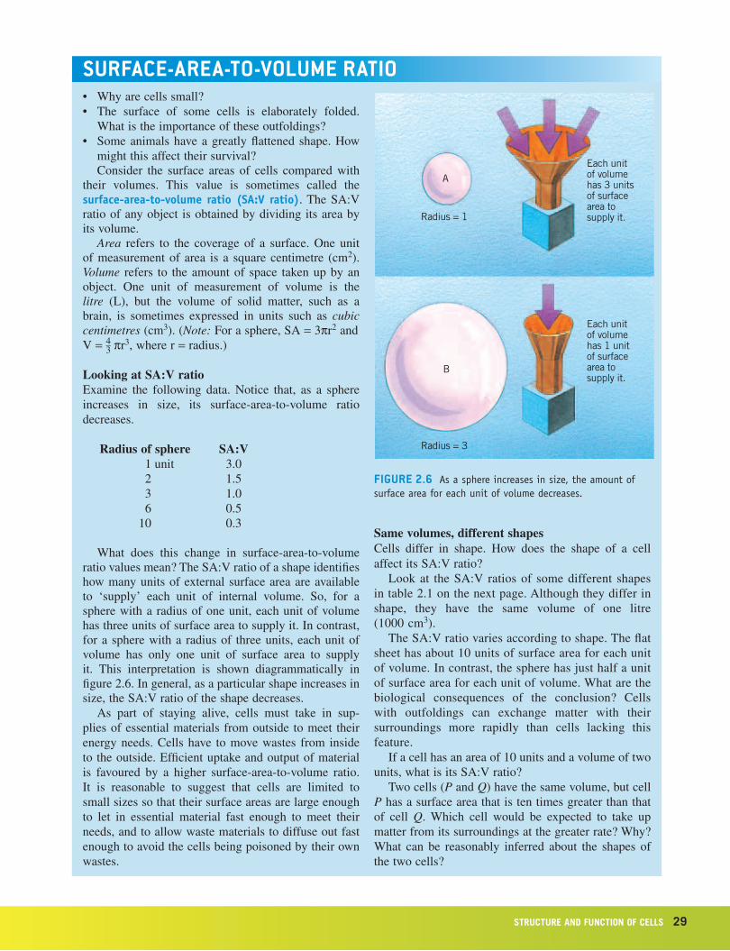
What does this change in surface-area-to-volume ratio values mean? The SA:V ratio of a shape identifies how many units of external surface area are available to ‘supply’ each unit of internal volume. So, for a sphere with a radius of one unit, each unit of volume has three units of surface area to supply it. In contrast, for a sphere with a radius of three units, each unit of volume has only one unit of surface area to supply it. This interpretation is shown diagrammatically in figure 2.6. In general, as a particular shape increases in size, the SA:V ratio of the shape decreases.
As part of staying alive, cells must take in sup-plies of essential materials from outside to meet their energy needs. Cells have to move wastes from inside to the outside. Efficient uptake and output of material is favoured by a higher surface-area-to-volume ratio. It is reasonable to suggest that cells are limited to small sizes so that their surface areas are large enough to let in essential material fast enough to meet their needs, and to allow waste materials to diffuse out fast enough to avoid the cells being poisoned by their own wastes.
A
B
Radius = 1
Radius = 3
Each unitof volumehas 3 unitsof surface area tosupply it.
Each unitof volumehas 1 unitof surface area tosupply it.
Same volumes, different shapesCells differ in shape. How does the shape of a cell affect its SA:V ratio?
Look at the SA:V ratios of some different shapes in table 2.1 on the next page. Although they differ in shape, they have the same volume of one litre (1000 cm3).
The SA:V ratio varies according to shape. The flat sheet has about 10 units of surface area for each unit of volume. In contrast, the sphere has just half a unit of surface area for each unit of volume. What are the biological consequences of the conclusion? Cells with outfoldings can exchange matter with their surroundings more rapidly than cells lacking this feature.
If a cell has an area of 10 units and a volume of two units, what is its SA:V ratio?
Two cells (P and Q) have the same volume, but cell P has a surface area that is ten times greater than that of cell Q. Which cell would be expected to take up matter from its surroundings at the greater rate? Why? What can be reasonably inferred about the shapes of the two cells?
30 Nature of biology book 1
A cell has many outfoldings on its surface. How would these outfoldings affect its surface area as com-pared with a cell with a ‘smooth’ surface?
SummaryAs a structure increases in size, its surface-area-to-volume ratio (SA:V) decreases.
Various shapes differ in their SA:V ratios, with this ratio being highest in flattened shapes and lowest in spheres.
The size of a cell is limited by the SA:V ratio, since these ratios influence the rate of entry and exit of sub-stances into and out of cells.
free passage: diffusionDiffusion is the net movement of a substance, typically in solution, from a region of high concentration of the substance to a region of low concentration. The process of diffusion does not require energy.
Figure 2.7 shows a representation of this process for dissolved substance X. At all times, molecules of X are in random movement. At first, some molecules col-lide with and cross the plasma membrane into the cell (see figure 2.7a). As long as substance X is more concentrated outside the cell than inside, more collisions causing molecules of X to move from outside to inside occur than collisions from the opposite direction. As a result, a net movement of molecules of substance X occurs from outside to inside and the concentration of X inside the cell rises (figure 2.7b). Eventually, the numbers of collisions occurring on both sides of the membrane become equal. At that time (figure 2.7c), the number of molecules of X passing into the cell is equal to the number passing out. Diffusion stops at the stage when the concentration of substance X is equal on the two sides of the membrane.
One special case of diffusion is known as osmosis. The process of osmosis occurs when a net movement of water molecules occurs by diffusion across a cell membrane either into or out of a cell. Read the box on page 31, which outlines the movement of water into and out of a cell when it is placed in a strong sugar solution (figure 2.8a) and pure water (figure 2.8b) respectively.
Substances that can dissolve readily in water are termed hydrophilic, or ‘water-loving’. Some substances that have a low water solubility or do not dissolve in water are able to dissolve in or mix uniformly with lipid. These substances are termed lipophilic (sometimes called hydrophobic). Examples of lipophilic substances include alcohol and ether. Lipophilic substances can cross plasma membranes readily. This observation provides indirect evidence for the presence of lipid in the structure of the plasma membrane. The rapid absorption of substances, such as alcohol across plasma membranes, appears to be related to the ability of alcohol to mix with lipid.
StruCture aNd fuNCtioN of CellS 31
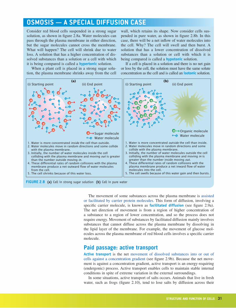
oSMoSiS — a SpeCial diffuSion CaSeConsider red blood cells suspended in a strong sugar solution, as shown in figure 2.8a. Water molecules can pass through the plasma membrane in either direction, but the sugar molecules cannot cross the membrane. What will happen? The cell will shrink due to water loss. A solution that has a higher concentration of dis-solved substances than a solution or a cell with which it is being compared is called a hypertonic solution.
When a plant cell is placed in a strong sugar solu-tion, the plasma membrane shrinks away from the cell
wall, which retains its shape. Now consider cells sus-pended in pure water, as shown in figure 2.8b. In this case, there will be a net inflow of water molecules into the cell. Why? The cell will swell and then burst. A solution that has a lower concentration of dissolved substances than a solution or cell with which it is being compared is called a hypotonic solution.
If a cell is placed in a solution and there is no net gain or loss by the cell, the solution must have the same solute concentration as the cell and is called an isotonic solution.
figure 2.8 (a) Cell in strong sugar solution (b) Cell in pure water
Sugar moleculeWater molecule
1. Water is more concentrated inside the cell than outside.2. Water molecules move in random directions and some collide
with the plasma membrane.3. Initially, the number of water molecules inside the cell
colliding with the plasma membrane and moving out is greater than the number outside moving in.
4. These differential rates of random collisions with the plasma membrane produce a net outward flow of water molecules from the cell.
5. The cell shrinks because of this water loss.
(i) Starting point (ii) End point
Organic moleculeWater molecule
1. Water is more concentrated outside the cell than inside.2. Water molecules move in random directions and some
collide with the plasma membrane.3. Initially, the number of water molecules outside the cell
colliding with the plasma membrane and moving in is greater than the number inside moving out.
4. These differential rates of random collisions with the plasma membrane produce a net inward flow of water molecules into the cell.
5. The cell swells because of this water gain and then bursts.
(i) Starting point (ii) End point(a) (b)
The movement of some substances across the plasma membrane is assisted or facilitated by carrier protein molecules. This form of diffusion, involving a specific carrier molecule, is known as facilitated diffusion (see figure 2.9a). The net direction of movement is from a region of higher concentration of a substance to a region of lower concentration, and so the process does not require energy. Movement of substances by facilitated diffusion mainly involves substances that cannot diffuse across the plasma membrane by dissolving in the lipid layer of the membrane. For example, the movement of glucose mol-ecules across the plasma membrane of red blood cells involves a specific carrier molecule.
Paid passage: active transportActive transport is the net movement of dissolved substances into or out of cells against a concentration gradient (see figure 2.9b). Because the net move-ment is against a concentration gradient, active transport is an energy-requiring (endergonic) process. Active transport enables cells to maintain stable internal conditions in spite of extreme variation in the external surroundings.
In some situations, active transport of salts occurs. Animals that live in fresh water, such as frogs (figure 2.10), tend to lose salts by diffusion across their
32 Nature of biology book 1
skin-cell plasma membranes into the surrounding fresh water. Energy in the form of adenosine triphosphate (ATP) is used to transport salt molecules against a concentration, from the surrounding water where salt concentration is low, across plasma membranes into cells where the salt concentration is very high.
This process involves a carrier protein for each substance that is actively transported. If the carrier protein for a particular substance is defective, the organism may show a disorder. In human beings, a defect in the carrier protein involved in the active transport of chloride ions (Cl–) has been found to be the cause of the inherited disorder cystic fibrosis.
ATP
Salts
Salts
Diffusion
figure 2.10 To balance the loss ofsaltsthatoccursfromfrogskincellsbydiffusion,energyisusedtodriveactivetransportofsaltsfromaregionoflowconcentrationin the surrounding water,acrossplasmamembranes,intothefrogskincells,whichhaveahighconcentrationofsalts.
Some bacteria thrive in highly salty water where other organisms cannot sur-vive (see table 2.2). How do these halophytic (‘salt-loving’) bacteria maintain a stable internal environment?
Salt molecules do not readily cross the plasma membrane. A net movement of water molecules occurs down the concentration gradient from inside the cell to outside. However, the bacteria have an efficient mechanism for active trans-port of water. Water molecules are actively transported into the cell at a rate that compensates for the loss of water by osmosis, so that the internal conditions in the bacterial cell remain stable. Energy is needed to power this ‘water pump’. Placed in the same very salty conditions, cells of other organisms would shrivel and dehydrate.
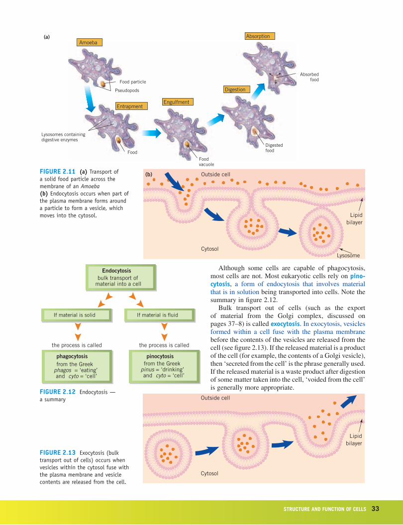
bulk transportSolid particles can be taken into a cell. For example, one kind of white blood cell is able to engulf a disease-causing bacterial cell and enclose it within a lysosome sac where it is destroyed. Unicellular protists, such as Amoeba and Paramecium, obtain their energy for living in the form of relatively large ‘food’ particles, which they engulf and enclose within a sac where the food is digested (see figure 2.11a).
Note how part of the plasma membrane encloses the material to be trans-ported and then pinches off to form a membranous vesicle that moves into the cytosol (figure 2.11b). This process of bulk transport of material into a cell is called endocytosis. When the material being transported is a solid food particle, the type of endocytosis is called phagocytosis.
most cells are not. Most eukaryotic cells rely on pino-cytosis, a form of endocytosis that involves material that is in solution being transported into cells. Note the summary in figure 2.12.
Bulk transport out of cells (such as the export of material from the Golgi complex, discussed on pages 37–8) is called exocytosis. In exocytosis, vesicles formed within a cell fuse with the plasma membrane before the contents of the vesicles are released from the cell (see figure 2.13). If the released material is a product of the cell (for example, the contents of a Golgi vesicle), then ‘secreted from the cell’ is the phrase generally used. If the released material is a waste product after digestion of some matter taken into the cell, ‘voided from the cell’ is generally more appropriate.
Functionsemi-rigid, protective structure deposited by the cell outside the cell membrane
The plasma membrane forms the exterior of animal cells. However, in plants, fungi and bacteria, a rigid cell wall lies outside the plasma membrane. The absence of a cell wall is characteristic of organisms in Kingdom Animalia.
Composition of cell wallThe cell wall varies in composition between plants, fungi and bacteria (see table 2.3).
Type of organism Compounds present in cell wall
plant include cellulose
fungus include chitin
bacterium include complex polysaccharides
In some flowering plants, the original or primary cell wall in certain tissues becomes thickened and strengthened by the addition of lignin to form secondary cell walls. This process provides great elastic strength and support, allowing certain plants to develop as woody shrubs or trees.
Cells have a complex internal organisation and are able to carry out many functions. The control centre of the cells of animals, plants, algae and fungi is the nucleus (see figure 2.24, page 44). The nucleus in these cells forms a distinct spherical structure that is enclosed within a double membrane, known as the nuclear envelope. Cells that have a membrane-bound nucleus are called eukaryotic cells. The regular presence of a nucleus in living cells was first identified in 1831 by a Scottish botanist, Robert Brown (1773–1858) (see pages 9–10).
Cells of organisms from Kingdom Monera, such as bacteria, contain the gen-etic material (DNA), but it is not enclosed within a distinct nucleus. Cells that lack a nuclear envelope are called prokaryotic cells.
A light microscope view reveals that the nucleus of a eukaryotic cell con-tains stained substance called chromatin, which is made of the genetic mat-erial deoxy ribonucleic acid (DNA). The DNA is usually dispersed within the nucleus. During the process of cell reproduction, however, the DNA becomes organised into a number of rod-shaped chromosomes (refer to chapter 4, pages 82–4). The nucleus also contains one or more large inclusions known as nucleoli, which are composed of ribonucleic acid (RNA).
Textbook diagrams often show a cell as having a single nucleus. This is the usual situation, but it is not always the case. Your bloodstream contains very large numbers of mature red blood cells, each with no nucleus. However, at an earlier stage, as immature cells located in your bone marrow, each of these cells did have a nucleus. Some liver cells have two nuclei.
Function site of production of much of the ATP required by a cell
Living cells use energy all the time. The usable energy supply for cells is chemical energy present in a compound known as ATP (adenosine triphos-phate) (see figure 2.14). The ATP supplies in living cells are continually being used up and must be replaced.
ATP is produced during cellular respiration (or just simply respiration). In eukaryotic cells, most of this process occurs in organelles known as mitochon-dria (singular: mitochondrion), which form part of the cytoplasm. Mitochondria cannot be resolved using an LM, but can be seen with an electron microscope. Each mitochondrion has an outer membrane and a highly folded inner mem-brane. Mitochondria are not present in prokaryote cells.
The role of mitochondria in respiration is discussed further in chapter 3.Prokaryotes obtain their energy from a range of sources. This will be
ribosomes: protein factoriesProkaryotes Eukaryotes
Ribosomes present present
Function site of protein synthesis
Living cells make proteins by linking amino acid building blocks into long chains. Human red blood cells manufacture haemoglobin, an oxygen- transporting protein; pancreas cells manufacture insulin, a small protein that is an important hormone; liver cells manufacture many protein enzymes, such as catalase; stomach cells produce digestive enzymes, such as pepsin; muscle cells manufacture the contractile proteins actin and myosin.
Ribosomes are the organelles where production of proteins occurs. These organelles, which are part of the cytoplasm, can be seen only through a TEM (see figures 2.15 and 2.16, page 37). Chemical testing shows that ribosomes are composed of protein and ribonucleic acid (RNA).
Ribosomes are not enclosed by a membrane. The structures of prokaryotic and eukaryotic ribosomes are almost identical and function in a similar way. Although ribosomes are free within prokaryotic cells, in eukaryotes many are attached to membranous internal channels, called endoplasmic reticulum, within the cell (see page 37).
odd faCtManybiologistsagreewiththehypothesisthat,thousandsofmillionsofyearsago,mitochondriawerefree-livingorganisms,likebacteria.Thishypothesissuggeststhattheseorganismsbecameassociated with larger cells toformamutuallybeneficialarrangement.ThisideaissupportedbythefactthatmitochondriacontainsmallamountsofthegeneticmaterialDNA.Thesizeofamitochondrionisabout 1.5µmby0.5µm.Thisissimilartothedimensionsofatypicalbacterialcell.
StruCture aNd fuNCtioN of CellS 37
endoplasmic reticulum: transport within cellsProkaryotes Eukaryotes
Endoplasmic reticulum absent present
Function series of membranous channels for transport
Transport of substances within cells occurs through a system of channels known as the endoplasmic reticulum (ER). Figure 2.16 shows part of this system of channels in a cell. The channel walls are formed by membranes.
Endoplasmic reticulum with ribosomes attached is known as rough endo-plasmic reticulum. Without ribosomes, the term smooth endoplasmic is used.
golgi complex: export from cellsProkaryotes Eukaryotes
Golgi complex absent present
Functionstacks of membranous sacs that package
materials for transport
The proteins made by some cells are kept inside those cells. Examples are contractile proteins made by muscle cells and the haemoglobins made by red blood cells. Other cells, however, produce proteins that are released for use outside the cells. The digestive enzyme, pepsin, is produced by cells lining the stomach and released into the stomach cavity; the protein hormone, insulin, is made by pancreatic cells and released into the bloodstream.
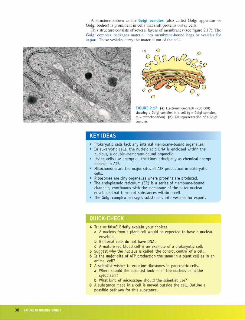
A structure known as the Golgi complex (also called Golgi apparatus or Golgi bodies) is prominent in cells that shift proteins out of cells.
This structure consists of several layers of membranes (see figure 2.17). The Golgi complex packages material into membrane-bound bags or vesicles for export. These vesicles carry the material out of the cell.
Function principal site of digestion within a cell
The human hand is a marvellous living tool that allows a person to grasp objects, manipulate and investigate them. Typically, a human hand has five digits that are separated from each other along their length. This is not always the case — a rare condition, known as syndactyly (pronounced sin-dack-till-ee), in which the fingers are fused, can occur. How does this happen?
During human embryonic development, the hands appear first as tiny buds with no separate digits (see figure 2.18). The separation of the fingers nor-mally occurs on about the 52nd day of development (see figure 2.19). This separation involves the ‘programmed death’ of groups of cells between the fingers. The process of programmed cell death is called apoptosis. If this programmed cell death does not occur, the fingers and toes form but they remain fused.
Animal cells have sac-like structures surrounded by a membrane and filled with a fluid containing dissolved digestive enzymes. These fluid-filled sacs are known as lysosomes. Lysosomes can release their enzymes within the cell, causing the death of the cell. This process of controlled ‘self-destruction’ of cells is important in development; lysosomes appear to play a role in the con-trolled death of zones of cells in embryonic human hands and feet so that the fingers become separated.
Lysosomes contain digestive enzymes and are the principal sites for diges-tion of large molecules and unwanted structures within a cell.
Solar-powered cars have travelled across Australia. The power source for these cars is not the chemical energy present in petrol but the radiant energy of sun-light trapped and converted to electrical energy by solar cells. Use of solar cells is becoming more common in Australian households and it is not unusual to see solar cells on a roof.
Solar cells are a relatively new technology. However, hundreds of millions of years ago, some bacteria and all algae and then land plants developed the ability to capture the radiant energy of sunlight and to transform it to chemical energy present in organic molecules, such as sugars. The remarkable organelles present in some cells of plants and algae that carry out this function are known as chloroplasts (see figure 2.20a). The complex process of converting sunlight energy to chemical energy present in sugar is known as photosynthesis.
Chloroplasts can be easily seen through an LM. They are green in colour owing to the presence of light-trapping pigments known as chlorophylls. Each chloroplast has an outer membrane and also has an intricate internal structure
figure 2.18 Notethewebbingbetween the fingers and toes inanearlyembryoat6weeksdevelopment.
Photosynthesis is discussed further in chapter 3, pages 70–2.
40 Nature of biology book 1
consisting of many folded membrane layers, called grana, that provide a large surface area where chlorophylls are located. Stroma is fluid between the grana.
Prokaryotic cells do not have chloroplasts. Some kinds of bacteria, however, possess pigments that enable them to capture the radiant energy of sunlight and use that energy to make sugars from simple inorganic material. These are known as photosynthetic bacteria.
The length of a typical chloroplast is 5 to 10 µm. In comparison, the length of a mitochondrion is about 1.5 µm. In 1908, the Russian scientist Mereschkowsky suggested that chloroplasts were once free-living bacteria that later ‘took up residence’ in eukaryotic cells. Some evidence in support of this suggestion comes from the fact that a single chloroplast is very similar to a photosynthetic bacterial cell.
other membrane-bound structuresOther small membranous structures found in the cytosol of eukaryotic cells include the endosomes (animal cells only) and peroxisomes. (These are dealt with in more detail in Nature of Biology Book 2, Fourth edition.) Many plant cells also contain vacuoles, some very large that almost fill a cell. Vacuoles are filled with a fluid, mostly water, containing a number of different materials in solution, including plant pigments.
flagella and cilia: whipping aroundSome bacterial cells and other single-celled organisms have a whip-like struc-ture that is attached to the plasma membrane and protrudes through the cell
StruCture aNd fuNCtioN of CellS 41
wall (see figure 2.21). This structure is usually known as a flagellum (plural: flagella, from the Latin word meaning ‘whip’). What role might this structure serve?
The rotation of a flagellum results in the movement of the organism. Some bacteria have many flagella, such as the bacteria that cause typhoid (Salmonella typhosa). Other bacteria, such as species of Pseudomonas, have one flagellum or a cluster of several flagella at one end.
Many eukaryotic cells have one or many whip-like structures on their cell surfaces. When many such structures are present, they are termed cilia (singular: cilium, from the Latin word meaning ‘eyelash’); when only one or two are present, they are termed flagella (figure 2.21).
In eukaryotes, each cilium and flagellum is enclosed in a thin extension of the plasma membrane. Inside this extension of the membrane are fine protein filaments known as microtubules. In the human body, the cells lining the tra-chea or air passage have cilia that project into the cavity of the trachea. The synchronised movement of these cilia assists mucus to travel up the trachea to an opening at the back of the throat. Other human cells that have flagella include sperm cells.
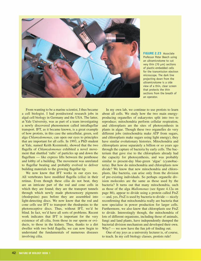
Dr Peter Beech, a cell biologist, carries out research on the replication of cells and their organelles. Figures 2.21 and 2.22 and figure 4.12a (page 86) show some of his results. Read what he has to say about his work.
biologiSt at worKAssociate Professor Peter Beech — cell biologistAssociate Professor Peter Beech is a Research Scientist in the School of Biological and Chemical Sciences at Deakin University in Melbourne. Peter writes:
Like many kids who watched Jacques Cousteau on television exploring the world’s oceans, I wanted to be a marine scientist. I spent summers at the beach won-dering about how I could get a job working with the sea. I was told ‘go to uni, study science and then see what grabs you’. It was good advice, and I quickly dis-covered that biology was indeed for me.
My first lab project was on identifying algal scales, the beautifully intricate cell coverings of many phytoplankton (figure 2.22). This work required an electron microscope, and I was thus irreversibly led into the world of the subcellular, where I could see scales being made, as well as the other cellular organelles — many of which are also found in our own cells.
My PhD was on how certain phytoplankton made their scales and deposited them on the cell surface, as well as how they made their flagella. Flagella are the whip-like appendages that beat to propel cells through the water — sperm tails are flagella. I was not the first to realise that, by looking at protists (as algae and many other mostly unicellular eukaryotes are known), we could learn a lot about cells. Many protists are ide-ally suited to laboratory culture and experimentation. Phytoplankton, for example, are unicells that have all they need to get by in life on their own. Often all that
is needed to grow them in the lab is light and clean sea water or pond water.
figure 2.22 AtransmissionelectronmicrographofbodyscalesmadebyanalgalcellofChrysochromulina pringsheimii.Thescalesandtheirintricatepatternsareconstructedofpolysaccharidefibrilsandaremadeinsidethecell.Pr=proximalsideofthescale, Di =distalside.
figure 2.21 Thaumatomastix,acolourlessmarineprotist.Notethetwoflagella,F1 and F2.Thescalesandspinesthatcovertheentirecellaremadeofsilica.
42 Nature of biology book 1
From wanting to be a marine scientist, I thus became a cell biologist. I had postdoctoral research jobs in algal cell biology in Germany and the USA. The latter, at Yale University, was as part of a team investigating a newly discovered phenomenon called intraflagellar transport. IFT, as it became known, is a great example of how protists, in this case the unicellular, green, soil alga Chlamydomonas, can open our eyes to principles that are important for all cells. In 1993, a PhD student at Yale, named Keith Kozminski, showed that the two flagella of Chlamydomonas exhibited a novel move-ment that shuttled ‘rafts’ of particles up and down the flagellum — like express lifts between the penthouse and lobby of a building. The movement was unrelated to flagellar beating and probably evolved to deliver building materials to the growing flagellar tip.
We now know that IFT works in our eyes too. All vertebrates have modified flagella (cilia) in their retinas. Even though these cilia do not beat, they are an intricate part of the rod and cone cells in which they are found; they are the transport tunnels through which newly made photosensory pigments (rhodopsins) pass before they are assembled into light-detecting discs. We now know that the rod and cone cells use IFT to transport the rhodopsins to the photoreceptive discs. Thus, without IFT, we’d be blind. In fact, we’d have all sorts of problems. Recent work indicates that IFT is important for the very existence of all cilia, from those in our sperm or ovi-ducts, to those in the kidney. Thus, thanks to a dirt dweller with two bold flagella, we can now begin to understand the fundamentals of numerous diseases involving cilia.
In my own lab, we continue to use protists to learn about all cells. We study how the two main energy-producing organelles of eukaryotes split into two to reproduce; mitochondria perform cellular respiration, and chloroplasts are the sites of photosynthesis in plants in algae. Though these two organelles do very different jobs (mitochondria make ATP from sugars, and chloroplasts make sugars using light energy), they have similar evolutionary histories. Mitochondria and chloroplasts arose separately a billion or so years ago through the capture of bacteria by early cells. The bac-terium that gave rise to the chloroplasts already had the capacity for photosynthesis, and was probably similar to present-day blue-green ‘algae’ (cyanobac-teria). But how do mitochondria and chloroplasts now divide? We know that new mitochondria and chloro-plasts, like bacteria, can arise only from the division of pre-existing individuals. So perhaps organelle div-ision molecules are the same as those used by the bacteria? It turns out that many mitochondria, such as those of the alga Mallomonas (see figure 4.12a on page 86), appear to divide using a protein called FtsZ — and, yes, FtsZ is used by bacteria to divide — nicely reconfirming that mitochondria really are bacteria that now specialise in power production for larger cells. Furthermore, we also know that chloroplasts use FtsZ to divide. Interestingly though, the mitochondria of lots of different organisms, including those of animals, fungi and land plants, have independently dumped the bacterial division mechanism and developed their own. Why? — we now have the fun job of finding out.
One of my joys as a university lecturer is, of course, to teach. In my cell biology classes, protists rule!
figure 2.23 Associate ProfessorPeterBeechusinganultramicrotometocutverythin(70µm)sectionsofplastic-embeddedcellsforthetransmissionelectronmicroscope.Thedarklineprojectingdownfromtheultramicrotomeisasideviewofathin,clearscreenthat protects the thin sectionsfromthebreathofanoperator.
StruCture aNd fuNCtioN of CellS 43
Putting it all togetherThe cell is both a unit of structure and a unit of function. Organelles within one cell do not act in isolation, but interact with each other. The normal functioning of each kind of cell depends on the combined actions of its various organelles, including plasma membrane, nucleus, mitochondria, ribosomes, endoplasmic reticulum and Golgi complex.
In some cells, the plasma membrane is very highly folded. This folding expands the surface area across which materials move into or out of cells while the internal volume remains unchanged. This produces an increase in the sur-face-area-to-volume ratio (SA:V) of cells.
Consider a cell that produces a specific protein for use outside the cell. Table 2.4 identifies the parts of a cell involved in this process.
Structure Function
plasma membrane
structure that controls the entry of raw materials, such as amino acids, into the cell
nucleus organelle that has coded instructions for making the protein
ribosomeorganelle where amino acids are linked, according to instructions, to build the protein
mitochondrionorganelle where ATP is formed; provides an energy source for the protein-manufacturing activity
endoplasmic reticulum
channels through which the newly made protein is moved within the cell
Golgi complexorganelle that packages the protein into vesicles for transport across the plasma membrane and out of the cell
Figure 2.24 shows the typical structures of an animal and a plant cell, including the organelles involved in the processes outlined in table 2.4. Examine the two cells. Note the presence of protein filaments in each cell. These give a cell shape; they form a kind of ‘internal skeleton’ for the cell and also provide a system for movement during, for example, mitosis (see chapter 4, page 82 onwards).
table 2.4 Partsofacellinvolvedin producing a specific protein
44 Nature of biology book 1
figure 2.24 Compare (a)ananimalcellwith (b)aplantcell.Whatorganellesarefoundinbothofthecells?Whatorganellesare unique to either plant oranimalcells?Whatotherdifferencesinstructurearethere between the two cells?
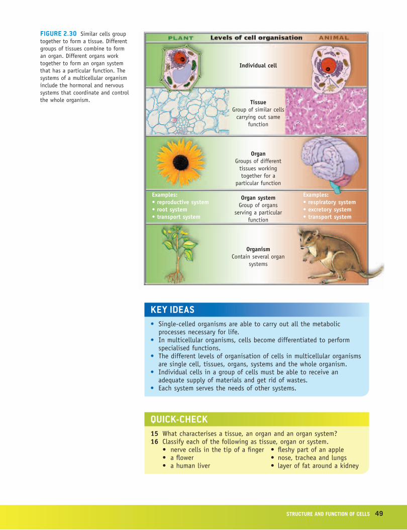
Cells in multicellular organisms: levels of organisationUnicellular organisms must carry out all the metabolic processes necessary for life. They are complex cells capable of independent existence. In contrast, multi cellular organisms have millions of cells that depend on each other for sur-vival. During development of a multicellular organism, groups of cells become specialised to perform particular functions that serve the whole organism. Specialised cells have fewer functions than those found in a unicellular organism but the functions they have are very highly developed. In addition, each group of specialised cells must coordinate with other specialised cells. We will consider the different levels of organisation that interact to ensure proper functioning for the whole organism.
tissuesWhen cells that are specialised in an identical way aggregate to perform a common function, they are called a tissue. Different kinds of tissue (see figure 2.25) serve different functions in an organism. For example, car-diac muscle is a particular kind of muscle tissue found only in the heart. Epidermal tissue is a general name for any tissue that forms a discrete layer around a structure. It may be a layer of plant cells forming the outermost cellular layer of leaves or it may be the outer layers of human skin.
You will recall from pages 29–30 that the surface-area-to-volume-ratio (SA:V) of a cell is important in determining the cell’s efficiency in moving materials across its membrane and that the higher the SA:V ratio of a cell, the more efficient it is in carrying out those functions. The need for small cells can be graphically demonstrated with regard to groups of cells (figure 2.26, page 47). Exchange of materials between tissues and their environments has the potential to be far more efficient if the tissue is made up of many small cells rather than fewer larger cells.
This potential for efficiency of small cells becomes a reality only if each of the cells in a group of cells is close to a delivery mechanism, capable of pro-viding material to and removing material from the cells (figure 2.27, page 47). A mass of small cells without a delivery system has no advantage over a single large cell.
Total surface area(height × width × numberof sides × number of cells)
Total volume(height × width × length ×
number of cells)
Surface-area-to-volumeratio
(surface area volume)
organsIn multicellular organisms, groups of different tissues often work together to ensure that a particular function is successfully performed (figure 2.28). A col-lection of such tissues is called an organ. Your stomach is an organ. Tissues of the stomach include an epithelium, smooth muscle cells and blood (see figure 2.28a). Other organs include your heart, brain and kidneys. A plant leaf is an organ. Tissues of a leaf include an epithelium, vascular tissue and parenchyma tissue (see figure 2.28b). Other plant organs include its root, stem and flower.
organ systemsYour digestive system comprises various organs that work together to ensure that the food you eat is digested and that the nutrients it contains are absorbed and
Food Wastes
OxygenCarbondioxide
Wastes and carbon dioxide
Food and oxygen
figure 2.27 For the inner cells ofatissuetooperateasefficientlyastheoutercells,theymusthaveadeliverysystemthattransportsfoodandgastothemandtakesawaywastes.Inmanyanimals,thedeliverysystemisthebloodcirculatorysystem.
transported to all cells of your body. This organisation is called an organ system. Your digestive system commences with your mouth and includes organs such as your teeth, oesophagus, stomach, intestines and liver (figure 2.29). Once digested food has been absorbed by cells lining the intestine, it is transported by the blood circulatory system throughout the body. This system links with the respiratory system where it picks up oxygen, also for delivery.
As blood delivers nutrients and oxygen to all tissues, it collects nitrogenous and gaseous wastes for delivery to the excretory systems of the body.
Because plants do not move from place to place, their energy needs are far less than mobile animals. Hence, plants lack the equivalent of complex organ systems such as the respiratory and digestive systems of animals. Green plants produce their own food through photosynthesis and this process also delivers oxygen directly to some cells. Other cells rely on diffusion to receive oxygen. The extensive root system of a plant ensures that it absorbs sufficient water to meet the plant’s requirements. An extensive vascular system delivers that water throughout the plant; however, there is relatively little difference in the structure of the various parts of a plant vascular system compared with differences found in systems of an animal.
We will consider some of the organ systems of animals and plants in greater detail in later chapters. A summary of the levels of organisation in multicellular organisms is shown in figure 2.30, page 49.
a Use at least eight of the key words above to make a concept map relating to the organelles observed in the cytosol of a plant cell. You may use other words in drawing your map.
b Use at least six of the key words above to make a concept map relating to the movement of substances across a cell membrane. You may use other words in drawing your map.
2 Applying your understanding ➡ Identify five locations in a typical cell where membranes are found. Describe how membranes in these various locations assist in the function of cells.
3 Communicating understanding ➡ Substances can enter or exit a cell through various processes.
a Prepare a table with the following headings:
Name of process Energy cost
Identify the processes by which material crosses the cell membrane and complete the table.
b Identify one other useful heading and add it and the relevant information to your table.
4 Analysing data and drawing conclusions ➡ In a series of six experi-ments, animal cells and plant cells were placed in solutions of different concentrations.
Solution 1: distilled waterSolution 2: same concentration as the cytosol of the cells
52 Nature of biology book 1
Solution 3: higher concentration than the cytosol of the cells
The initial appearance of the cells was as shown in figure 2.31a. After sev-eral minutes in the solutions the cells appeared as shown in figure 2.31b.
Which solution had been used in each of the experiments? Explain what has happened to the cell in each experiment.
5 Communicating understanding ➡ Where are the following in a eukaryotic cell?
a control centre of a cellb site of control of entry or exit of substances to or from a cellc energy source for celld internal transport systeme site of packaging for export from cellf ‘self-destruct button’ for cell
6 Applying your understanding ➡ a List the following in order of decreasing size from largest to smallest.
i cell ii tissue iii mitochondrioniv nucleus v nucleolus vi iribosome
b List the following in order from outside to inside a leaf cell. i nuclear envelope ii cell wall iii plasma membrane iv cytosol v nucleolus
7 Analysing information and drawing conclusions ➡ Suggest possible explanations for each of the following observations.
a Flight muscle fibres of bats contain very large numbers of mitochondria.b One kind of cell has a very prominent Golgi complex, while another kind
of cell appears to lack this organelle.c Chromosomes were seen in many cells of the root tip tissue of a flow-
ering plant.d After being soaked in water, a limp lettuce leaf becomes crisp.
StruCture aNd fuNCtioN of CellS 53
8 Communicating ideas ➡ Discuss the validity of each of the following statements.
a A tissue contains groups of cells where each group has quite a different function.
b Delivery mechanisms are important if a group of small cells is to operate more effectively than one large cell.
c The surface-area-to-volume ratio of a cell influences the rate at which substances can enter or exit the cell.
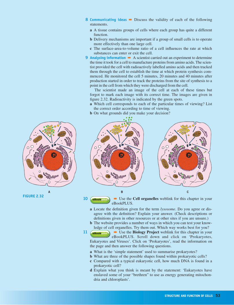
9 Analysing information ➡ A scientist carried out an experiment to determine the time it took for a cell to manufacture proteins from amino acids. The scien-tist provided the cell with radioactively labelled amino acids and then tracked them through the cell to establish the time at which protein syn thesis com-menced. He monitored the cell 5 minutes, 20 minutes and 40 minutes after production started in order to track the proteins from the site of synthesis to a point in the cell from which they were discharged from the cell.
The scientist made an image of the cell at each of these times but forgot to mark each image with its correct time. The images are given in figure 2.32. Radioactivity is indicated by the green spots.a Which cell corresponds to each of the particular times of viewing? List
the correct order according to time of viewing. b On what grounds did you make your decision?
A B Cfigure 2.32
10 ➡ Use the Cell organelles weblink for this chapter in your eBookPLUS.
a Locate the definition given for the term lysosome. Do you agree or dis-agree with the definition? Explain your answer. (Check descriptions or definitions given in other resources or at other sites if you are unsure.)
b The website provides a number of ways in which you can test your know-ledge of cell organelles. Try them out. Which way works best for you?
11 ➡ Use the Biology Project weblink for this chapter in your eBookPLUS. Scroll down and click on ‘Prokaryotes,
Eukaryotes and Viruses’. Click on ‘Prokaryotes’, read the information on the page and then answer the following questions.
a What is the ‘simple statement’ used to summarise prokaryotes?b What are three of the possible shapes found within prokaryotic cells?c Compared with a typical eukaryotic cell, how much DNA is found in a
prokaryotic cell?d Explain what you think is meant by the statement: ‘Eukaryotes have
enslaved some of your “brethren” to use as energy generating mitochon-dria and chloroplasts’.