95
How Animals Move Chapter 20
| Date post: | 23-Dec-2015 |
| Category: |
Documents |
| Upload: | redge-derrick-marcelino |
| View: | 215 times |
| Download: | 0 times |

How Animals Move
Chapter 20

20.1 Impacts/IssuesBulking Up Muscles
Exercise makes muscles bigger, not by adding cells but by adding proteins to existing cells
Certain hormones and other molecules regulate this process• Testosterone and human growth hormone
increase muscle growth• Myostatin slows muscle growth

Effects of Myostatin
A normal whippet and one homozygous for a mutation that prevents myostatin production

Video: Pumping up muscles

20.2 The Skeletal System
Muscles bring about movement by applying contractile force against body fluids or structural elements, such as bones
Three categories of skeletal systems are common in animals – hydrostatic skeletons, exoskeletons, and endoskeletons

Three Types of Skeletons
Hydrostatic skeleton (earthworm)• Fluid-filled chamber that muscles act on,
redistributing the fluid
Exoskeleton (fly)• Hard external parts that muscles attach to
Endoskeleton (humans, other vertebrates)• Hard internal parts that muscles attach to

The Human Skeleton
The human skeleton consists of skull bones, a vertebral column, a rib cage, a pelvic girdle, a pectoral girdle, and paired limbs
The vertebral column consists of individual segments called vertebrae, with intervertebral disks between them

The Vertebral Column
Vertebral column • The backbone
Vertebrae • Bones of the backbone
Intervertebral disk • Cartilage disk between two vertebrae

Functions of the Vertebral Column
The spinal cord runs through the vertebral column and connects with the brain through a hole in the base of the skull
The shape of the human backbone is an evolutionary adaptation to upright walking
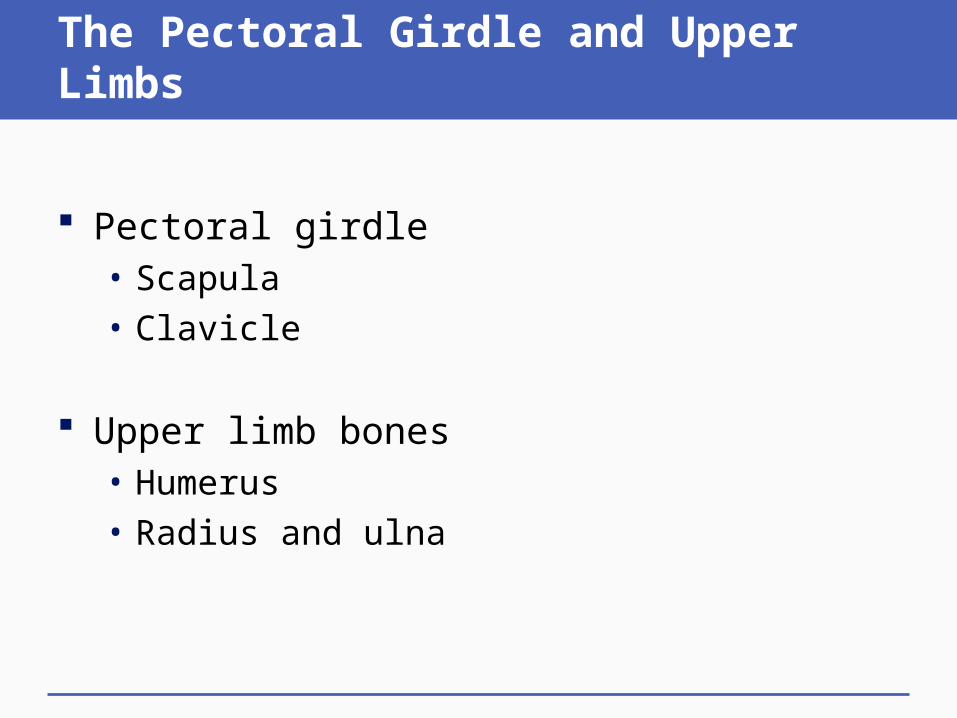
The Pectoral Girdle and Upper Limbs
Pectoral girdle • Scapula• Clavicle
Upper limb bones• Humerus• Radius and ulna

The Pelvic Girdle and Lower Limbs
Pelvic girdle• Six fused bones
Lower limb bones• Femur• Tibia and fibula• Patella

The Human Skeleton
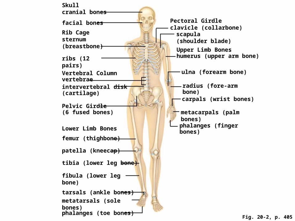
Fig. 20-2, p. 405
Skull cranial bones
facial bones Pectoral Girdle clavicle (collarbone)
scapula (shoulder blade)
Rib Cage sternum (breastbone)
Upper Limb Bones humerus (upper arm bone)ribs (12 pairs)
Vertebral Column vertebrae
ulna (forearm bone)
intervertebral disk (cartilage)
radius (fore-arm bone)
Pelvic Girdle (6 fused bones)
carpals (wrist bones)
metacarpals (palm bones)
Lower Limb Bones phalanges (finger bones)
femur (thighbone)
patella (kneecap)
tibia (lower leg bone)
fibula (lower leg bone)
tarsals (ankle bones)
metatarsals (sole bones)
phalanges (toe bones)

Animation: Human skeletal system

Bone Structure and Function
Bones are collagen-rich, mineralized organs, wrapped in connective tissue
Bones function in mineral storage, movement, and protection and support of soft organs• Ongoing mineral deposits and removals help
maintain blood levels of calcium and phosphorus, and also adjust bone strength
• Some bones are sites of blood cell formation
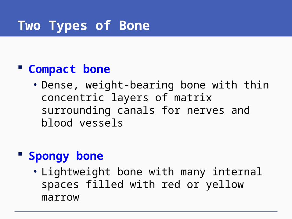
Two Types of Bone
Compact bone • Dense, weight-bearing bone with thin concentric
layers of matrix surrounding canals for nerves and blood vessels
Spongy bone• Lightweight bone with many internal spaces filled
with red or yellow marrow
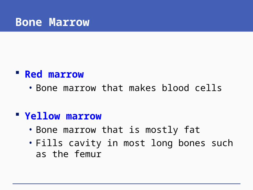
Bone Marrow
Red marrow • Bone marrow that makes blood cells
Yellow marrow • Bone marrow that is mostly fat• Fills cavity in most long bones such as the femur

Structure of a Femur
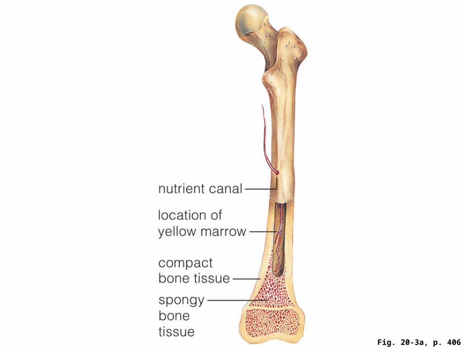
Fig. 20-3a, p. 406

Fig. 20-3a, p. 406
nutrient canal
location of yellow marrow
compact bone tissue
spongy bone tissue

Fig. 20-3b, p. 406
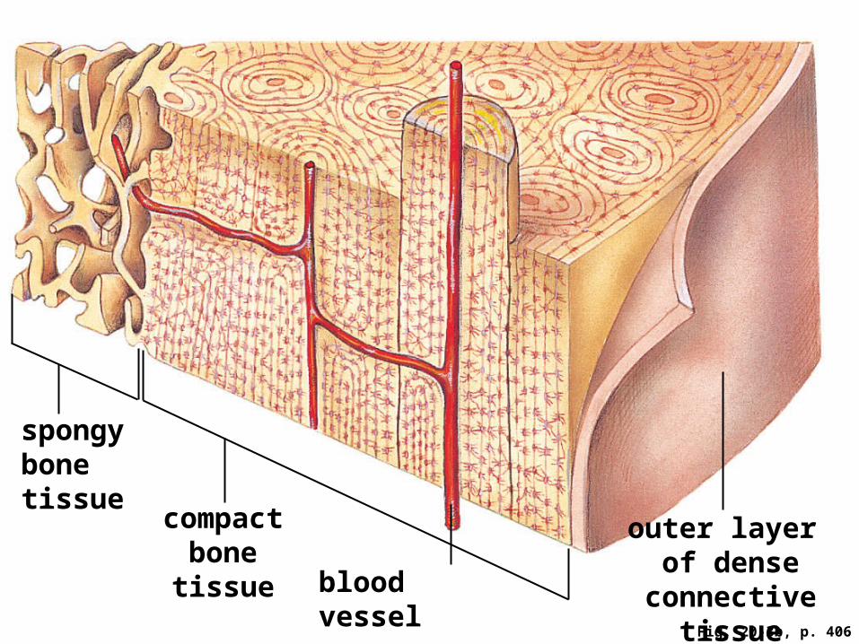
Fig. 20-3b, p. 406
spongy bone tissue
compact bone tissue
outer layer of dense
connective tissueblood vessel

Fig. 20-3c, p. 406

Fig. 20-3c, p. 406
space occupied by living bone cell
central canal

Animation: Structure of a femur
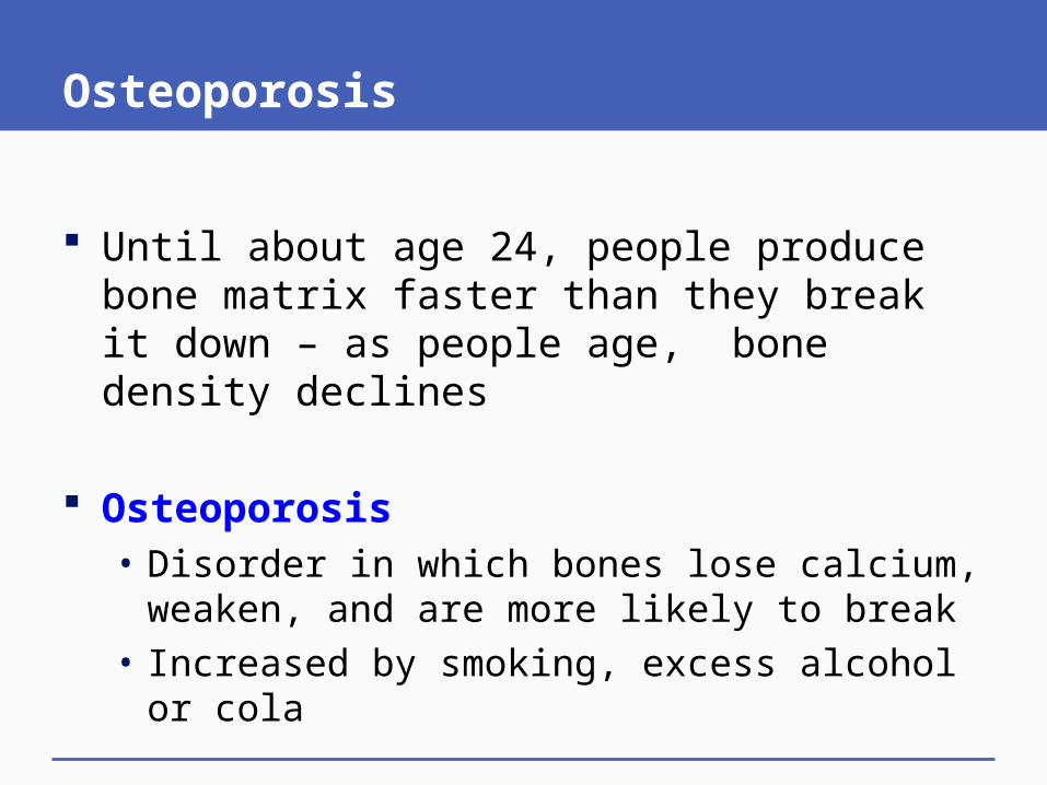
Osteoporosis
Until about age 24, people produce bone matrix faster than they break it down – as people age, bone density declines
Osteoporosis • Disorder in which bones lose calcium, weaken,
and are more likely to break • Increased by smoking, excess alcohol or cola

Where Bones Meet – Skeletal Joints
Joint • Region where bones meet and interact
Different joints have different movements• Ball-and-socket joint (shoulder, hip)• Gliding joints (wrists, ankles)• Hinge joints (elbows, knees)

Fibrous and Cartilaginous Joints
Fibrous joints hold bones tightly in place; cartilaginous joints let them move a bit
Fibrous joint • Joint where dense connective tissue holds bones
firmly in place (cranial bones)
Cartilaginous joint • Joint where pads of cartilage hold bones together
and provide cushioning, as between vertebrae

Synovial Joints
Synovial joints allow the most motion; ligaments connect bones at synovial joints
Synovial joint • Joint such as the knee that is lubricated by fluid
and allows movement of bones around the joint
Ligament • Dense connective tissue that holds bones
together at a joint

Knee Joint
A hinge-type synovial joint, held together by ligaments, stabilized by cartilage

Fig. 20-4, p. 407
femur
patella
cartilage
cruciate ligaments
menisci
tibia
fibula

Joint Injuries
Common joint injuries include sprained ankles, torn cruciate ligaments, and dislocations
Sprain • Ligaments of a joint are injured
Dislocation • Bones of a joint are out of place

Arthritis
Arthritis • Chronic inflammation and associated pain and
swelling of a joint
Two types of arthritis:• Osteoarthritis typically occurs in old age when
cartilage is worn down• Rheumatoid arthritis is an autoimmune disorder
which attacks all synovial joints

Animation: Fly wing action

Animation: Vertebrate skeletons

Animation: Long bone formation

Video: ABC News: Taller and taller
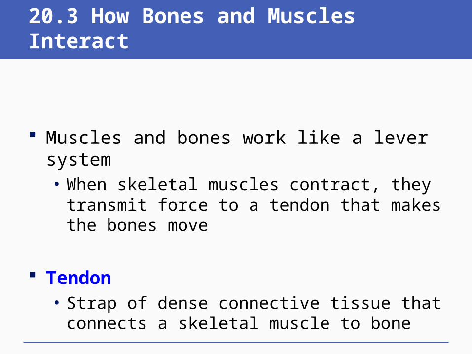
20.3 How Bones and Muscles Interact
Muscles and bones work like a lever system • When skeletal muscles contract, they transmit
force to a tendon that makes the bones move
Tendon • Strap of dense connective tissue that connects a
skeletal muscle to bone

How Skeletal Muscles Move
Muscles can only pull on bones, they cannot push them
Skeletal muscles often work as opposing pairs• Action of one reverses the action of the other• Example: biceps and triceps

Opposition: Biceps and Triceps

Fig. 20-5, p. 408
1biceps
radius
2
triceps

Animation: Human skeletal muscles

Animation: Structure of a sarcomere

20.4 Skeletal Muscle Structure and Function
The internal organization of a skeletal muscle promotes a strong, directional contraction • Many myofibrils make up a skeletal muscle fiber • A myofibril consists of units of sarcomeres, lined
up along its length • Each sarcomere has parallel arrays of actin and
myosin filaments
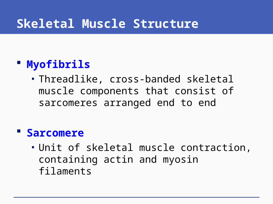
Skeletal Muscle Structure
Myofibrils • Threadlike, cross-banded skeletal muscle
components that consist of sarcomeres arranged end to end
Sarcomere • Unit of skeletal muscle contraction, containing
actin and myosin filaments

Skeletal Muscle Structure
Actin • Globular protein• Thin filaments of muscle fibers• Works with myosin to contract muscles
Myosin • Motor protein with a club-shaped head• Thick filaments of muscle fibers• Works with actin to contract muscles

Skeletal Muscle Structure

Fig. 20-6 (left), p. 409

Fig. 20-6 (left), p. 409
biceps brachii
triceps brachii deltoid
pectoralis major
trapezius
latissimus dorsi
rectus abdominis
gluteus maximus
biceps femoris
quadriceps femoris
gastrocnemius
Achilles tendon

Fig. 20-6 (a-c), p. 409

Fig. 20-6a, p. 409

Fig. 20-6a, p. 409
outer sheath of one skeletal muscle
one bundle of many muscle fibers in parallel inside the sheath
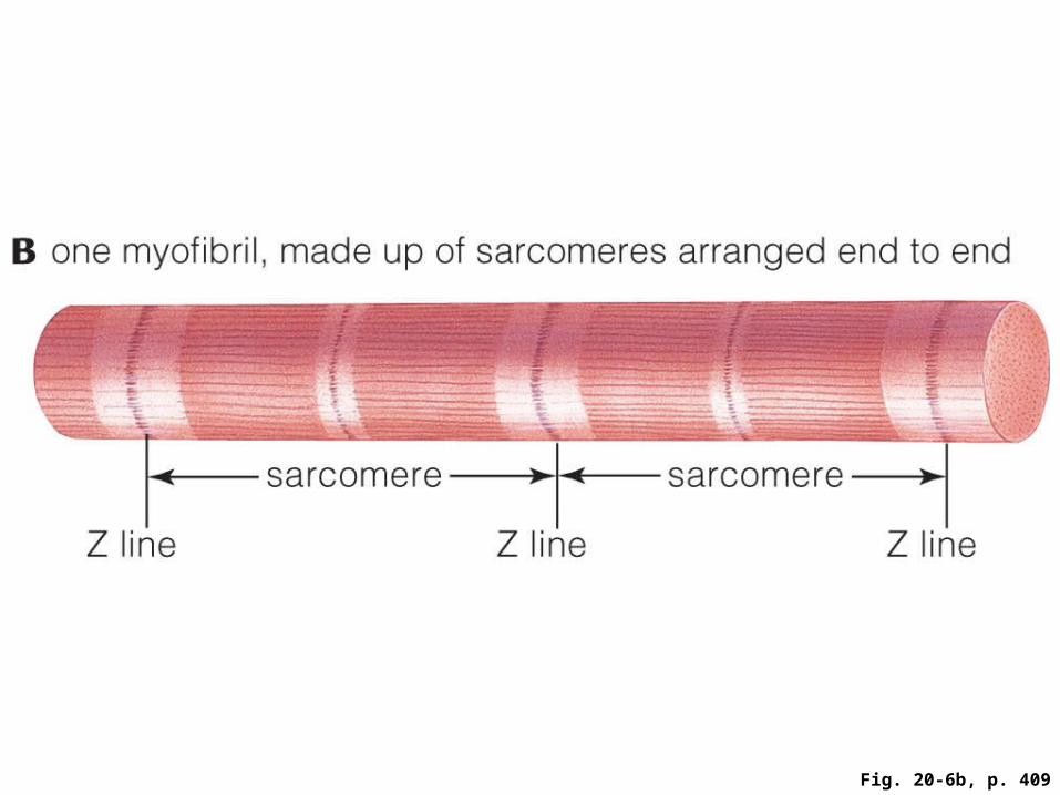
Fig. 20-6b, p. 409

Fig. 20-6b, p. 409
B one myofibril, made up of sarcomeres arranged end to end
sarcomere sarcomere
Z line Z line Z line

Fig. 20-6c, p. 409

Fig. 20-6c, p. 409
Z line Z line
C one sarcomere, with parallel actin and myosin filaments
actin myosin actin
Z line Z line

Animation: Structure of a skeletal muscle

Muscle Contraction
Skeletal muscles contract in response to signals from the nervous system
Sliding-filament model • Explains how interactions of actin and myosin
filaments shorten a sarcomere and bring about muscle contraction

How Sarcomeres Shorten
The sliding-filament model• Actin and myosin filaments lie close to each other• ATP activates myosin heads in thick filaments• Calcium is released; myosin binds to actin• Myosin heads tilt, sliding actin toward the center;
the sarcomere contracts• Binding of ATP releases myosin from actin; the
sarcomere relaxes

The Sliding Filament Model

Fig. 20-7a, p. 410

Fig. 20-7a, p. 410
actin myosin actin
Sarcomere between contractions

Fig. 20-7b, p. 410

Fig. 20-7b, p. 410
myosin head
one of many myosin-binding sites on actin
cross-bridge cross-bridge

Fig. 20-7c, p. 410

Fig. 20-7d, p. 410

Fig. 20-7d, p. 410
cross-bridge broken cross-bridge broken
Same sarcomere, contracted

Animation: Sliding filament model

Getting Energy For Contraction
Muscle fibers produce ATP needed for contraction by three pathways:• Dephosphorylation of creatine phosphate (lasts 5
to 10 seconds)• Aerobic respiration of glycogen (another 5 to 10
minutes), then of blood glucose and fatty acids (as long as oxygen is available)
• Lactate fermentation (when oxygen is no longer available)

Three Metabolic Pathways of ATP

Fig. 20-8a, p. 411
pathway 1 dephosphorylation of creatine phosphate
ADP + Pi
creatine
pathway 2 aerobic respiration
pathway 3 lactate fermentation
glucose from bloodstream and from glycogen breakdown in cellsoxygen

Fig. 20-8a, p. 411
pathway 1 dephosphorylation of creatine phosphate
ADP + Pi
creatine
pathway 3 lactate fermentation
pathway 2 aerobic respiration
glucose from bloodstream and from glycogen breakdown in cellsoxygen
Stepped Art

Fig. 20-8b, p. 411

Animation: Energy sources for contraction

Animation: Opposing muscle action

Animation: Troponin and tropomyosin

Animation: Muscle contraction overview

20.5 Properties of Whole Muscles
Motor unit • One motor neuron and all muscle fibers that form
junctions with its endings• All fibers of a motor unit contract at the same time• Repeated stimulation of a motor unit results in a
strong, sustained contraction• Brief stimulation causes a muscle twitch
Muscle twitch • Brief muscle contraction and relaxation

Stimulation of a Motor Unit

Fig. 20-9, p. 411
Fo
rce relaxation starts
stimulus
A A single, brief stimulus causes a twitch.
sustained contraction
twitch
Fo
rce
repeated stimulationTime
B Repeated stimulation results in a sustained contraction with several times the force of a twitch.
contraction

Animation: Types of contractions

Muscle Tension
Muscle tension is a mechanical force caused by muscle contraction• Opposed by a load (weight of object or gravity)
Muscle tension • Force exerted by a contracting muscle• Affected by number of fibers recruited
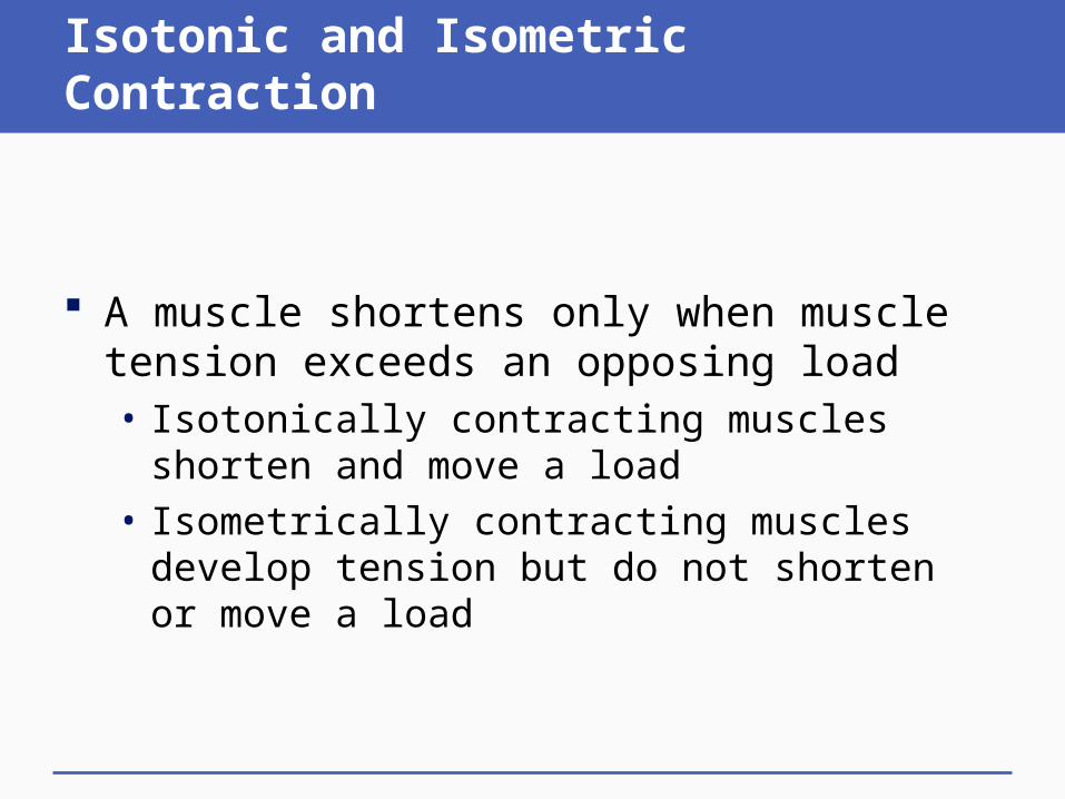
Isotonic and Isometric Contraction
A muscle shortens only when muscle tension exceeds an opposing load• Isotonically contracting muscles shorten and
move a load• Isometrically contracting muscles develop tension
but do not shorten or move a load

Isotonic and Isometric Contraction
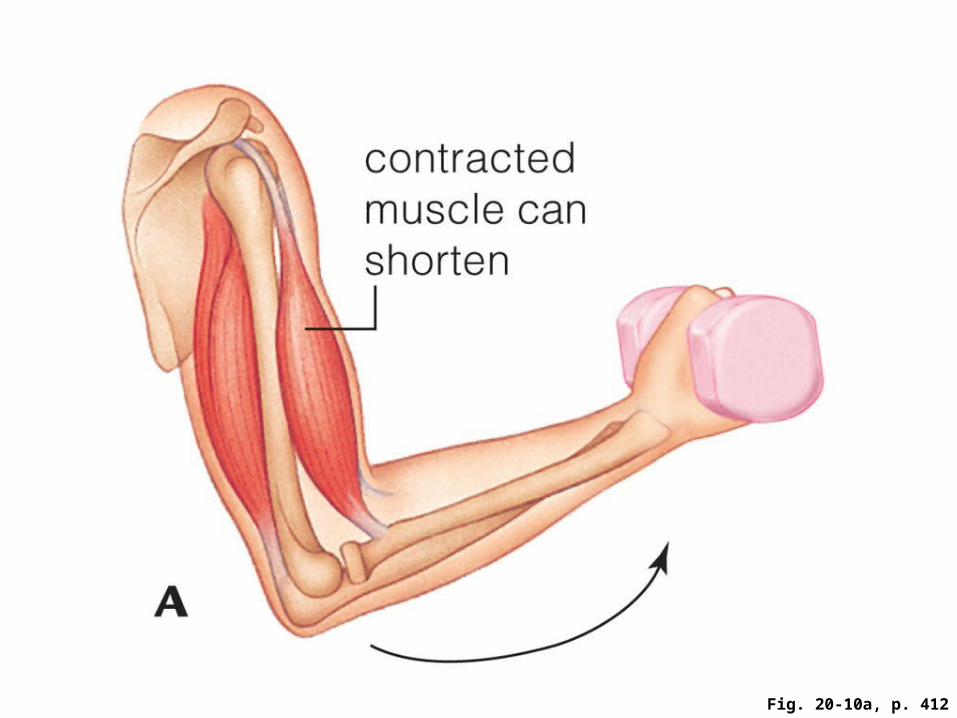
Fig. 20-10a, p. 412

Fig. 20-10a, p. 412
contracted muscle can shorten

Fig. 20-10b, p. 412

Fig. 20-10b, p. 412
contracted muscle cannot shorten

Muscles and Exercise
Aerobic exercise increases blood supply and number of mitochondria – which makes muscles more resistant to muscle fatigue
Strength training stimulates formation of more actin and myosin, but muscles fatigue rapidly
Muscle fatigue • Decrease in a muscle’s ability to contract despite
ongoing stimulation

Muscles and Aging
Muscle strength decreases with age• Muscles shrink; number of muscle fibers declines• Injuries take longer to heal
Strength training and aerobic exercise are helpful at any age• Slows loss of muscle tissue, improves circulation• Also good for the brain

Impaired Muscle Contraction
Some genetic disorders affect muscle structure and impair muscle function • Duchenne muscular dystrophy (X-linked)
Some diseases and toxins affect motor neurons• Poliovirus kills motor neurons• Tetanus, caused by toxins of Clostridium tetani,
kills by locking skeletal muscles in contraction• Amyotrophic lateral sclerosis (ALS)

Muscular Dystrophy
Normal skeletal muscle and muscle with muscular dystrophy

Tetanus

20.6 Impacts/Issues Revisited
Research on drugs that inhibit myostatin activity may help slow muscle loss resulting from muscular dystrophy, ALS, or even normal aging

Digging Into Data:Building Stronger Bones


