Chapter 20: Chapter 20: The Knee and The Knee and Related Structures Related Structures Jennifer Doherty-Restrepo, MS, LAT, Jennifer Doherty-Restrepo, MS, LAT, ATC ATC Academic Program Director, Entry- Academic Program Director, Entry- Level ATEP Level ATEP Florida International University Florida International University Acute Care and Injury Prevention Acute Care and Injury Prevention
Transcript
Chapter 20: Chapter 20: The Knee and The Knee and
Related StructuresRelated StructuresJennifer Doherty-Restrepo, MS, LAT, ATCJennifer Doherty-Restrepo, MS, LAT, ATC
Academic Program Director, Entry-Level ATEPAcademic Program Director, Entry-Level ATEPFlorida International UniversityFlorida International University
Acute Care and Injury PreventionAcute Care and Injury Prevention
Functional AnatomyFunctional Anatomy Movement of the knee requires flexion, Movement of the knee requires flexion,
extension, rotation and the arthrokinematic extension, rotation and the arthrokinematic motions of rolling and glidingmotions of rolling and gliding
Rotational component involves the “screw Rotational component involves the “screw home mechanism”home mechanism” As the knee extends it externally rotates As the knee extends it externally rotates
because the medial femoral condyle is larger because the medial femoral condyle is larger than the lateralthan the lateral
Provides increased stability to the kneeProvides increased stability to the knee
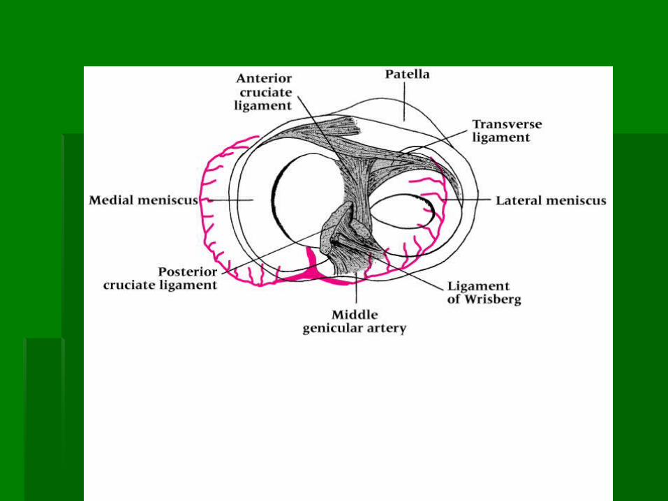
Capsular ligaments Capsular ligaments Taut during full extension and relaxed with flexionTaut during full extension and relaxed with flexion
Allows rotation to occurAllows rotation to occur Deeper capsular ligaments remain taut to keep rotation Deeper capsular ligaments remain taut to keep rotation
in checkin check
PCL prevents excessive internal rotation, guides PCL prevents excessive internal rotation, guides the knee in flexion, and acts as drag during initial the knee in flexion, and acts as drag during initial glide phase of flexionglide phase of flexion
ACL stops excessive internal rotation, stabilizes the ACL stops excessive internal rotation, stabilizes the knee in full extension, and prevents hyperextensionknee in full extension, and prevents hyperextension
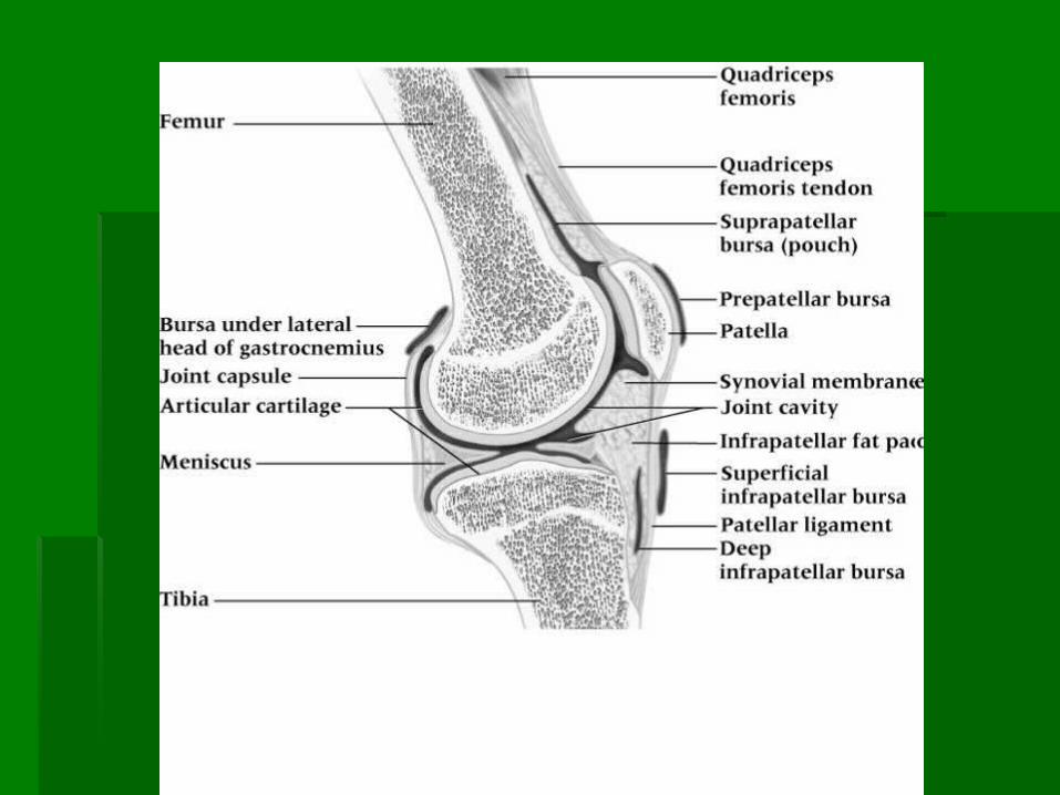
Patella Patella Aids knee during extension, providing a Aids knee during extension, providing a
mechanical advantagemechanical advantage Distributes compressive stress on the femur by Distributes compressive stress on the femur by
increasing contact between patellar tendon and increasing contact between patellar tendon and femurfemur
Protects patellar tendon against frictionProtects patellar tendon against friction When moving from extension to flexion the When moving from extension to flexion the
patella glides laterally and further into trochlear patella glides laterally and further into trochlear groovegroove
Kinetic ChainKinetic Chain Directly affected by motions and forces occurring at Directly affected by motions and forces occurring at
the foot, ankle, lower leg, thigh, hip, pelvis, and the foot, ankle, lower leg, thigh, hip, pelvis, and spinespine
With the kinetic chain, forces must be absorbed and With the kinetic chain, forces must be absorbed and distributeddistributed
If body is unable to manage the imposed forces, If body is unable to manage the imposed forces, breakdown in the kinetic chain occursbreakdown in the kinetic chain occurs
Knee is very susceptible to injury resulting from the Knee is very susceptible to injury resulting from the absorption of forcesabsorption of forces
Assessment of the Assessment of the Knee JointKnee Joint
Determine MOI - This is critical!!!Determine MOI - This is critical!!! History: Acute InjuryHistory: Acute Injury
Past historyPast history Position of body at time of injury?Position of body at time of injury? Did the knee collapse?Did the knee collapse? Did you hear or feel anything?Did you hear or feel anything? Could you move your knee immediately after Could you move your knee immediately after
injury or was it locked?injury or was it locked? Did swelling occur?Did swelling occur? Where was the painWhere was the pain
History: Recurrent or Chronic InjuryHistory: Recurrent or Chronic Injury What is your major complaint?What is your major complaint? When did you first notice the condition?When did you first notice the condition? Is there recurrent swelling?Is there recurrent swelling? Does the knee lock or catch?Does the knee lock or catch? Is there severe pain?Is there severe pain? Grinding or grating?Grinding or grating? Does it ever feel like giving way?Does it ever feel like giving way? What does it feel like when ascending and descending What does it feel like when ascending and descending
stairs?stairs? What past treatment have you undergone?What past treatment have you undergone?
ObservationObservation Walking, half squatting, going up and down stairsWalking, half squatting, going up and down stairs Swelling, ecchymosisSwelling, ecchymosis Leg alignmentLeg alignment
Genu valgum and genu varumGenu valgum and genu varum Hyperextension and hyperflexionHyperextension and hyperflexion Patella alta and bajaPatella alta and baja Patella rotated inward or outwardPatella rotated inward or outward Tibial torsion, femoral anteversion and retroversion Tibial torsion, femoral anteversion and retroversion
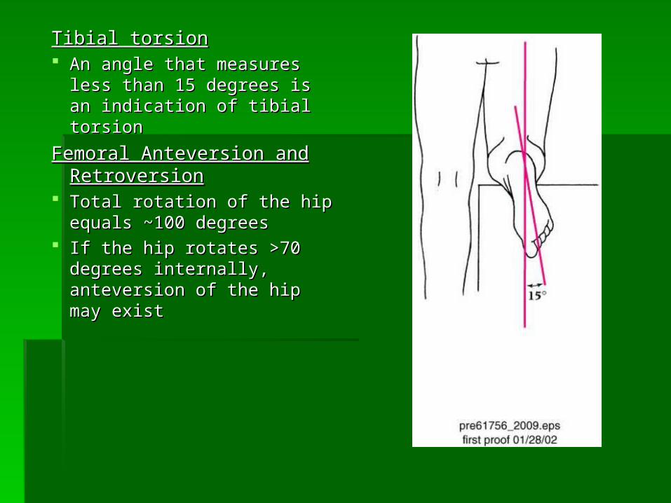
Tibial torsionTibial torsion An angle that measures less An angle that measures less
than 15 degrees is an than 15 degrees is an indication of tibial torsionindication of tibial torsion
Femoral Anteversion and Femoral Anteversion and RetroversionRetroversion
Total rotation of the hip Total rotation of the hip equals ~100 degreesequals ~100 degrees
If the hip rotates >70 degrees If the hip rotates >70 degrees internally, anteversion of the internally, anteversion of the hip may existhip may exist
Observation cont.Observation cont. Knee Symmetry or AsymmetryKnee Symmetry or Asymmetry
Do the knees look symmetrical? Do the knees look symmetrical? Is there obvious swelling? Is there obvious swelling? Atrophy?Atrophy?
Leg Length DiscrepancyLeg Length Discrepancy Anatomical or functionalAnatomical or functional Anatomical differences can potentially Anatomical differences can potentially
cause problems in all weight bearing jointscause problems in all weight bearing joints Functional differences can be caused by Functional differences can be caused by
pelvic rotations or mal-alignment of the pelvic rotations or mal-alignment of the spinespine
Intracapsular swellingIntracapsular swelling May be referred to as joint effusionMay be referred to as joint effusion Swelling within the joint that is caused by Swelling within the joint that is caused by
synovial fluid and blood is called synovial fluid and blood is called hemarthrosishemarthrosis Sweep maneuver – sign of joint effusionSweep maneuver – sign of joint effusion Ballotable patella - sign of joint effusionBallotable patella - sign of joint effusion
Extracapsular swellingExtracapsular swelling Localized over the injured structure Localized over the injured structure May ultimately migrate down to foot and ankleMay ultimately migrate down to foot and ankle
Palpation - SwellingPalpation - Swelling
Special Tests: Special Tests: Knee Knee InstabilityInstability
Valgus Stress TestValgus Stress Test Used to assess the Used to assess the
integrity of MCL integrity of MCL Testing at 0 degrees Testing at 0 degrees
Testing at 30 degrees Testing at 30 degrees of flexion isolates the of flexion isolates the ligamentsligaments
Special Tests: Special Tests: Knee InstabilityKnee Instability
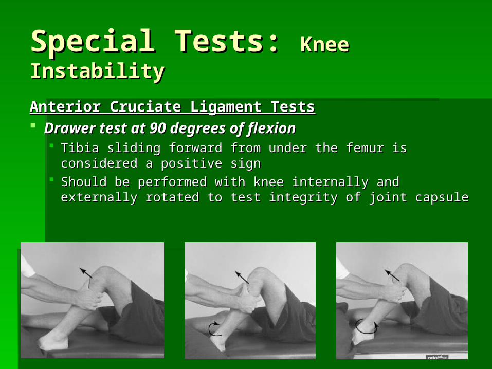
Anterior Cruciate Ligament TestsAnterior Cruciate Ligament Tests Drawer test at 90 degrees of flexionDrawer test at 90 degrees of flexion
Tibia sliding forward from under the femur is considered a Tibia sliding forward from under the femur is considered a positive sign positive sign
Should be performed with knee internally and externally Should be performed with knee internally and externally rotated to test integrity of joint capsulerotated to test integrity of joint capsule
Special Tests: Special Tests: Knee InstabilityKnee Instability
Avoids painful flexion immediately after injuryAvoids painful flexion immediately after injury Reduces hamstring involvementReduces hamstring involvement At 30 degrees of flexion an attempt is made to translate the tibia anteriorly on the At 30 degrees of flexion an attempt is made to translate the tibia anteriorly on the
femurfemur A positive test indicates damage to the ACLA positive test indicates damage to the ACL
Variations for the Lachman Drawer TestVariations for the Lachman Drawer Test May be necessary if athlete is large or May be necessary if athlete is large or
examiner’s hands are smallexaminer’s hands are small
Variations include:Variations include: Rolled towel under the femurRolled towel under the femur Leg off the table with athlete supineLeg off the table with athlete supine Athlete prone on table with knee and lower Athlete prone on table with knee and lower
leg just off tableleg just off table
Special Tests: Special Tests: Knee InstabilityKnee Instability
Pivot Shift TestPivot Shift Test Used to determine anterolateral Used to determine anterolateral
rotary instabilityrotary instability Position starts with knee extended Position starts with knee extended
and leg internally rotatedand leg internally rotated The thigh and knee are then The thigh and knee are then
flexed with a valgus stress applied flexed with a valgus stress applied to the kneeto the knee
Reduction of the tibial plateau Reduction of the tibial plateau (producing a clunk) is a positive (producing a clunk) is a positive signsign
Jerk TestJerk Test Reverses direction of the pivot shiftReverses direction of the pivot shift Moves from position of flexion to extensionMoves from position of flexion to extension Without an ACL the tibia will sublux at 20 Without an ACL the tibia will sublux at 20
degrees of flexiondegrees of flexion
Special Tests: Special Tests: Knee InstabilityKnee Instability
Flexion-Rotation Drawer TestFlexion-Rotation Drawer Test Knee is taken from a position of 15 Knee is taken from a position of 15
degrees of flexion degrees of flexion Tibia is subluxed anteriorly with femur Tibia is subluxed anteriorly with femur
externally rotatedexternally rotated
Knee is moved into 30 degrees of flexion Knee is moved into 30 degrees of flexion where tibia rotates posteriorly and femur where tibia rotates posteriorly and femur internally rotatesinternally rotates
Special Tests: Special Tests: Knee InstabilityKnee Instability
Knee is flexed at 90 degrees and a Knee is flexed at 90 degrees and a posterior force is applied to determine posterior force is applied to determine translation of the tibia posteriorlytranslation of the tibia posteriorly
Positive sign indicates a PCL deficient Positive sign indicates a PCL deficient kneeknee
Special Tests: Special Tests: Knee InstabilityKnee Instability
With the athlete supine, the leg is lifted by With the athlete supine, the leg is lifted by the great toethe great toe
If the tibia externally rotates and slides If the tibia externally rotates and slides posteriorly there may be a PCL injury and posteriorly there may be a PCL injury and damage to the posterolateral corner of the damage to the posterolateral corner of the capsulecapsule
Special Tests: Special Tests: Knee InstabilityKnee Instability
Special Tests: Special Tests: Knee InstabilityKnee Instability
Posterior Sag Test (Godfrey’s test)Posterior Sag Test (Godfrey’s test) Athlete is supine with both knees flexed to 90 Athlete is supine with both knees flexed to 90
degreesdegrees Lateral observation is required to determine Lateral observation is required to determine
extent of posterior sag while comparing extent of posterior sag while comparing bilaterallybilaterally
Instrument Assessment of the Instrument Assessment of the Cruciate LigamentsCruciate Ligaments
A number of devices are A number of devices are available to quantify AP available to quantify AP displacement of the kneedisplacement of the knee KT-2000 arthrometerKT-2000 arthrometer Stryker knee laxity tester Stryker knee laxity tester GenucomGenucom
Test can be taken pre & Test can be taken pre & post-operatively and post-operatively and throughout rehabilitationthroughout rehabilitation
McMurray’s Meniscal TestMcMurray’s Meniscal Test Used to determine displaced meniscal tearUsed to determine displaced meniscal tear Leg is moved into flexion and extension Leg is moved into flexion and extension
while knee is internally and externally while knee is internally and externally rotated in conjunction with valgus and rotated in conjunction with valgus and varus stressesvarus stresses
A positive test is found when clicking and A positive test is found when clicking and popping are feltpopping are felt
Special Tests: Special Tests: Meniscal TestsMeniscal Tests
Special Tests: Special Tests: Meniscal TestsMeniscal Tests
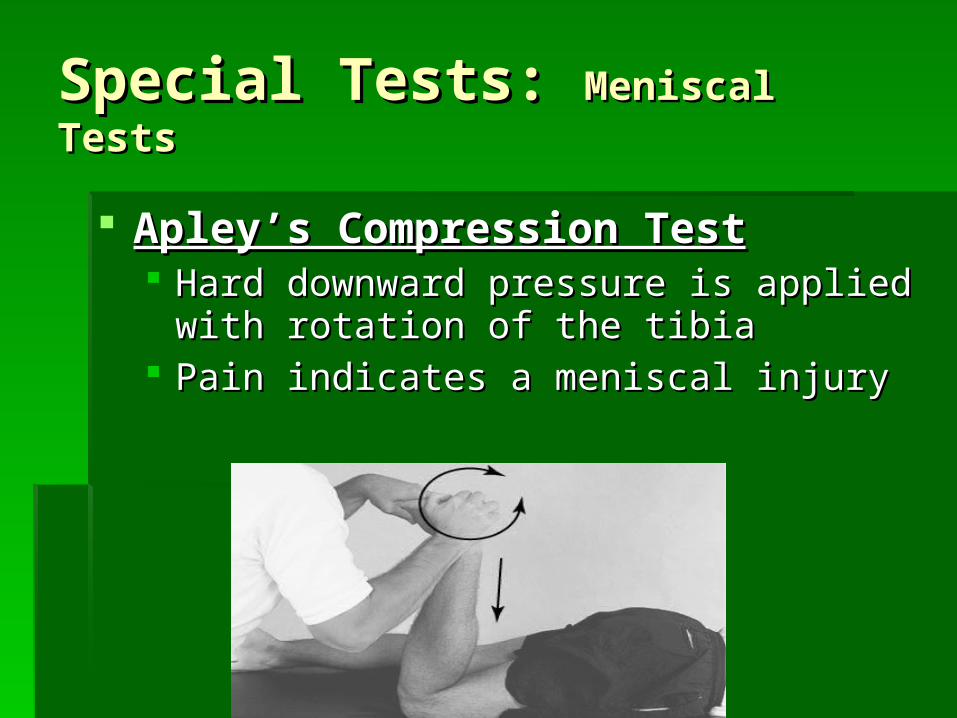
Apley’s Compression TestApley’s Compression Test Hard downward pressure is applied with Hard downward pressure is applied with
rotation of the tibiarotation of the tibia Pain indicates a meniscal injuryPain indicates a meniscal injury
Special Tests: Special Tests: Meniscal TestsMeniscal Tests
Apley’s Distraction TestApley’s Distraction Test Traction is applied with rotation of the tibiaTraction is applied with rotation of the tibia Pain will occur if there is damage to the Pain will occur if there is damage to the
capsule or ligamentscapsule or ligaments No pain will occur if there is a meniscal No pain will occur if there is a meniscal
injuryinjury
Girth MeasurementsGirth Measurements Changes in girth may result due to atrophy, Changes in girth may result due to atrophy,
swelling, and conditioningswelling, and conditioning Circumferential measures to determine Circumferential measures to determine
deficits and gains during rehabilitation deficits and gains during rehabilitation
Subjective RatingsSubjective Ratings Used to determine patient’s perception of Used to determine patient’s perception of
pain, stability, and functional performancepain, stability, and functional performance
Other Special TestsOther Special Tests
Functional ExaminationFunctional Examination Assess walking, running, turning, cutting, etcAssess walking, running, turning, cutting, etc Co-contraction test, vertical jump, single leg Co-contraction test, vertical jump, single leg
hop tests, and the duck walkhop tests, and the duck walk Resistive strength testingResistive strength testing
Other Special TestsOther Special Tests
The Q - AngleThe Q - Angle Line which bisect the patella relative to the ASIS and Line which bisect the patella relative to the ASIS and
the tibial tuberclethe tibial tubercle Normal angle is 10Normal angle is 10°° for males and 15 for males and 15 °° for females for females Elevated angles often lead to pathological conditions Elevated angles often lead to pathological conditions
associated with improper patella trackingassociated with improper patella tracking
The A - AngleThe A - Angle Patellar orientation relative to the tibial tuberclePatellar orientation relative to the tibial tubercle Quantitative measure of the patellar realignment after Quantitative measure of the patellar realignment after
rehabilitationrehabilitation An angle greater than 35An angle greater than 35°° is often correlated is often correlated
associated with patellofemoral pathomechanicsassociated with patellofemoral pathomechanics
Other Special TestsOther Special Tests
Patella Grinding TestPatella Grinding Test Determines integrity of patellar cartilage on the Determines integrity of patellar cartilage on the
undersurface of patellaundersurface of patella
Patella Compression TestPatella Compression Test Determines integrity of patellar cartilage on the Determines integrity of patellar cartilage on the
undersurface of patellaundersurface of patella
Apprehension TestsApprehension Tests Patella pushed laterally to determine presence Patella pushed laterally to determine presence
of subluxation/dislocation of subluxation/dislocation
Special Tests: Special Tests: PatellaPatella
Recognition and Recognition and Management of Management of Specific InjuriesSpecific Injuries
MOI = severe blow or outward twistMOI = severe blow or outward twist Grade I: Signs and Symptoms Grade I: Signs and Symptoms
Little fiber tearing or stretchingLittle fiber tearing or stretching Stable valgus testStable valgus test Little or no joint effusionLittle or no joint effusion Some joint stiffness and point tenderness on Some joint stiffness and point tenderness on
laterallateral aspect of the knee aspect of the knee Relatively normal ROMRelatively normal ROM
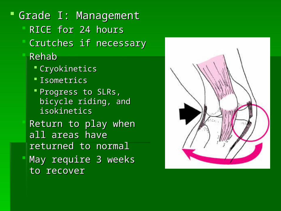
Grade I: ManagementGrade I: Management RICE for 24 hoursRICE for 24 hours Crutches if necessaryCrutches if necessary RehabRehab
CryokineticsCryokinetics Isometrics Isometrics Progress to SLRs, bicycle Progress to SLRs, bicycle
riding, and isokineticsriding, and isokinetics
Return to play when all Return to play when all areas have returned to areas have returned to normalnormal
May require 3 weeks to May require 3 weeks to recoverrecover
Grade II: Signs and SymptomsGrade II: Signs and Symptoms Complete tear of deep capsular ligament and Complete tear of deep capsular ligament and
partial tear of MCLpartial tear of MCL No gross instability; laxity at 5-15 degrees of No gross instability; laxity at 5-15 degrees of
flexionflexion Slight swellingSlight swelling Moderate to severe joint tightness Moderate to severe joint tightness Decreased ROMDecreased ROM Pain along medial aspect of kneePain along medial aspect of knee
Grade II: ManagementGrade II: Management RICE for 48-72 hoursRICE for 48-72 hours Crutch use until acute inflammation phase has Crutch use until acute inflammation phase has
resolvedresolved Possibly a brace or casting prior to the initiation of Possibly a brace or casting prior to the initiation of
ROM activitiesROM activities Modalities 2-3 times daily for painModalities 2-3 times daily for pain Gradual progression from isometrics (quad exercises) Gradual progression from isometrics (quad exercises)
Grade III: Signs and Symptoms Grade III: Signs and Symptoms Complete tear of supporting ligamentsComplete tear of supporting ligaments Complete loss of medial stabilityComplete loss of medial stability Minimum to moderate swellingMinimum to moderate swelling Immediate pain followed by acheImmediate pain followed by ache Loss of motion due to effusion and Loss of motion due to effusion and
hamstring guardinghamstring guarding Positive valgus stress testPositive valgus stress test
Grade III: ManagementGrade III: Management RICERICE Conservative non-operative versus surgical Conservative non-operative versus surgical
approachapproach Limited immobilization (with a brace)Limited immobilization (with a brace) Progressive weight bearing and increased Progressive weight bearing and increased
ROM over 4-6 week periodROM over 4-6 week period Rehab would be similar to Grade I & II Rehab would be similar to Grade I & II
injuriesinjuries
MOI = Varus force usually with the tibia MOI = Varus force usually with the tibia internally rotatedinternally rotated
Direct blow is rare MOIDirect blow is rare MOI If severe enough damage may also If severe enough damage may also
occur to occur to Cruciate ligamentsCruciate ligaments ITBITB MeniscusMeniscus Bony fragments may result as wellBony fragments may result as well
Signs and SymptomsSigns and Symptoms Pain and tenderness over LCLPain and tenderness over LCL Swelling and effusion around the LCLSwelling and effusion around the LCL Joint laxity with varus testingJoint laxity with varus testing May cause irritation of the peroneal nerveMay cause irritation of the peroneal nerve
ManagementManagement Same as MCL injury managementSame as MCL injury management
MOI = tibia externally rotated with a valgus force MOI = tibia externally rotated with a valgus force Occasionally the result of hyperextension resulting Occasionally the result of hyperextension resulting
from a direct blowfrom a direct blow Research is quite extensive in regards to impact of Research is quite extensive in regards to impact of
femoral notch, ACL size and laxity, mal-alignments femoral notch, ACL size and laxity, mal-alignments (Q-angle), and faulty biomechanics(Q-angle), and faulty biomechanics
Extrinsic factors may include, conditioning, skill Extrinsic factors may include, conditioning, skill acquisition, playing style, equipment, preparation timeacquisition, playing style, equipment, preparation time
May also involve damage to other structures including May also involve damage to other structures including meniscus, capsule, and MCLmeniscus, capsule, and MCL
Signs and SymptomsSigns and Symptoms Experience pop with severe pain and disabilityExperience pop with severe pain and disability Positive anterior drawer and Lachman’s Positive anterior drawer and Lachman’s Rapid swelling at the joint lineRapid swelling at the joint line Other ACL tests may also be positiveOther ACL tests may also be positive
ManagementManagement RICE; use of crutchesRICE; use of crutches Arthroscopy may be necessary to determine extent of injuryArthroscopy may be necessary to determine extent of injury Surgical repairSurgical repair
Without surgery, joint degeneration may resultWithout surgery, joint degeneration may result Surgery may involve joint reconstruction with grafts (tendon), transplantation Surgery may involve joint reconstruction with grafts (tendon), transplantation
of external structuresof external structures Also requires 4-6 months of rehabAlso requires 4-6 months of rehab
MOI = fall on bent knee (most common)MOI = fall on bent knee (most common) Most at risk during 90 degrees of flexionMost at risk during 90 degrees of flexion Injury may result due to a rotational forceInjury may result due to a rotational force
Signs and SymptomsSigns and Symptoms Feel a pop in the back of the kneeFeel a pop in the back of the knee Tenderness and relatively little swelling in Tenderness and relatively little swelling in
the popliteal fossathe popliteal fossa Laxity with posterior sag testLaxity with posterior sag test
Appropriate for grade I and II injuries Appropriate for grade I and II injuries Focus on quad strengtheningFocus on quad strengthening
Post-operative rehabPost-operative rehab Surgery will require 6 weeks of Surgery will require 6 weeks of
immobilization in extension immobilization in extension Full weight bearing on crutchesFull weight bearing on crutches ROM after 6 weeks ROM after 6 weeks PRE at 4 monthsPRE at 4 months
Most common MOI is rotary force with Most common MOI is rotary force with knee flexed or extendedknee flexed or extended
Tears may be longitudinal, oblique, or Tears may be longitudinal, oblique, or transverse transverse
Medial meniscus is more commonly Medial meniscus is more commonly injured due to ligamentous attachments injured due to ligamentous attachments and decreased mobilityand decreased mobility Also more prone to disruption through Also more prone to disruption through
torsional and valgus forcestorsional and valgus forces
Meniscal LesionsMeniscal Lesions
Signs and SymptomsSigns and Symptoms Effusion developing over 48-72 hoursEffusion developing over 48-72 hours Pain in joint line Pain in joint line Loss of motionLoss of motion Intermittent locking and giving wayIntermittent locking and giving way Pain with squattingPain with squatting Portions of meniscus may become Portions of meniscus may become
detached causing locking, giving way, or detached causing locking, giving way, or catching within the jointcatching within the joint
If chronic injury, recurrent swelling or If chronic injury, recurrent swelling or muscle atrophy may occurmuscle atrophy may occur
ManagementManagement No locking but indications of a tear are presentNo locking but indications of a tear are present
Further diagnostic testing may be requiredFurther diagnostic testing may be required
If locking occurs, anesthesia may be necessary If locking occurs, anesthesia may be necessary to unlock the joint to unlock the joint Possible arthroscopic surgery Possible arthroscopic surgery
Healing dependent on location of tearHealing dependent on location of tear Menisectomy Menisectomy
Partial weight bearing, quick return to activityPartial weight bearing, quick return to activity
Repaired meniscusRepaired meniscus Requires immobilization, gradual return to activity Requires immobilization, gradual return to activity
over the course of 12 weeksover the course of 12 weeks
MOI = irritation of the plica MOI = irritation of the plica Often associated with chondromalaciaOften associated with chondromalacia
Signs and SymptomsSigns and Symptoms Possible history of knee pain/injuryPossible history of knee pain/injury Recurrent episodes of painful pseudo-lockingRecurrent episodes of painful pseudo-locking Possible snapping and poppingPossible snapping and popping Pain with stairs and squattingPain with stairs and squatting Little or no swellingLittle or no swelling No ligamentous laxityNo ligamentous laxity
ManagementManagement Treat conservatively w/ RICE and NSAID’s if the result of traumaTreat conservatively w/ RICE and NSAID’s if the result of trauma Recurrent conditions may require surgeryRecurrent conditions may require surgery
Knee PlicaKnee Plica
MOI = twisting, sudden cutting, or direct blowMOI = twisting, sudden cutting, or direct blow Signs and SymptomsSigns and Symptoms
Hear a snap Hear a snap Feeling of giving wayFeeling of giving way Immediate swelling Immediate swelling Considerable painConsiderable pain
ManagementManagement Diagnosis confirmed through arthroscopic examDiagnosis confirmed through arthroscopic exam Surgery used to replace fragments in order to avoid joint Surgery used to replace fragments in order to avoid joint
degeneration and arthritis degeneration and arthritis
MOI = partial or complete separation of MOI = partial or complete separation of articular cartilage and subchondral bonearticular cartilage and subchondral bone
Exact cause is unknown but may include: Exact cause is unknown but may include: Blunt trauma, Blunt trauma, Possible skeletal or endocrine abnormalities, Possible skeletal or endocrine abnormalities, Prominent tibial spine impinging on medial Prominent tibial spine impinging on medial
femoral condyle, or femoral condyle, or Impingement due to patellar facetImpingement due to patellar facet
Signs and SymptomsSigns and Symptoms Aching pain and point tendernessAching pain and point tenderness Recurrent swelling Recurrent swelling Possible lockingPossible locking Possible quadriceps atrophy Possible quadriceps atrophy
ManagementManagement Rest and immobilization for childrenRest and immobilization for children Surgery may be necessary in teenagers and adults Surgery may be necessary in teenagers and adults
Drilling to stimulate healing, pinning, or bone graftsDrilling to stimulate healing, pinning, or bone grafts
MOI = repeated traumaMOI = repeated trauma May result due to osteochondritis dissecans, meniscal May result due to osteochondritis dissecans, meniscal
fragments, synovial tissue damage, or cruciate ligaments fragments, synovial tissue damage, or cruciate ligaments injuryinjury
Signs and SymptomsSigns and Symptoms May become lodged and cause locking or poppingMay become lodged and cause locking or popping Pain Pain Sensation of instabilitySensation of instability
ManagementManagement If not surgically removed it can lead to conditions causing If not surgically removed it can lead to conditions causing
joint degenerationjoint degeneration
Loose Bodies Loose Bodies
MOI = direct blow MOI = direct blow Signs and SymptomsSigns and Symptoms
Severe painSevere pain Acute inflammationAcute inflammation Loss of movement Loss of movement SwellingSwelling
If not resolved within a week then a chronic condition may exist (synovitis or If not resolved within a week then a chronic condition may exist (synovitis or bursitis) bursitis)
EcchymosisEcchymosis Possible capsular damagePossible capsular damage
ManagementManagement RICE RICE Progress to normal activity following return of ROM Progress to normal activity following return of ROM Padding for protectionPadding for protection
Joint ContusionsJoint Contusions
MOI = compression due to a direct blowMOI = compression due to a direct blow Signs and SymptomsSigns and Symptoms
Local pain and possible shooting nerve painLocal pain and possible shooting nerve pain Numbness and paresthesia Numbness and paresthesia Added pressure may exacerbate conditionAdded pressure may exacerbate condition Generally resolves quickly Generally resolves quickly
In the event it does not resolve, it could result in drop footIn the event it does not resolve, it could result in drop foot
ManagementManagement RICE RICE Return to play once symptoms resolve and no weakness is Return to play once symptoms resolve and no weakness is
presentpresent Padding for fibular headPadding for fibular head
Peroneal Nerve Peroneal Nerve ContusionContusion
MOI = acute, chronic, or recurrent swellingMOI = acute, chronic, or recurrent swelling Prepatellar = continued kneelingPrepatellar = continued kneeling Infrapatellar = overuse of patellar tendonInfrapatellar = overuse of patellar tendon
Signs and SymptomsSigns and Symptoms Localized swelling that results in ballotable patellaLocalized swelling that results in ballotable patella Swelling in popliteal fossa may indicate a Baker’s cystSwelling in popliteal fossa may indicate a Baker’s cyst
Associated with burse over the semimembranosus or medial head of Associated with burse over the semimembranosus or medial head of gastrocnemiusgastrocnemius
Commonly painless and causing little disabilityCommonly painless and causing little disability May progress and should be treated accordinglyMay progress and should be treated accordingly
ManagementManagement Eliminate causeEliminate cause RICE and NSAID’sRICE and NSAID’s Aspiration and steroid injection if chronicAspiration and steroid injection if chronic
BursitisBursitis
MOI = direct or indirect trauma MOI = direct or indirect trauma Semi-flexed position with forceful contraction, which may occur Semi-flexed position with forceful contraction, which may occur
while falling, jumping or runningwhile falling, jumping or running
Signs and SymptomsSigns and Symptoms Hemorrhaging and joint effusion Hemorrhaging and joint effusion Possible capsular tearing, separation of bone fragments, and Possible capsular tearing, separation of bone fragments, and
possible quadriceps tendon tearing due to bone fragmentspossible quadriceps tendon tearing due to bone fragments
ManagementManagement X-ray necessary for confirmation X-ray necessary for confirmation RICE and splinting if fracture suspectedRICE and splinting if fracture suspected ReferRefer Possible immobilize for 2-3 monthsPossible immobilize for 2-3 months
Patellar FracturePatellar Fracture
MOI = deceleration with simultaneous cutting in MOI = deceleration with simultaneous cutting in opposite direction (valgus force)opposite direction (valgus force) Quad pulls the patella out of alignmentQuad pulls the patella out of alignment Repetitive subluxation will impose stress to medial Repetitive subluxation will impose stress to medial
restraintsrestraints
Signs and SymptomsSigns and Symptoms SubluxationSubluxation
Pain, swelling, restricted ROM, and palpable tenderness over Pain, swelling, restricted ROM, and palpable tenderness over adductor tubercleadductor tubercle
Dislocations Dislocations Total loss of functionTotal loss of function
Patella Subluxation or Patella Subluxation or DislocationDislocation
ManagementManagement Reduction Reduction
Performed by flexing hip, moving patella medially, and slowly extending the Performed by flexing hip, moving patella medially, and slowly extending the kneeknee
Following reduction, immobilize for at least 4 weeks Following reduction, immobilize for at least 4 weeks Use crutches Use crutches Isometric exercises Isometric exercises
After immobilization period, horseshoe pad with elastic wrap should After immobilization period, horseshoe pad with elastic wrap should be used to support patellabe used to support patella
Rehab focuses on strengthening the muscles around the knee, thigh, Rehab focuses on strengthening the muscles around the knee, thigh, and hip and hip
Possible surgery to release tight structuresPossible surgery to release tight structures Improve postural and biomechanical factorsImprove postural and biomechanical factors
MOI = becomes wedged between the tibia and MOI = becomes wedged between the tibia and patellapatella Irritated by chronic kneeling, pressure, or traumaIrritated by chronic kneeling, pressure, or trauma
Signs and SymptomsSigns and Symptoms Capillary hemorrhaging and swellingCapillary hemorrhaging and swelling Chronic irritation may lead to scarring and Chronic irritation may lead to scarring and
calcificationcalcification Pain below the patellar ligament during knee Pain below the patellar ligament during knee
extensionextension May display weakness, mild swelling, and stiffness May display weakness, mild swelling, and stiffness
during movementduring movement
Infrapatellar Fat PadInfrapatellar Fat Pad
ManagementManagement Rest Rest
Avoid irritating activities until inflammation has subsidedAvoid irritating activities until inflammation has subsided
Utilize therapeutic modalities for inflammationUtilize therapeutic modalities for inflammation Heel lift to prevent irritation during extensionHeel lift to prevent irritation during extension Hyperextension taping to prevent full extensionHyperextension taping to prevent full extension
MOI = softening and deterioration of the MOI = softening and deterioration of the articular cartilagearticular cartilage
Three stages:Three stages: Swelling and softening of cartilageSwelling and softening of cartilage Fissure of softened cartilageFissure of softened cartilage Deformation of cartilage surfaceDeformation of cartilage surface
Often associated with abnormal trackingOften associated with abnormal tracking Abnormal patellar tracking may be due to genu Abnormal patellar tracking may be due to genu
Signs and SymptomsSigns and Symptoms Pain with walking, running, stairs, and squattingPain with walking, running, stairs, and squatting Possible recurrent swellingPossible recurrent swelling Grating sensation with flexion and extensionGrating sensation with flexion and extension Pain at inferior border during palpationPain at inferior border during palpation
RICE, NSAID’s, isometrics, orthotics to correct dysfunctionRICE, NSAID’s, isometrics, orthotics to correct dysfunction
Surgical possibilitiesSurgical possibilities Altering muscle attachmentsAltering muscle attachments Shaping and smoothing of surfacesShaping and smoothing of surfaces DrillingDrilling Elevating tibial tubercleElevating tibial tubercle
MOI = lateral deviation of patella while tracking in femoral MOI = lateral deviation of patella while tracking in femoral groovegroove May result due to tight structures, pronation, increased Q angle, May result due to tight structures, pronation, increased Q angle,
insufficient medial musculatureinsufficient medial musculature Signs and SymptomsSigns and Symptoms
Tenderness at lateral facet of patella Tenderness at lateral facet of patella Swelling associated with irritation of synoviumSwelling associated with irritation of synovium Dull ache in center of kneeDull ache in center of knee Patellar compression will elicit pain and crepitusPatellar compression will elicit pain and crepitus Apprehension when patella is forced laterallyApprehension when patella is forced laterally
ManagementManagement Correct imbalances (strength and flexibility)Correct imbalances (strength and flexibility) McConnell tapingMcConnell taping Lateral retinacular release if conservative measures failLateral retinacular release if conservative measures fail
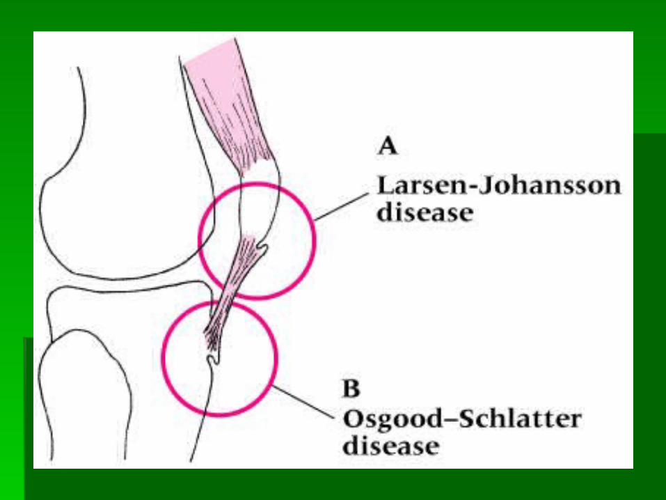
Osgood Schlatter’s is apophysitis at the tibial Osgood Schlatter’s is apophysitis at the tibial tubercle tubercle MOI = repeated avulsion of patellar tendonMOI = repeated avulsion of patellar tendon Bony callus develops enlarging the tibial tubercleBony callus develops enlarging the tibial tubercle Resolves with agingResolves with aging
Larsen Johansson is the result of excessive Larsen Johansson is the result of excessive pulling on the inferior pole of the patellapulling on the inferior pole of the patella
Signs and SymptomsSigns and Symptoms SwellingSwelling Hemorrhaging Hemorrhaging Gradual degeneration of the apophysis due to Gradual degeneration of the apophysis due to
impaired circulationimpaired circulation Pain with kneeling, jumping, and runningPain with kneeling, jumping, and running Point tendernessPoint tenderness
ManagementManagement ConservativeConservative Reduce stressful activity Reduce stressful activity Possible castingPossible casting Ice before and after activityIce before and after activity IsometericsIsometerics
MOI = sudden or repetitive extensionMOI = sudden or repetitive extension Jumping or kicking places tremendous strain on patellar or Jumping or kicking places tremendous strain on patellar or
quadriceps tendonquadriceps tendon Signs and SymptomsSigns and Symptoms
Pain and tenderness at inferior pole of patellaPain and tenderness at inferior pole of patella 3 phases: 3 phases:
1) pain after activity, 1) pain after activity, 2) pain during and after activity,2) pain during and after activity, 3) pain during and after activity that may become constant3) pain during and after activity that may become constant
Patellar TendinitisPatellar Tendinitis (Jumper’s or Kicker’s Knee)(Jumper’s or Kicker’s Knee)
MOI = sudden, powerful quad contractionMOI = sudden, powerful quad contraction Rare unless a chronic inflammatory condition exists Rare unless a chronic inflammatory condition exists
resulting in tissue degenerationresulting in tissue degeneration Occurs primarily at point of attachmentOccurs primarily at point of attachment
Signs and SymptomsSigns and Symptoms Palpable defectPalpable defect Lack of knee extensionLack of knee extension Considerable swelling and pain (initially)Considerable swelling and pain (initially)
ManagementManagement Surgical repair is neededSurgical repair is needed Proper conservative treatment of jumper’s knee can Proper conservative treatment of jumper’s knee can
minimize chances of occurringminimize chances of occurring
Patellar Tendon RupturePatellar Tendon Rupture
MOI = repetitive/overuse conditions attributed to MOI = repetitive/overuse conditions attributed to mal-alignment and structural asymmetriesmal-alignment and structural asymmetries
Signs and SymptomsSigns and Symptoms IT Band Friction SyndromeIT Band Friction Syndrome
Irritation at band’s insertion Irritation at band’s insertion Commonly seen in individual that have genu varum or Commonly seen in individual that have genu varum or
pronated feetpronated feet Pes Anserine Tendinitis or BursitisPes Anserine Tendinitis or Bursitis
Result of excessive genu valgum and weak vastus medialisResult of excessive genu valgum and weak vastus medialis Often occurs due to running with one leg higher than the Often occurs due to running with one leg higher than the
other other Running on a slope or crowned roadRunning on a slope or crowned road
ManagementManagement Correction of mal-alignmentsCorrection of mal-alignments Ice before and after activityIce before and after activity Utilize proper warm-up and stretching techniquesUtilize proper warm-up and stretching techniques Avoidance of aggravating activitiesAvoidance of aggravating activities NSAID’s NSAID’s OrthoticsOrthotics
Giving way of knee Giving way of knee Result of… Result of…
Weak quadricepsWeak quadriceps Chronic instability of ligamentous structuresChronic instability of ligamentous structures Torn meniscusTorn meniscus Loose bodies within the kneeLoose bodies within the knee Subluxating patellaSubluxating patella ChondromalaciaChondromalacia Due to painDue to pain
The Collapsing KneeThe Collapsing Knee
Prevention of Knee Prevention of Knee InjuriesInjuries
Total body conditioning is requiredTotal body conditioning is required Strength, flexibility, cardiovascular and Strength, flexibility, cardiovascular and
muscular endurance, agility, speed and muscular endurance, agility, speed and balancebalance
Muscles around joint must be conditioned Muscles around joint must be conditioned to maximize stabilityto maximize stability Flexibility and strengtheningFlexibility and strengthening
Must avoid abnormal muscle action Must avoid abnormal muscle action through flexibilitythrough flexibility
ACL Prevention ProgramsACL Prevention Programs Focus on strength, neuromuscular control, Focus on strength, neuromuscular control,
and balanceand balance Series of different programs which address Series of different programs which address
balance board training, landing strategies, balance board training, landing strategies, plyometric training, and single leg plyometric training, and single leg performanceperformance
Can be implemented in rehabilitation and Can be implemented in rehabilitation and preventative training programspreventative training programs
Shoe TypeShoe Type Change in football footwear has drastically Change in football footwear has drastically
reduced the incidence of knee injuriesreduced the incidence of knee injuries Shoes with more short cleats does not allow Shoes with more short cleats does not allow
foot to become fixed foot to become fixed Still allows for control during running and cuttingStill allows for control during running and cutting
Functional and Prophylactic Functional and Prophylactic Knee BracesKnee Braces Used to protect MCLUsed to protect MCL Used to prevent further Used to prevent further
damage to grade 1 and grade damage to grade 1 and grade 2 ACL sprains 2 ACL sprains
Used to protect the ACL Used to protect the ACL following surgeryfollowing surgery
Can be custom molded and Can be custom molded and designed to control rotational designed to control rotational forcesforces
Knee Joint Knee Joint RehabilitationRehabilitation General Body ConditioningGeneral Body Conditioning
Must be maintained with non-weight bearing activitiesMust be maintained with non-weight bearing activities Weight BearingWeight Bearing
Initial crutch use, non-weight bearingInitial crutch use, non-weight bearing Gradual progression to weight bearing while wearing Gradual progression to weight bearing while wearing
Used to reduce arthrofibrosisUsed to reduce arthrofibrosis Patellar mobilization is key following surgeryPatellar mobilization is key following surgery CPM unitsCPM units
FlexibilityFlexibility Must be regained, maintained, and improvedMust be regained, maintained, and improved
Muscular StrengthMuscular Strength Progression of isometrics, isotonics, Progression of isometrics, isotonics,
isokinetics, and plyometricsisokinetics, and plyometrics Incorporate eccentric muscle actionIncorporate eccentric muscle action Open vs. closed kinetic chain exercisesOpen vs. closed kinetic chain exercises
Neuromuscular ControlNeuromuscular Control Loss of control is generally due to pain and Loss of control is generally due to pain and
swellingswelling Through exercise and balance equipment Through exercise and balance equipment
proprioception can be enhanced and regainedproprioception can be enhanced and regained
BracingBracing Variety of braces Variety of braces
Some used to control for specific injuries while Some used to control for specific injuries while others are designed for specific forces, stability, others are designed for specific forces, stability, and providing resistanceand providing resistance
Typically worn for 3-6 weeks after surgeryTypically worn for 3-6 weeks after surgery Used to limit ROM for a period of time Used to limit ROM for a period of time
Functional ProgressionFunctional Progression Gradual return to sports specific skillsGradual return to sports specific skills Progress with weight bearing, move into Progress with weight bearing, move into
walking and running, and then onto sprinting walking and running, and then onto sprinting and change of directionand change of direction
Return to ActivityReturn to Activity Based on healing process Based on healing process
Sufficient time for healing must be allowedSufficient time for healing must be allowed
Objective criteria should include…Objective criteria should include… Strength assessmentStrength assessment ROM measuresROM measures Functional performance testsFunctional performance tests
SummarySummary
Review anatomyReview anatomy AssessmentAssessment
History, observation, palpationHistory, observation, palpation Special TestsSpecial Tests