Enhanced Permeability and Retention (EPR) Effectfor Anticancer Nanomedicine Drug Targeting
Khaled Greish
Abstract
Effective cancer therapy remains one of the most challenging tasks to the scientific community, withlittle advancement on overall cancer survival landscape during the last two decades. A major limitationinherent to most conventional anticancer chemotherapeutic agents is their lack of tumor selectivity. Oneway to achieve selective drug targeting to solid tumors is to exploit abnormalities of tumor vasculature,namely hypervascularization, aberrant vascular architecture, extensive production of vascular permeabilityfactors stimulating extravasation within tumor tissues, and lack of lymphatic drainage. Due to their largesize, nano-sized macromolecular anticancer drugs administered intravenously (i.v.) escape renal clearance.Being unable to penetrate through tight endothelial junctions of normal blood vessels, their concentra-tion builds up in the plasma rendering them long plasma half-life. More importantly, they can selectivelyextravasate in tumor tissues due to its abnormal vascular nature. Overtime the tumor concentration willbuild up reaching several folds higher than that of the plasma due to lack of efficient lymphatic drainagein solid tumor, an ideal application for EPR-based selective anticancer nanotherapy. Indeed, this selectivehigh local concentration of nano-sized anticancer drugs in tumor tissues has proven superior in therapeu-tic effect with minimal side effects in both preclinical and clinical settings.
Anticancer chemotherapy is one of the most notorious drugsknown to human. The sole purpose of their use is cell killing, atask that usually achieved with high efficacy but little precision.Most anticancer chemotherapeutic agents used in clinic targetdividing cells, regardless of its nature whether a dividing tumorcell or active intestinal epithelial cells, with same potency. The
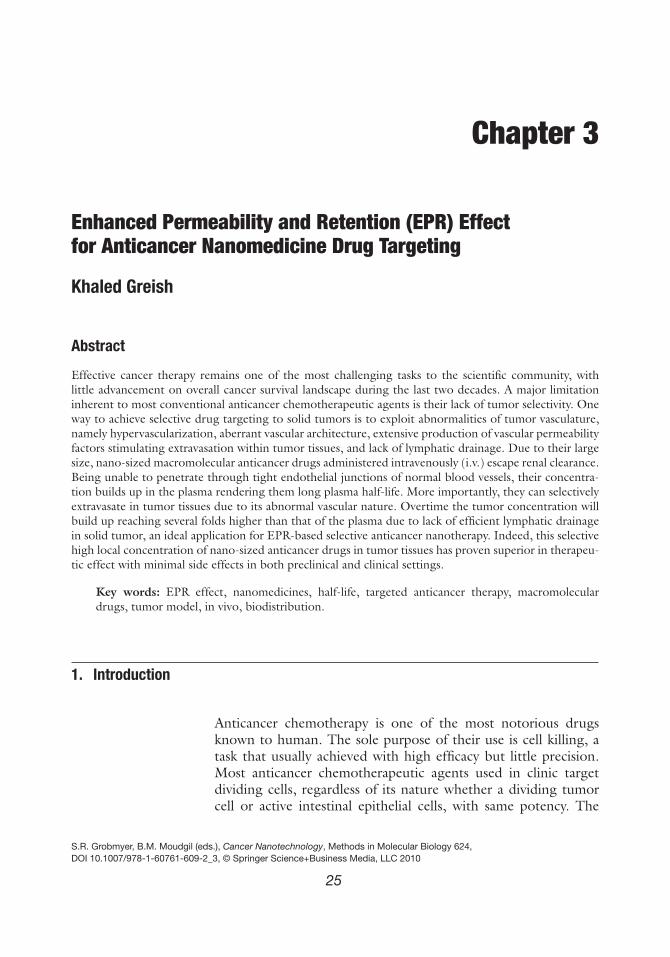
term of maximum tolerated dose thus is not based on the amountneeded to cure the disease, conversely on the amount needednot to incapacitate the host. Alternatively, anticancer targetingproved to be a more effective approach with minimum toxicity.On the gross level, tumor vasculature proved to be a potentialtarget for cancer treatment. Tumor vascular typically comprised ofpoorly aligned defective endothelial cells with wide fenestrations(up to 4 μm), lacking smooth muscle layer or innervations, withrelatively wide lumen and impaired receptor function for vasoac-tive mediators especially angiotensin II; further they lack func-tional lymphatics (1–5). In addition, hyperproduction of vascularmediators, such as vascular endothelial growth factor, bradykinin,nitric oxide peroxynitrite, prostaglandins, and matrix metallopro-teinases (6–10), contributes greatly to this hyperpermeability intumor tissues. EPR effect involves two major components, firstaltered biodistribution, where the nano-size drug shows differ-ential accumulation in the tumor tissues reaching higher concen-tration than that in the plasma or other organs (Fig. 3.1A), thiseffect is time dependent and can be reproduced in tumors of dif-ferent size as shown in Fig. 3.1B. EPR effect is mainly the func-tion of molecular weight with molecules ranging 40–800 kD canexhibit preferential tumor targeting (Fig. 3.1C).
The other aspect of EPR effect is increased plasma half-life of the nano-size drugs as their size exceeded the limit ofrenal excretion threshold, limiting their clearance (Fig. 3.1D). Aspharmacological effect and plasma concentration are parallel, thisphenomenon results in prolonged therapeutic effect in additionto targeting (11, 12). The gain expected from EPR effect is in themagnitude of 7- to 10-fold higher tumor concentration comparedto equivalent doses of the same drug at low molecular weightform (13). EPR is a passive phenomenon that can be actively aug-mented by the following techniques related to the abnormalitiesof tumor vasculature.
1.1. Hypertension In normal blood vessels, the smooth muscle layer responds tovascular mediators such as bradykinin, acetylcholine, NO, andcalcium via the receptors on smooth muscle cells of blood ves-sels, and hence helps maintain constant blood flow volume(Fig. 3.2 a B). Raising the systolic blood pressure from 100to 150 mm Hg for 15 min by infusing angiotensin II (AT II)into tumor-bearing rats caused 2- to 6-fold selective increasein tumor blood flow; blood flow in normal organs and tissuesremained constant regardless of the induced blood pressure (14).When 51Cr-labeled-SMANCS or 51Cr-albumin was injected i.v.under AT-II-induced hypertension, 3-fold higher accumulationwas achieved in tumor tissue compared to normotensive condi-tion. Additionally, the amount of drug delivered to normal organssuch as kidney and bone marrow was reduced because of the
EPR Effect for Anticancer Nanomedicine Drug Targeting 27
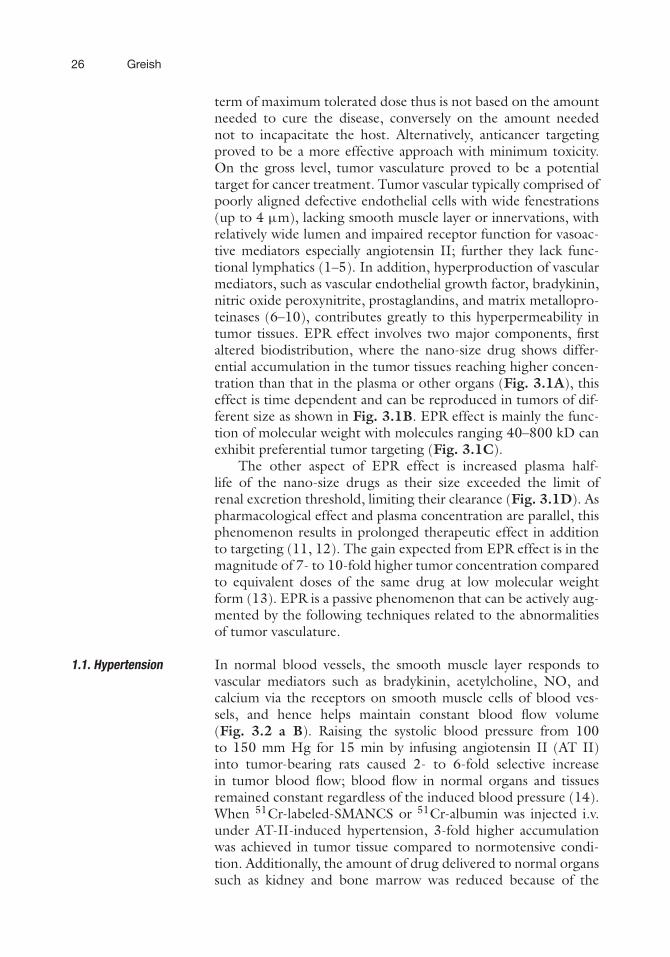
Fig. 3.1. Selective accumulation of Evans blue dye bound to albumin (70 kDa) in S 180 tumor in DdY mice. a, Bloodconcentration versus tumor, liver, kidney, and muscle. The graph represents the result of methods in Section 3.2. b,EBD accumulation in small tumor (A), large tumor (B ), (C ) shows the retention of the dye at 24 h and (D ) is crosssection of (B ), the central region of the tumor is necrotic and avascular, and hence does not facilitate the uptake ofnano-size EBD. c, The accumulation of 125I-labeled N-(2-hydroxypropyl) methacrylamide (HPMA) copolymer in the solidtumor tissues. Note that large macromolecules, but not smaller ones, manifest progressive accumulation. d, The relationbetween molecular weight, AUC, tumor accumulation, and renal clearance. (Reproduced from 27 with permission fromImperial College Press.)
vasoconstriction occurring in normal organs under AT-II-inducedhypertension. However, mitomycin C (334.3 Da) did not pro-duce the same effect under the same experimental conditions(15). Better therapeutic effect could be demonstrated in the clin-ical setting, by using this method with SMANCS/Lipiodol fortumors such as cholangiocarcinoma, metastatic liver cancer, andrenal cancer (16).
1.2. Bradykinin (BK) BK-generating cascade is normally activated in tumor tissues andwas found to be involved in accumulation of malignant asciticand pleural fluids in cancer patients. Various human and rodentsolid tumors express excessive levels of BK receptor B2 (7–9).BK is degraded by many peptidases, especially angiotensin-converting enzyme (ACE). Therefore, ACE inhibitors can causelocal increase of BK levels at the tumor site. On the basis ofthese data, we used the ACE inhibitors enalapril and temocapril
28 Greish
Fig. 3.2. Diagrammatic representation of EPR effect. (A) Shows the diffusion of a low molecular weight (a) and highmolecular weight drug (b). Note progressive accumulation of macromolecular drug in the tumor tissues with time bythe EPR effect. (B) Vascular leakage in relation to AT-II. Note that under AT-II-induced hypertensive state (B-a), vaso-constriction in the normal blood vessels occurs due to the presence of smooth muscle layer, while tumor vessels (B-b),passively dilate due to the absence of smooth muscle layer leading to enhanced extravasations of macromolecular drugsand hence augmentation of EPR effect. (Reproduced from 26 with permission from Elsevier.)
to inhibit BK degradation, this resulted in elevated BK level andfurther enhancement of the EPR effect (17). ACE inhibitors thusincrease delivery of macromolecular drugs to tumors, even undernormotensive conditions (Fig. 3.3).
1.3. Nitric Oxide (NO) NO plays a key role in angiogenesis, cell proliferation, and EPReffect. NO is synthesized by NO synthase (NOS) from L-arginine.Consequently, inhibition of NO generation by NOS inhibitors asN-monomethyl-L-arginine was found to suppress tumor growth(7, 8, 10). NOS inhibitor irreversibly attenuated blood flow inR3230Ac rat mammary adenocarcinoma (18). In addition, wefound that pro-matrix metalloproteinases (proMMPs) are acti-vated by peroxynitrite (ONOO–), which is produced extensivelyin tumor and inflammatory tissues (19). Clinically, we have uti-lized the vasodilator effect of NO to increase the delivery of thenanomedicine drug SMANCS to tumor site in hepatocellular car-cinoma by local injection of isosorbide dinitrate (ISDN) into thetumor-feeding artery (Fig. 3.4).
1.4. PhotodynamicTherapy (PDT)
When a photosensitizer is administrated systemically or locallyand subsequently activated by illumination with visible light, thisleads to the generation of reactive oxygen species. This effect hasactive role in enhancing tumor vascular permeability (20, 21).The concentration of nano-size FITC-dextran was 5-fold higher
EPR Effect for Anticancer Nanomedicine Drug Targeting 29
Fig. 3.3. Enhancement of permeability in the skin of guinea pig after SC injection ofvarious doses of BK and the 56 kDa protease which induces kallikrein–kinin system. EBDwas injected intravenously after 10 min of SC injection. The results show the enhance-ment of permeability at 6 h after injection of EBD.
Fig. 3.4. The effect of local injection of the vasodilator isosorbide dinitrate (ISDN) on the diameter of the proper hepaticartery (arrow): (a) before ISDN and (b) after ISDN injection. Note the 187% increase in the vessel diameter (b), thistechnique was successfully used in the clinic to enhance SMANCS targeting into hepatic tumors.
30 Greish
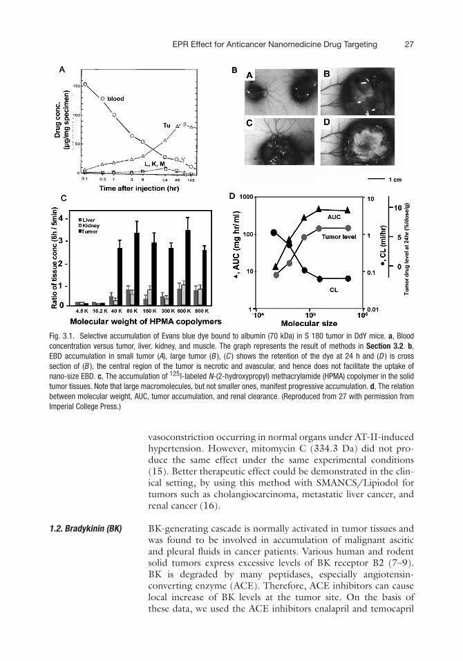
in orthotropic MatLyLu rat prostate tumors treated with thephotosensitizer verteporfin, at 15 min following light irradiation,compared to nonirradiated control group (21). Photosensitiza-tion causes endothelial cell microtubule depolymerization andinduces the formation of actin stress fibers. Thus, endothelial cellswere found to retract, leading to the formation of intercellulargaps, which result in enhanced vascular permeability. In addition,endothelial cell damage leads to the establishment of thrombo-genic sites within the vessel lumen, and this initiates a physiologi-cal cascade of responses including platelet aggregation, the releaseof vasoactive molecules, leukocyte adhesion, and increases in vas-cular permeability (20). We have used the photosensitizer ZnPPto enhance tumor permeability and could achieve 3-fold higheraccumulation of the Evans blue dye in tumor tissue after 5 minirradiation with ambient light (Fig. 3.5).
CA B
Fig. 3.5. Enhancement of the EPR effect by photodynamic therapy (PDT). (a), EBD concentration in S-180 murine sar-coma without PDT. (b) 15 min after injection of 5 mg/kg ZnPP photosensitizer, animals were injected with EBD 10 mg/kg.After another 30 min of ZnPP injection, animals were irradiated for 5 min at 50.000 Lux, 4 h after irradiation animalswere scarified and EBD was quantified. (c), ZnPP-mediated photoactivity enhanced the EPR effect by 3-fold magnitude.
2. Materials
2.1. Establishment ofIn Vivo Tumor Model
1. Mouse sarcoma S-180 cells, ATCC Number TIB-66 (Man-assas, VA).
2. Male DdY mice, 6 weeks old weighing 30–35 g (SLC, Inc.,Shizuoka, Japan).
1. Inject 5 × 10⁶ S-180 cells in 1 mL PBS into the right lowerquadrant of the abdomen of DdY mice using 27-G needle.
2. Observe animals for 7–10 days, this is a proper time toaspirate the resulting ascetic fluid of tumor; longer timewill result in bloody aspirate.
3. Euthanize animal using CO2 for 2 min.4. Place the animal on its side and insert G18 needle mounted
10 mL syringe into the most gravity-dependent point andaspirate cells.
5. Cells to be diluted with five times cold sterile PBS and cen-trifuged at 800 rpm at 4◦C for 5 min to remove proteinsand blood from cancer cells, repeat for two times or untilthe cell precipitates become clear.
6. Repeat Steps 1–5 for another extra passage before inducingSC tumor to insure high tumorgenic ability of cells.
7. Count a small sample of cell (5 μL) using trypan blue dyein hemocytometer.
8. Prepare 2 × 10⁶ cells per 200 μL PBS and keep in ice.9. Anesthetize animals using 3–4% isoflurane for 2–3 min in
induction chamber of the vapor anesthesia system, thenmove animals to warm heated blanket, and use nose conefor anesthesia.
32 Greish
10. Shave two bilateral dorsal sites where tumor injection isintended.
11. Lay anesthetized animal ventrally, raise a dorsal skin foldwith a tweezers, clean it with gauze soaked in 70% ethanol,and at near horizontal angel, insert the 27 G needlemounted on 1-mL syringe, inject cell slowly to preventleakage from injection site. The development of SC blebindicates successful injection.
12. Follow the animal daily for 7–10 days, at this time usuallyanimal will develop tumors of 5–7 mm diameter which areoptimal for EPR-related studies.
3.2. Demonstration ofEPR Effect in TumorTissues
1. Dissolve Evans blue dye (EBD) in DW at final concentra-tions of 1 mg/mL, and then adjust the concentration to givefinal dose of 10 mg/kg of mice weight in 0.2 mL.
2. As the tumor reaches the diameter of 7 mm, immobilize theanimal in immobilization chamber and inject 0.2 EBD insaline into the tail vein.
3. At each time point of evaluation, euthanize animals in CO2chamber for 2 min.
4. Place the animal ventrally and through midline incision,expose inferior vena cava, collect 0.2 mL of blood, and mixinstantly with 2.8 mL of Isoton II, followed by centrifuga-tion at 800 rpm for 5 min, then read by spectrophotometerat 620 nm.
5. After blood collection, cut the diaphragm and expose theheart, grasp the heart with tweezers, and using 20-mLsyringe with 24-G needle, inject 20 mL of saline slowly intothe left side of the heart to remove the dye from the bloodcomponent of any organ.
6. Expose the dorsal skin having the tumor, visually documentthe EBD color in the tumor tissue in contrast to surroundingskin using camera, remove the tumor from the base usingsurgical scissor, weigh, and add to 3 mL formamide.
7. Collect sample from other tissue similarly, weigh, and add to3 mL formamide.
8. Incubate at 60◦C water bath with shaking for 48 h toextract EBD.
9. Read by spectrophotometer at 620 nm and convert the read-ing into μg EBD/mg tissue using standard curve of EBD.
3.3. PreparingSMA-Micelles
1. Dissolve SMA powder in water at pH 5.0 at 10 mg/mL.2. Dropwise add doxorubicin solution in DW at a concentra-
tion of 10 mg/mL.
EPR Effect for Anticancer Nanomedicine Drug Targeting 33
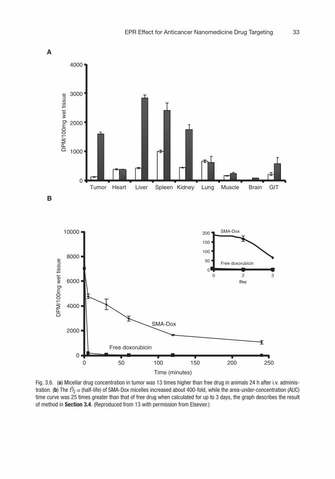
Fig. 3.6. (a) Micellar drug concentration in tumor was 13 times higher than free drug in animals 24 h after i.v. adminis-tration. (b) The t1/2 α (half-life) of SMA-Dox micelles increased about 400-fold, while the area-under-concentration (AUC)time curve was 25 times greater than that of free drug when calculated for up to 3 days, the graph describes the resultof method in Section 3.4. (Reproduced from 13 with permission from Elsevier.)
34 Greish
3. Add water soluble carbodiimide (EDAC) 10 mg/mL. Thesolution will precipitate.
4. Wash precipitates by centrifugation at 8,000 rpm for 5 mintwice, and then reconstitute in pH 7, leave for 60 min tototally solubilize the micelles.
5. Wash and concentrate the micelle to one-tenth of the origi-nal volume by ultrafiltration with the Amicon ultrafiltration,repeat three times.
6. Measure concentration of the resulting micelle through UV,comparing it to standard curve of the absorbance of freedoxorubicin.
3.4.Pharmacokineticsand Drug Distribution
1. Use animal model as described in Section 3.1.2. Divide animals into two groups, control the group to receive
radio-labeled free doxorubicin, and test the group to receiveradio-labeled SMA-Dox micelles. Inject 5 mg/kg of doxoru-bicin equivalent through the tail vein in a 0.2-mL volumesaline.
3. At scheduled time points, euthanize mice in CO2 and collectblood samples from the inferior vena cava as in Step 4 ofSection 3.2.
4. Inject 20 mL of saline to remove blood components fromthe blood vessels in the tissues as in Step 5 of Section 3.2,then collect and weigh tumor tissues, liver, kidney, gastroin-testinal tract (GIT), heart, lung, brain, and muscles.
5. Mix each 100 mg sample of tissue with 1 mL of Soluene-350, solubilize the mixtures by incubation at 60◦C for 4 h,after which add to 10 mL of Hionic Fluor LSC cocktail.
6. Measure the radioactivity of plasma and various tissues byusing a beta liquid scintillation counter (Fig. 3.6).
4. Notes
1. Both xenogeneic and syngeneic animal models can be used;syngeneic model is preferred as it ensures natural tumorvessels development accounting for role of immunomodu-lators secreted by infiltrating neutrophils and macrophageson the tumor vasculature (22, 23).
2. Cancer cells should be passaged in animals twice beforestarting the final experiment.
3. Cell number needed to establish tumor in animals can varyfrom one tumor cell line to the other, usually ranges from1 to 5 million cell per injection site.
EPR Effect for Anticancer Nanomedicine Drug Targeting 35
4. Number of animal per experiment is chosen according tothe time points, many points needed to be selected toreflect both the pharmacokinetics of free drug and thenano-sized drug (5, 30,120, 480, 720 min, and 1–2 days)are example of the time points that can be used.
5. Tumor size of 5–7 mm in diameter (7–10) must be used;larger tumor size tends to have poor vascularization astumor necrosis develops centrally with collapse of the frag-ile tumor vasculature (Fig. 3.1B, D). Animals havingunder- or over-grown tumors should be removed.
6. Metastatic models can be used, however, metastatic tumortends to show poor EPR effect due to its more derangedvessel development (24).
7. When injecting the tumor cells subcutaneously, avoid thehind limb of the animal as rapid growth of the tumor canaffect animal mobility.
8. The use of SMA-micelles is experimental example; it can besubstituted with any nano-sized drug of molecular weightabove 40 kDa (Fig. 3.1C).
9. The injected dose calculation should be based on the activeingredient (cargo) dose, not on the total gram value ofinjected material including the polymer carrier.
10. Many macromolecular drug combine with plasma proteinchanging their in vivo molecular weight and biologicalproperties, this phenomenon is species specific (25).
11. In the previous experimental examples, UV and radioac-tivity detection methods were used, in addition fluores-cence properties could also be used. However, radioactivitydetection is the most accurate and time efficient.
12. Ideal experiment should delineate the concentration of thecarrier polymer from the drug cargo, double labeling ofboth the carrier polymer and the drug carrier can addressthis issue.
13. Expected gain of EPR effect is ∼7- to 10-fold higher tumorconcentration compared to free low molecular weightdrug, combining passive EPR targeting with hypertension,ISDN, PDT, or ACE inhibitor can result in up to 30-foldhigher tumor accumulation (26).
14. EPR effect is not limited to pharmacokinetics and biodistri-bution, the main benefit is more effective anticancer ther-apy with fewer side effects. In vivo anticancer assay andtoxicity study complement the study of EPR effect.
15. EPR effect can also similarly be demonstrated in inflamma-tory tissue, however, less retention time is always observedcompared to tumor tissues (11).
36 Greish
Acknowledgments
The author gratefully acknowledges the support of Prof. HiroshiMaeda. The EPR effect was first described and extensively studiedby Prof. Maeda’s group. Techniques related to EPR effect,described in this chapter, were developed by Maeda’s group inthe Department of Microbiology, Kumamoto University, Japan.
References
1. Folkman, J. (1971) Tumor angiogenesis:therapeutic implications. N Engl J Med 285,1182–1186.
2. Skinner, S. A., Tutton, P. J., and O’Brien,P. E. (1990) Microvascular architectureof experimental colon tumors in the rat.Cancer Res 50, 2411–2417.
3. Yuan, F., Salehi, H. A., Boucher, Y., Vasthare,U. S., Tuma, R. F., and Jain, R. K. (1994)Vascular permeability and microcirculationof gliomas and mammary carcinomas trans-planted in rat and mouse cranial windows.Cancer Res 54, 4564–4568.
4. Folkman, J. (1995) Angiogenesis in cancer,vascular, rheumatoid and other disease. NatMed 1, 27–31.
5. Hashizume, H., Baluk, P., Morikawa, S.,McLean, J. W., Thurston, G., Roberge, S.,Jain, R. K., and McDonald, D. M. (2000)Openings between defective endothelial cellsexplain tumor vessel leakiness. Am J Pathol156, 1363–1380.
6. Noguchi, Y., Wu, J., Duncan, R., Strohalm,J., Ulbrich, K., Akaike, T., and Maeda, H.(1998) Early phase tumor accumulation ofmacromolecules: a great difference in clear-ance rate between tumor and normal tissues.Jpn J Cancer Res 89, 307–314.
7. Wu, J., Akaike, T., and Maeda, H. (1998)Modulation of enhanced vascular permeabil-ity in tumors by a bradykinin antagonist, acyclooxygenase inhibitor, and a nitric oxidescavenger. Cancer Res 58, 159–165.
8. Maeda, H., Akaike, T., Wu, J., Noguchi,Y., and Sakata, Y. (1996) Bradykinin andnitric oxide in infectious disease and cancer.Immunopharmacology 33, 222–230.
9. Maeda, H., Matsumura, Y., and Kato, H.(1988) Purification and identification of[hydroxyprolyl3]bradykinin in ascitic fluidfrom a patient with gastric cancer. J BiolChem 263, 16051–16054.
10. Doi, K., Akaike, T., Horie, H., Noguchi,Y., Fujii, S., Beppu, T., Ogawa, M., andMaeda, H. (1996) Excessive production of
nitric oxide in rat solid tumor and its impli-cation in rapid tumor growth. Cancer 77,1598–1604.
11. Matsumura, Y. and Maeda, H. (1986) Anew concept for macromolecular therapeu-tics in cancer chemotherapy: mechanism oftumoritropic accumulation of proteins andthe antitumor agent smancs. Cancer Res 46,6387–6392.
12. Seymour, L. W., Miyamoto, Y., Maeda, H.,Brereton, M., Strohalm, J., Ulbrich, K., andDuncan, R. (1995) Influence of molecularweight on passive tumour accumulation of asoluble macromolecular drug carrier. Eur JCancer 31A, 766–770.
13. Greish, K., Sawa, T., Fang, J., Akaike, T., andMaeda, H. (2004) SMA-doxorubicin, a newpolymeric micellar drug for effective target-ing to solid tumours. J Control Release 97,219–230.
14. Suzuki, M., Hori, K., Abe, I., Saito, S.,and Sato, H. (1981) A new approach tocancer chemotherapy: selective enhancementof tumor blood flow with angiotensin II. JNatl Cancer Inst 67, 663–669.
15. Li, C. J., Miyamoto, Y., Kojima, Y., andMaeda, H. (1993) Augmentation of tumourdelivery of macromolecular drugs withreduced bone marrow delivery by elevatingblood pressure. Br J Cancer 67, 975–980.
16. Greish, K., Fang, J., Inutsuka, T., Nagamitsu,A., and Maeda, H. (2003) Macromoleculartherapeutics: advantages and prospects withspecial emphasis on solid tumour targeting.Clin Pharmacokinet 42, 1089–1105.
17. Tanaka, S., Akaike, T., Wu, J., Fang, J., Sawa,T., Ogawa, M., Beppu, T., and Maeda, H.(2003) Modulation of tumor-selective vascu-lar blood flow and extravasation by the stableprostaglandin 12 analogue beraprost sodium.J Drug Target 11, 45–52.
18. Meyer, R. E., Shan, S., DeAngelo, J.,Dodge, R. K., Bonaventura, J., Ong, E. T.,and Dewhirst, M. W. (1995) Nitric oxidesynthase inhibition irreversibly decreases
EPR Effect for Anticancer Nanomedicine Drug Targeting 37
perfusion in the R3230Ac rat mammary ade-nocarcinoma. Br J Cancer 71, 1169–1174.
19. Okamoto, T., Akaike, T., Sawa, T.,Miyamoto, Y., van der Vliet, A., and Maeda,H. (2001) Activation of matrix metallopro-teinases by peroxynitrite-induced proteinS-glutathiolation via disulfide S-oxide for-mation. J Biol Chem 276, 29596–29602.
20. Dougherty, T. J., Gomer, C. J., Hender-son, B. W., Jori, G., Kessel, D., Korbelik,M., Moan, J., and Peng, Q. (1998) Pho-todynamic therapy. J Natl Cancer Inst 90,889–905.
21. Chen, B., Pogue, B. W., Luna, J. M., Hard-man, R. L., Hoopes, P. J., and Hasan, T.(2006) Tumor vascular permeabilization byvascular-targeting photosensitization: effects,mechanism, and therapeutic implications.Clin Cancer Res 12, 917–923.
22. Nozawa, H., Chiu, C., and Hanahan, D.(2008) Infiltrating neutrophils mediate theinitial angiogenic switch in a mouse modelof multistage carcinogenesis. Proc Natl AcadSci USA 33, 12493–12498.
23. Furuya, M. and Yonemitsu, Y. (2008) Can-cer neovascularization and proinflammatory
microenvironments. Curr Cancer Drug Tar-gets 4, 253–265.
24. Nagamitsu, A., Inuzuka, T., Greish, K.,and Maeda, H. (2007) SMANCS Dynamictherapy for various advanced solid tumorsand promising clinical effects enhanced drugdelivery by hydrodynamic modulation withvascular mediators, particularly angiotensinII, during arterial infusion. DDS 22,510–522.
25. Pahlman, I. and Gozzi, P. (1999) Serumprotein binding of tolterodine and itsmajor metabolites in humans and severalanimal species. Biopharm Drug Dispos 2,91–99.
26. Iyer, A. K., Khaled, G., Fang, J., and Maeda,H. (2006) Exploiting the enhanced perme-ability and retention effect for tumor target-ing. Drug Discov Today 11, 812–818.
27. Greish, K., Arun, I., Fang, J., and Maeda,H. (2006) Enhanced permeability and reten-tion (EPR) effect and tumor-selective deliv-ery of anticancer drugs. In Delivery ofProtein and Peptide Drugs in Cancer. V.P. Torchilin(Ed.), Imperial College Press,London, 37–52.


![Excretion [2015]](https://static.documents.pub/doc/80x56/55d39c87bb61eb05278b46dd/excretion-2015-55d47f0693bf7.jpg)


