11 ROS: Negative for vision, thyroid, cardiac, gas- trointestinal, renal, or musculoskeletal problems. WHAT DO YOU WANT TO DO NEXT? Physical Examination Vitals: T 98.8, HR 105, BP 100/60, RR 22, O 2 sat 99%, Weight 14 kg. General: Well appearing, well developed, playful with parents, shy with examiner. Caucasian, brown hair. Eyes: Normal brown irides. Ears: Normal pinna, canals, tympanic membranes. No pits or tags. Does not turn toward voice consistently. Nasal Cavity: Normal, healthy pink mucosa. Oral Cavity/Oropharynx: No evidence of trismus. Tongue appears normal with good strength. Tonsils 1+. Palate elevates normally. Larynx: Strong voice, no stridor. Neck: Bilateral level 1–2 lymphadenopathy <1 cm in size, well circumscribed, mobile, nontender. No thyroid lesions. Cranial Nerves II–VII, IX–XII: Normal. Speech: About 20% is intelligible. He uses mainly single words. He responds to some single word commands. Neurologic: Runs, jumps, draws. A 3-year-old male was recently adopted. His adoptive parents are concerned about his speech development. WHAT ELSE WOULD YOU LIKE TO KNOW? History HPI: This 3-year-old Caucasian male was adopted 3 months ago. Although he is interac- tive and happy, his adoptive parents are concerned that he speaks fewer than 10 poorly understandable words and has no dual word phrases. He has not had any ear infections over their short time together. He runs, jumps, and can throw a ball overhand. He draws circles and squares. He has had good growth and weight gain. His prior home was English speaking. PMH: He was born full term, with no known complications. He has no known history of medical problems, but information from his prior home is limited. PSH: None known Meds: None All: NKDA FH: The birth family’s medical history is unavailable. SH: He apparently was removed from his original home due to child neglect. CHAPTER 3 Practice Cases Andrew McCall, Philip Song, Michael Moore, Kevin Emerick, Theodore Chen, Mauro Ruffy, Iee Ching Anderson, and Jennifer J. Shin CASE A
Transcript
11
ROS: Negative for vision, thyroid, cardiac, gas-trointestinal, renal, or musculoskeletal problems.
What do you Want to do next?
Physical Examination
Vitals: T 98.8, HR 105, BP 100/60, RR 22, O2 sat 99%, Weight 14 kg.
General: Well appearing, well developed, playful with parents, shy with examiner. Caucasian, brown hair.
Eyes: Normal brown irides.
Ears: Normal pinna, canals, tympanic membranes. No pits or tags. Does not turn toward voice consistently.
Nasal Cavity: Normal, healthy pink mucosa.
Oral Cavity/Oropharynx: No evidence of trismus. Tongue appears normal with good strength. Tonsils 1+. Palate elevates normally.
Larynx: Strong voice, no stridor.
Neck: Bilateral level 1–2 lymphadenopathy <1 cm in size, well circumscribed, mobile, nontender. No thyroid lesions.
Cranial Nerves II–VII, IX–XII: Normal.
Speech: About 20% is intelligible. He uses mainly single words. He responds to some single word commands.
Neurologic: Runs, jumps, draws.
A 3-year-old male was recently adopted. His adoptive parents are concerned about his speech development.
What else Would you like to knoW?
History
HPI: This 3-year-old Caucasian male was adopted 3 months ago. Although he is interac-tive and happy, his adoptive parents are concerned that he speaks fewer than 10 poorly understandable words and has no dual word phrases. He has not had any ear infections over their short time together. He runs, jumps, and can throw a ball overhand. He draws circles and squares. He has had good growth and weight gain. His prior home was English speaking.
PMH: He was born full term, with no known complications. He has no known history of medical problems, but information from his prior home is limited.
PSH: None known
Meds: None
All: NKDA
FH: The birth family’s medical history is unavailable.
SH: He apparently was removed from his original home due to child neglect.
chapter 3
Practice CasesAndrew McCall, Philip Song, Michael Moore, Kevin Emerick, Theodore Chen, Mauro Ruffy, Iee Ching Anderson, and Jennifer J. Shin
CasE a
12 Otolaryngology Prep and PracticeCH
aPTE
R 3
Prac
tice
Case
s
What is your impression of his speech capacity?
speech Level
This child’s speech is clearly delayed. At 3 years of age, he has not met the usual language milestones achieved for this age group. A 3-year-old child can typically follow 2-word commands, put at least 2 words together, and about 75% of expressive speech is intelligible.
What type of evaluation is appropriate?
Diagnostic Evaluation
3-year-old male with speech delay.
1. Audiogram
His audiogram shows bone conduction curves beyond the limit of the audiometer. Air-conduction pure-tone averages are 88 dB in the left ear and 87 dB in the right ear. Tympanometry is normal.
2. Evaluation by speech therapist
A speech therapist can determine the age equiva-lence of the child’s speech and help coordinate a plan to maximize speech development with the residual and rehabilitated hearing.
A speech therapist evaluates this child and sends the following report:
Receptive language: Receptive language skills were severely delayed and scattered. In the 6- to 9-month developmental range, he recognizes family members’ names, responds to “no” most of the time, gestures “come up” or “want up.” He also attends to pictures and waves “bye-bye.” In the 9- to 12-month range, he gives objects with prompts, performs a routine activity upon request, looks at familiar objects when signed, vocalizes with response to requests, and follows simple commands. In the 12- to 15-month range, he follows one-step commands during play, maintains attention to pictures and responds to “give me” command.
Expressive language: His expressive language skills were judged to be severely delayed. He communicates with gestures, signs, and vocalizations. In the 6- to 9-month range, he vocalizes different syllables, in response to objects that move, and during games. He will also shout/vocalize to gain attention and may imitate syllables. In the 9- to 12-month range, he vocalizes with intent and with a desire for a change in activity. He imitates consonant/vowel combinations and says 1 to 2 words. He does not yet say “mama” or “dada” meaningfully, use a word to call an adult, or imitate the name of familiar objects. In the 12- to 15-month range, he shakes his head “no,” varies his pitch when vocalizing, takes turns vocalizing with other children, combines vocalizations, and gestures to obtain a desired object.
3. Consultations and related testing
A genetics evaluation, including testing for connexin mutation, is negative. An ophthalmologic exam is normal, as is an EKG, and a urinalysis. A temporal bone CT scan is obtained.
What do the cuts from the ct scan images beloW shoW?
Figure 3a–1a
Practice Cases 13CH
aPTER 3Practice Cases
The temporal bone CT scan images reveal normal anatomy, including a well pneumatized mastoid, normal facial nerve anatomy, normal cochlea, and normal internal auditory canal. An MRI may also be obtained if there is concern for eighth nerve aplasia.
noW it is clear that you have a 3-year-old male With severe to profound sensorineural hearing loss. What Would be an appropriate next step in management?
Management
Parents of a child with severe to profound hearing impairment may be offered several choices of com-munication. Options include
1. Oral-Auditory Method. This method focuses on the development and production of speech, using lipreading and the maximal use of residual and rehabilitated hearing. This includes the use of hearing amplification, FM systems, and cochlear implantation. The goal is for the child to integrate into the hearing community and use of manual communication is not encouraged.
2. Manual Communication Method. This method focuses on communication through visual means, and utilizes sign language, finger spelling, manual English, and/or other gestures for communication. Use of hearing and verbal speech is not encouraged.
3. Cued Speech Method. This method focuses on speakers simultaneously using lip reading and hand gestures for communication.
4. Total Communication. The method focuses on developing all of the above techniques to communicate.
the adoptive parents of this child Would like to pursue the oral-auditory method. What is the appropriate next step in management?
A trial of hearing aids is an appropriate first step. Children with severe or profound sensorineural hearing loss may benefit from hearing aids. A hear-ing aid trial of 3 to 6 months is appropriate, during which speech is encouraged and monitored.
This child uses aids for 4 months and the par-ents enroll him in a school program for deaf and hard of hearing children with focus on oral-auditory communication. He undergoes follow-up audiologic and speech evaluations and you receive the follow-ing reports:
Figure 3a–1C
Figure 3a–1B
14 Otolaryngology Prep and PracticeCH
aPTE
R 3
Prac
tice
Case
s
1. Audiologic evaluation
Aided audiograms at 1 month (Figure 3A–2A) and 4 months (Figure 3A–2B) following hearing aid implementation.
range. In the 18- to 21-month range, he understands the meaning of action words, chooses familiar objects upon request, identifies pictures when named and understands commands “sit down” and “come here.” In the 24- to 27-month range, he is able to point to four action words in pictures and recognize family members sign his name.
Expressive language: He is now using approximately 50 signs and combining two signs together in the 18- to 21-month range. For example, daddy eat and school bus. He is now producing the /ch/ and /sh/ sounds and will say “pa.” He is trying to imitate more often. He initiated vowel sounds, /sh/, /ch/, and “ba.” He had difficulty producing the /m/ sound but was able to put his lips together. He often vocalized during the assessment, babbled occasionally, and said “bye” while waving. He was also observed pretending to sing and said “babababa” with various intonation. He repeated “ahah” for lala. He does not yet sign two words frequently, refer to himself by name, use new signs regularly or use 3 signs together occasionally in the 21- to 24-month range. His expressive language is about 90% sign, 10% oral.
Intelligibility: Reduced but could not test due to insufficient language skills.
Articulation: Could not test due to insufficient language skills.
Fluency: Could not test due to insufficient language skills.
Voice resonance: Could not test due to insufficient language skills.
What are the options for hearing rehabilitation at this point?
The child has improved his receptive speech, but has made very limited progress on expressive speech measures. Options at this stage include continuing the amplification trial with close follow-up or begin-ning the cochlear implant evaluation process.
Figure 3a–2a
Figure 3a–2B
2. Speech evaluation
Receptive language: He now completes two requests with one object and understands 50 words, in the 15- to 18-month developmental
Practice Cases 15CH
aPTER 3Practice Cases
Consideration for cochlear implantation is a multifaceted process. Cochlear implantation is FDA approved for pediatric patients 12 months of age and older with bilateral severe-to-profound stable sensorineural hearing loss without adequate benefit from hearing aids. Children should also be enrolled in aural/oral education program that will foster oral communication, and have parents who are moti-vated to complete the mapping and training sessions needed postoperatively. Children should also have favorable anatomy, with a present cochlear nerve, adequate facial recess access, and a cochlea that will accept the implant electrode.
The patient has continued limitation in oral expressive speech. The parents and the cochlear implantation team decide to proceed.
hoW is a cochlear implantation performed?
The patient is positioned with the head turned so the operative ear is accessible at about the height of the shoulder when the patient is supine. A facial nerve
monitor is utilized. A postauricular incision is made in preparation to begin with a simple mastoidectomy and drilling a well in which to secure the receiver/stimulator and magnet portion of the implant. The facial recess is then drilled until the oval window and round window niche are seen. The oval win-dow can be used to help confirm the location of the round window niche, which typically lies about 2 mm inferior to the footplate. The niche’s overhang is then drilled to gain access to the anterior inferior aspect, where the cochleostomy is performed. The implant is then opened and the monopolar cautery removed from the field. The receiver/stimulator is then secured in the well and the implant electrode is introduced and a fascial plug is placed to seal the cochleostomy. Some surgeons confirm its position with the use of an intraoperative plain film. Measur-ing electrically evoked compound action potentials can be performed, and is often useful in pediatric cochlear implant programming postoperatively. The wound is closed in layers. Some surgeons leave a drain. A mastoid dressing is placed.
759
Chapter 12
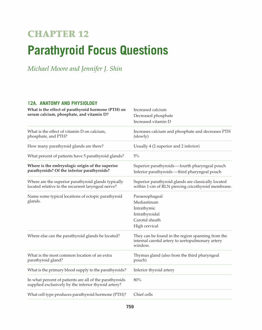
Parathyroid Focus QuestionsMichael Moore and Jennifer J. Shin
12A. ANATOMY ANd PHYSIOLOGYWhat is the effect of parathyroid hormone (PTH) on serum calcium, phosphate, and vitamin D?
Increased calciumDecreased phosphateIncreased vitamin D
What is the effect of vitamin D on calcium, phosphate, and PTH?
Increases calcium and phosphate and decreases PTH (slowly)
How many parathyroid glands are there? Usually 4 (2 superior and 2 inferior)
What percent of patients have 5 parathyroid glands? 5%
Where is the embryologic origin of the superior parathyroids? Of the inferior parathyroids?
Superior parathyroids — fourth pharyngeal pouchInferior parathyroids — third pharyngeal pouch
Where are the superior parathyroid glands typically located relative to the recurrent laryngeal nerve?
Superior parathyroid glands are classically located within 1-cm of RLN piercing cricothyroid membrane.
Name some typical locations of ectopic parathyroid glands.
Where else can the parathyroid glands be located? They can be found in the region spanning from the internal carotid artery to aortopulmonary artery window.
What is the most common location of an extra parathyroid gland?
Thymus gland (also from the third pharyngeal pouch)
What is the primary blood supply to the parathyroids? Inferior thyroid artery
In what percent of patients are all of the parathyroids supplied exclusively by the inferior thyroid artery?
80%
What cell type produces parathyroid hormone (PTH)? Chief cells
760 Otolaryngology Prep and PracticeCH
APTE
R 12
Para
thyr
oid
Focu
s Qu
estio
ns
12B. METABOLIC
Hypocalcemia
How much calcium does an adult need? 1–3 g/day of elemental calcium (usually divided into 2 to 4 doses)
How much calcium does a child need? 45–65 mg/kg/day (usually divided in 4 doses)
How much elemental calcium does calcium carbonate contain?
40%
How much elemental calcium does calcium gluconate contain?
9%
How much elemental calcium does calcium citrate contain?
21%
What is calcitriol? 1,25 vitamin D
How do you correct serum calcium based on serum albumin?
What medications can cause hypocalcemia? I-131, cimetidine, cisplatin, digoxin, Amphotericin-B, and chronic alcohol abuse
What symptoms are associated with hypocalcemia? Neurological: Paresthesias (typically perioral or in the hands), fasciculations, muscle spasm, tetany, irritability, movement disorder, seizure, psychosisCardiovascular: Elongated QT interval, congestive heart failure, hypotensionVisual: Cataracts, optic neuritis, papilledemaPulmonary: BronchospasmGastrointestinal: Adominal cramping, biliary colicGenitourinary: Preterm labor
What is Chvostek’s sign? Tapping in the preauricular region just below zygoma elicits facial twitching on the ipsilateral side.
Can Chvostek’s sign be positive even if calcium levels are normal?
Yes, so it should be checked preoperatively
What is Trousseau’s sign? Inflation of a blood pressure cuff for a few minutes results in forearm muscle spasms on the ipsilateral side.
How do you treat acute severe hypocalcemia? 100–300 mg (10–30 mL) 10% calcium-gluconate in 150 mL D5 water over 10 minutesContinuous infusion at a rate of 0.5 mg/kg/hr
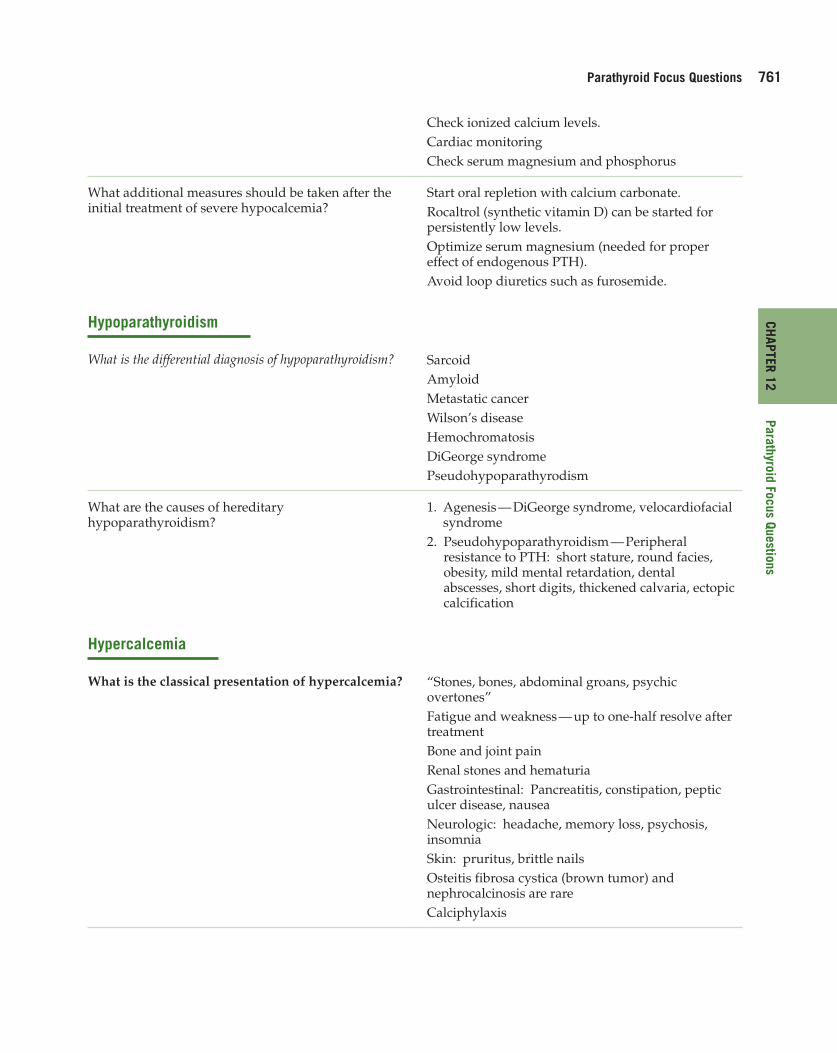
Parathyroid Focus Questions 761CH
APTER 12Parathyroid Focus Questions
Check ionized calcium levels.Cardiac monitoringCheck serum magnesium and phosphorus
What additional measures should be taken after the initial treatment of severe hypocalcemia?
Start oral repletion with calcium carbonate.Rocaltrol (synthetic vitamin D) can be started for persistently low levels.Optimize serum magnesium (needed for proper effect of endogenous PTH).Avoid loop diuretics such as furosemide.
Hypoparathyroidism
What is the differential diagnosis of hypoparathyroidism? SarcoidAmyloidMetastatic cancerWilson’s diseaseHemochromatosisDiGeorge syndromePseudohypoparathyrodism
What are the causes of hereditary hypoparathyroidism?
2. Pseudohypoparathyroidism — Peripheral resistance to PTH: short stature, round facies, obesity, mild mental retardation, dental abscesses, short digits, thickened calvaria, ectopic calcification
Hypercalcemia
What is the classical presentation of hypercalcemia? “Stones, bones, abdominal groans, psychic overtones”Fatigue and weakness — up to one-half resolve after treatmentBone and joint painRenal stones and hematuriaGastrointestinal: Pancreatitis, constipation, peptic ulcer disease, nauseaNeurologic: headache, memory loss, psychosis, insomniaSkin: pruritus, brittle nailsOsteitis fibrosa cystica (brown tumor) and nephrocalcinosis are rareCalciphylaxis
762 Otolaryngology Prep and PracticeCH
APTE
R 12
Para
thyr
oid
Focu
s Qu
estio
ns
What is the treatment of acute severe hypercalcemia? Saline hydrationFurosemide diuresis (avoid thiazides)Bisphosphonates (onset of action 24–48 hours)Calcitonin (immediate onset)Hemodialysis
What are the most common causes of primary hypercalcemia?
Primary hyperparathyroidism and malignancy constitute 90% of cases of hypercalcemia.
What is the differential diagnosis of hypercalcemia? Parathyroid diseaseMalignancy (direct bone destruction and tumor release of PTH-related peptid)Vitamin D intoxicationHigh bone turnoverRenal failureAluminum intoxicationLithium ingestionThiazide diuretics
What is familial hypocalciuric hypercalcemia? Should such patients undergo parathyroidectomy?
Familial hypocalcuric hypercalcemia is an autosomal dominant condition which is characterized by hypocalciuria (usually <50 mg/24 h), variable hypermagnesemia, and normal or minimally elevated levels of PTH. Subtotal parathyroid excision will not normalize the associated hypercalcemia. The condition has a favorable prognosis and does not require surgery. It is readily diagnosed with family history and urinary calcium clearance metrics.
Hyperparathyroidism
What are the different types of hyperparathyroidism?
Primary — Overproduction of PTH by parathyroid glands → high serum calcium and PTH, low serum phosphateSecondary — Increased PTH production due to renal failure or decreased GI absorption of calcium → high serum PTH with normal or low calciumTertiary — Persistent elevation of PTH after correction of the underlying cause of secondary hyperparathyroidism-parathyroid hyperplasia results
What is the most common cause of primary hyperparathyroidism?
Parathyroid adenoma (80–85%, approximately 5% have multiple adenomas)Other causes include parathyroid hyperplasia (15–20%) and parathyroid carcinoma (1%).
Parathyroid Focus Questions 763CH
APTER 12Parathyroid Focus Questions
What are the medical options for management of hyperparathyroidism?
Asymptomatic patients may be followed at intervals and managed medically.Oral calcium <1 g/dayEvery 6 months serum Ca, Cr, and PTH should be checked, along with a urine analysis.Measure a bone scan annuallyEstrogen repletion in postmenopausal womenLoop diuretics and hydration can help keep serum calcium levels lower; avoid thiazide diuretics (which increase serum calcium).Treat chronic renal failure
Name some potential ways to localize hyperactive parathyroid tissue?
UltrasoundCTMRIAngiography with or without selective venous samplingSestamibi scanSingle photon emission CT (SPECT)
How accurate is sestamibi scan for localizing parathyroid disease?
85–95% accurate in localizing a solitary adenoma
What causes false positives for parathyroid adenomas on sestamibi scan?
Thyroid nodules
What causes false negatives on sestamibi scan? Small adenomas and hyperplasia (four gland hypertrophy does not absorb enough radioactivity to be detectable)
Does sestamibi scan have high sensitivity, specificity, or both for solitary parathyroid adenomas?
High specificity but lower sensitivity
What other head and neck tissues demonstrate positive uptake on sestamibi scan?
Salivary tissueLacrimal glandsNasal mucosa
How is ultrasound for localizing parathyroid disease?
Normal parathyroid glands are not seen on ultrasound
n Sensitivity is 65–85% for adenomas; 30–90% for enlarged glands
n It provides less insight in patients with multinodular thyroid, short thick necks, or ectopic parathyroid glands
n May be useful for sestamibi scan-negative adenomas
What is the success rate for surgery for primary hyperparathyroidism?
95%
764 Otolaryngology Prep and PracticeCH
APTE
R 12
Para
thyr
oid
Focu
s Qu
estio
ns
What is the success rate for surgery for hyperparathyroidism secondary to renal failure?
50–85%
What is the appropriate management of a parathyroid adenoma?
Excision of the adenoma
When is it appropriate to perform only a unilateral neck exploration for a parathyroid adenoma?
When there is localization on preoperative imaging such as a sestamibi scan or ultrasoundWhen there is an appropriate drop of PTH when assessed by intraoperative level evaluation
What drop in PTH levels intraoperatively confirms that a solitary parathyroid adenoma has been successfully excised?
A drop of at least 50% from the preoperative level after 10 minutes. Should return to within the normal range
What is the appropriate surgery for four gland parathyroid hyperplasia?
Subtotal excision (remove three and one-half glands) with or without autotransplantation of one-half gland
When might you consider autotransplantation? Four gland hyperplasia, MEN type 1 and type 2a syndromes, and chronic renal failure
What are potential sites for autotransplantation? Sternocleidomastoid muscle or brachioradialis muscle
What is the failure rate for autotransplantation? Up to 50%
How much tissue should be autotransplanted? 20–30 mg in 1 to 2-mm slices
What are the potential complications of parathyroid surgery?
Failure: missed ectopic adenoma, incomplete resection in multigland diseaseHypocalcemia (20–30%) — wait for appearance of symptomsInjury to RLN (<1%)Hematoma
What happens with calcium levels after successful removal of a parathyroid adenoma?
PTH levels undetectable at 8 hoursPTH levels normalize by 30 hoursCalcium nadirs at 20 hoursCalcium levels normal on day 2–3
What is a brown tumor? Tumor arises in the setting of excess osteoclast activity — leads to fibrous tissue, woven bone, supporting vasculature but no matrix. It is radiolucent on a radiograph.
What is calciphylaxis? Vascular calcification and skin necrosis usually seen in end stage renal disease, leads to chronic nonhealing wounds, may require parathyroidectomy and hyperbaric oxygen in some cases.
References
1. Rodriquez S, Newlands S. Hyperparathyroidism. In: UTMB Grand Rounds Archive. Quinn FB, ed. 2006. 2. Parathyroid diseases. Norman Parathyroid Center, Parathyroid.com, 1996–2010. 3. Rosen FS, Pou A. Parathyroid Disease. University of Texas Medical Branch, 2002. http://www.utmb.edu/otoref/grnds/
parathyroid-2002-03/parathyroid-2002-03-slides.pdf 4. Parathyroid Glands. Minden Jog Fentartva, 2009. http://www.szote.u-szeged.hu/in1st/eloadasok2/endoc/angolPHP.pdf
Parathyroid Focus Questions 765CH
APTER 12Parathyroid Focus Questions
12C. MASSES — MALIGNANT
How does parathyroid malignancy usually present? Very high serum calcium levels (often over 13 mg/dL), palpable neck mass
What is the hyperparathyroidism-jaw tumor syndrome?
Severe hypercalcemia in a teenager due to multiple parathyroid adenomas; 10% develop parathyroid carcinoma.
What is the treatment of parathyroid malignancy? Surgery–Resection of cancer, ipsilateral thyroid lobectomy, skeletonization of RLN if not involved with tumor, and dissection of paratracheal nodesRadiation is only used for residual or recurrent disease or those who are not surgical candidates.
How often does parathyroid malignancy result in regional or distant metastases?
Regional/distant metastases in 25–30%
How often does local recurrence occur in parathyroid malignancy after surgery?
Local recurrence 30%
In addition to PTH, what is another common tumor marker for parathyroid malignancy?
Human chorionic gonadotropin
How is the histopathology of parathyroid hyperplasia different from that of a parathyroid adenoma?
Parathyroid hyperplasia classically lacks the rim of normal parathyroid tissue at the periphery that is often seen in parathyroid adenomas. They are, however, difficult to distinguish based on histopathology alone.
References
1. Rodriquez S, Newlands S. Hyperparathyroidism. In: UTMB Grand Rounds Archive. Quinn FB, ed. 2006. 2. Parathyroid diseases. Norman Parathyroid Center, Parathyroid.com, 1996–2010.
766 Otolaryngology Prep and PracticeCH
APTE
R 12
Para
thyr
oid
Focu
s Qu
estio
ns
12d. CONGENITAL
Multiple Endocrine Neoplasia Syndromes
What is the inheritance pattern for the multiple endocrine neoplasia (MEN) syndromes?
Autosomal dominant
What clinical conditions constitute (define) MEN 1? 1. Moderate-severe hyperparathyroidism in 85%2. Zollinger-Ellison syndrome (tumor in GI tract
with secretion of gastrin and development of ulcers), pancreatic disease
3. Prolactinomas and other pituitary tumors
What chromosome is affected in MEN 1? MEN 1 gene (chromosome 11) produces Menin (tumor suppressor)
How does hyperparathyroidism present and progress in MEN 1?
Early onset with multiple glands affected
How do MEN 1 patients do after surgery for hyperparathyroidism?
Postoperative hypoparathyroidism is more common (more extensive surgery) than with other surgeries for four gland hyperplasia.Successful subtotal parathyroidectomy is followed by recurrent hyperparathyroidism in 50% of cases at 10 years.
What clinical conditions constitute MEN 2A and what is the typical penetrance of each?
Mild hyperparathyroidism in 20–70%Medullary carcinoma 100%Pheochromocytoma 35–50%
What chromosome is affected in MEN 2A? The RET proto-oncogene, which is affected in 98% of cases, is on chromosome 10.
What causes the hyperparathyroidism in MEN 2A? Is it a single adenoma or multigland disease?
Usually it is a single adenoma but multigland hyperplasia may be seen.
How should the parathyroid glands be managed in MEN 2A?
Unless hyperparathyroidism is present, these glands are left in place. Often the glands are marked with clips or long, nonabsorbable sutures to allow for identification if reoperation is required.
What clinical conditions constitute MEN 2B? Aggressive, early onset medullary thyroid carcinoma, pheochromocytomas, marfanoid features, mucosal neuromas, and ganglioneuromatosis of the gastrointestinal tract
What chromosome is affected in MEN 2B? RET proto-oncogene (chromosome 10); it is a different mutation than in MEN 2A.
Parathyroid Focus Questions 767CH
APTER 12Parathyroid Focus Questions
Reference
1. Thyroid Carcinoma Task Force. AACE/AAES Medical/surgical guidelines for clinical practice: Management of thyroid carcinoma. Endocr Prac. May/June 2001; 7(3).
2. Rodriguez S, Newlands S. Quinn F. Ryan M. Hyperparathyroidism. University of Texas Medical Branch, 2006. http://www.utmb.edu/otoref/grnds/Hyperparathyroid-060208