Page 1
Chapter 3 Review of Literature
Dept. Of Pharmacognosy & Phytochemistry KLE University’s College of Pharmacy, Belgaum. 16
CHAPTER 3: REVIEW OF LITERATURE
"Genetics loads the Gun, Life style pulls the trigger". There are many diseases that
are caused due to genetical disorders and is one of the cause for diabetes mellitus60
.
3.1 DIABETES MELLITUS
Diabetes mellitus (DM) was recognized as early as 1500 B.C. by Egyptian
physicians who described it as a disease associated with "the passage of much
urine". The term "diabetes" was coined by the Greek physician Aretaeus, who
noticed that patients with diabetes had a disease that caused the siphoning of the
structural components of the body into the urine61
.
DM afflicts about 5% of the general population. Diabetes is a mysterious illness, a
statement made in antiquity by the physician Aerates of Cappadocia (81-138 AD) is still
valid today. At first Galen suspected that this illness was caused by a kidney complaint.
Avicenna alone has been credited with two additional discoveries, first, the mention
of further symptoms– besides the triad (polydypsia, polyuria and marasmus) known to
antiquity– namely physical, mental, sexual weakness, occurrence of carbuncles,
gangrene and secondly the alleged discovery of the sweetness of diabetic urine62
. The
study suggest that for the world as a whole, between the years 1995 and 2025, the adult
population will increase by 64%, prevalence of diabetes in adults will increase by 35%
and the number of people with diabetes will increase by 122%. For the developed
countries, there will be an 11% increase in the adult population, a 27% increase in the
prevalence of adult diabetes and a 42% increase in the number of people with diabetes.
For the developing countries, there will be an 82% increase in the adult population, a
48% increase in the prevalence of adult diabetes and a 170% increase in the number of
people with diabetes63
.
Page 2
Chapter 3 Review of Literature
Dept. Of Pharmacognosy & Phytochemistry KLE University’s College of Pharmacy, Belgaum. 17
In recent years, developed nations have witnessed an explosive increase in the
prevalence of DM predominantly related to lifestyle changes and the resulting surge in
obesity. The metabolic consequences of prolonged hyperglycemia and dyslipidemia,
including accelerated atherosclerosis, chronic kidney disease and blindness, pose an
enormous burden on patients with diabetes mellitus and on the public health system64
.
The number of patients with DM is markedly increasing worldwide. DM is
associated with impaired glucose metabolism that leads to an increase in free radical
production and increase in triglyceride and lipoprotein levels. Oxygen free radical can
initiate peroxidation of lipids, which in turn stimulates glycation of protein, inactivation
of antioxidant enzymes and play a role in the long-term complications of diabetes.
Therefore, among the various therapeutic strategies, combination of antihyperglycemic,
antihyperlipidemic and antioxidant activity can be beneficial in the prevention of DM
and its complications65
.
3.1.1 Disease profile
a) Definition
Diabetes is defined as a state in which homeostasis of carbohydrate, protein and
lipid metabolism is improperly regulated by insulin. This results primarily in elevated
fasting and postprandial blood glucose levels. In diabetic condition, dyslipidemia, lipid
abnormalities are the unbalanced metabolic states of diabetes66
. DM may present with
characteristic symptoms such as polyphagia, polydypsia, polyuria, blurring of vision and
weight loss. In its severe forms, ketoacidosis or a non-ketonic hyperosmolar state may
develop and lead to stupor, coma and in the absence of effective treatment to death67
.
b) Prevalence
There are two types of diabetes- Type-1 diabetes mellitus formerly known as
insulin dependent diabetes mellitus (IDDM) and Type-2 diabetes mellitus formerly
Page 3
Chapter 3 Review of Literature
Dept. Of Pharmacognosy & Phytochemistry KLE University’s College of Pharmacy, Belgaum. 18
known as non-insulin dependent diabetes (NIDDM). The vast majority of diabetic
patients are Type-2 diabetes mellitus.
Diabetes patients are 25 times more prone to blindness, 2 times more prone to heart
attacks, 2-6 times more prone to stroke and 17 times more prone to kidney damage as
compared to non diabetics68
.
c) Epidemiology
DM in humans is undergoing a remarkable upsurge in prevalence in the India.
Historically, the usual ratio for Type -1 to Type- 2 diabetes has been 1:20. Classically,
Type- 1 diabetes is described as an autoimmune disease in which a foreign protein is
incorporated into islet β cells, perhaps via viral infection. In response, the patient's
lymphocytes attack the foreign protein and inadvertently destroy the patient's β cells as
collateral damage. This leads to a state of absolute insulin deficiency.
The pathogenesis of Type- 2 diabetes is less well defined, however, it is invariably
associated with defective sensing of glucose signals by the β cell. It is often associated
with a state of insulin resistance, which means insulin that is secreted by the β cell and
bound to liver, muscle and fat cells is sub normally efficacious in carrying out its
metabolic actions69
.
The WHO has predicted that the global prevalence of Type-2 diabetes will be more
than from 135 million in 1995 to 300 million in 2025 and that this increase will affect
both industrialized and developing countries expecting the greatest increase in India,
from 19.4 to 57.2 million70
.
Page 4
Chapter 3 Review of Literature
Dept. Of Pharmacognosy & Phytochemistry KLE University’s College of Pharmacy, Belgaum. 19
d) Classification of diabetes mellitus71
Table No. 1: Classification of diabetes mellitus
Classes Examples
Type-1 diabetes Islet β-cell destruction, autoimmune, idiopathic
Type-2 diabetes Insulin resistance, insulin deficiency
Genetic defects of β-cell function Chromosome 20, HNF 4α, chromosome 7,
glucokinase
Genetic defects in insulin action Type A insulin resistance, lipoatrophic diabetes
Disease of the exocrine pancreas Pancreatitis, neoplasia, cystic fibrosis,
pancreatectomy
Endocrinopathies Cushing’s syndrome, hyperthyroidism
Drug- or chemical-induced Nicotinic acid, thiazides, glucosteroids
Infections Congenital rubella, cytomegalo virus
Uncommon forms of immune-
mediated diabetes
Insulin auto immune syndrome, Anti-insulin
receptor antibodies
Other genetic syndromes Down’s syndromes, Huntington’s chorea
The clinical staging reflects that diabetes progresses through several clinical stages
during its natural history. Moreover, individual subjects may move from stage to stage in
either direction. Persons who have, or who are developing, DM can be categorized by
stage according to the clinical characteristics, even in the absence of information
concerning the underlying etiology. The classification by etiological type results from
improved understanding of the causes of DM.
e) Different forms of Diabetes mellitus72
General: Type- 1 Diabetes mellitus (formerly called insulin dependent diabetes
mellitus or IDDM)
Autoimmune Type -1 diabètes mellitus (Type- 1A).
Non-autoimmune or idiopathic Type- 1 diabètes mellitus (Type- 1B)
Page 5
Chapter 3 Review of Literature
Dept. Of Pharmacognosy & Phytochemistry KLE University’s College of Pharmacy, Belgaum. 20
Fig. No. 1: Type- 1 Diabetes mellitus
Type- 2 Diabetes mellitus (formerly called non-insulin dependent diabetes mellitus
or NIDDM)
Fig. No. 2: Type- 2 Diabetes mellitus
Specific: Defined gene mutations
Maturity-onset diabetes of youth (MODY)
MODY 1, chromosome 20 – hepatic nuclear factor 4 gene mutations
Page 6
Chapter 3 Review of Literature
Dept. Of Pharmacognosy & Phytochemistry KLE University’s College of Pharmacy, Belgaum. 21
MODY 2, chromosome 7-glucokinase gene mutation
MODY 3, chromosome 12-hepatic nuclear factor 1 gene mutations
MODY 4, chromosome 13-pancreatic determining factor X-gene mutations
MODY X, unidentified gene mutation (s)
Maternally inherited diabetes and deafness-mitochondrial leucine tRNA gene
mutations
Other Specific forms of Diabetes:
1) Diseases of the exocrine pancreas
Fibrocalcaneous pancreatopathy, Pancreatitis, Trauma, Pancreatectomy,
Neoplasia, Cystic fibrosis, Haemochromatosis and others
2) Endocrinopathies
Cushing's syndrome, Acromegaly, Pheochromocytoma, Glucagonoma,
Hyperthyroidism, Somatostatinoma etc.
3) Infections
Congenital rubella, Coxsackie B, Cytomegalovirus, Mumps, Adenoviruse etc.
4) Drug or chemical induced diabetes mellitus
Nicotinic acid, Glucocorticoids, Thyroid hormone, -Adrenergic agonists, -
adrenergic agonists, Thiazides, Dilantin, Vacor, Interferon- therapy etc.
5) Other genetic syndromes sometimes associated with diabetes
Down's syndrome, Friedreich's ataxia, Huntington's chorea, Klinefelter's
syndrome, Laurence – Moon – Biedel syndrome, Porphyria, Prader willi
syndrome, Turner's syndrome, Wolfram's syndrome etc.
6) Associated with Pregnancy
Gestational Impaired Glucose Tolerance (GIGT)
Gestational Diabetes Mellitus (GDM)
Page 7
Chapter 3 Review of Literature
Dept. Of Pharmacognosy & Phytochemistry KLE University’s College of Pharmacy, Belgaum. 22
3.1.2 Insulin
The DM has been well known as a wasting disease due to insulin deficiency in
human beings. The pancreas secretes insulin. Carbohydrate metabolism is primarily
under the control of insulin. Insulin deficiency occurs in a person due to the
functional disorder of the pancreas.
a) The Endocrine part of the Islets of Langerhans:
The normal human adult pancreas contains on an average some 500,000 islets
of langerhans, distributed in scattered manner within the gland, comprising 1 to 3%
of the total tissue. Each group of cells of the endocrine part is surrounded by the
acini of the exocrine part, they look like islands and are hence termed as islets. The
distribution of islets is maximum in the tail and minimum in the head of the gland73
.
Three types of cells are found in the islets. These are called the (alpha),
(beta) and (delta) types. The cells are fewer in number about 20% and they exist
peripherally in the islets, while the most numerous cells (about 75% to 80%) are
situated centrally in the form of lumps.
The synthesis of two hormones insulin and glucagon takes place in the cells and
cells respectively in the islets of Langerhans. Both hormones play an important role in
carbohydrate metabolism. The function of the cells (about 5% in number) is not clearly
known. It is assumed that they may secrete serotonin but some others believe that gastrin
is secreted by these cells73
.
b) Chemistry:
It has minimum molecular weight of 5734. Insulin from different sources (eg.
pig, cattle, sheep and horses) shows minor differences in amino acid composition
and immunological activity. The nearest to human insulin in structure is insulin
from pig. Insulin is destroyed by action of digestive enzymes and is hence inactive
Page 8
Chapter 3 Review of Literature
Dept. Of Pharmacognosy & Phytochemistry KLE University’s College of Pharmacy, Belgaum. 23
when given (administered) by mouth. The biological action of the hormones can be
prolonged by combining it with protamine or globin (protamine zinc insulin and
globin insulin) or by altering the size of the crystals (ultralente insulin; large
crystals and slow acting)74
.
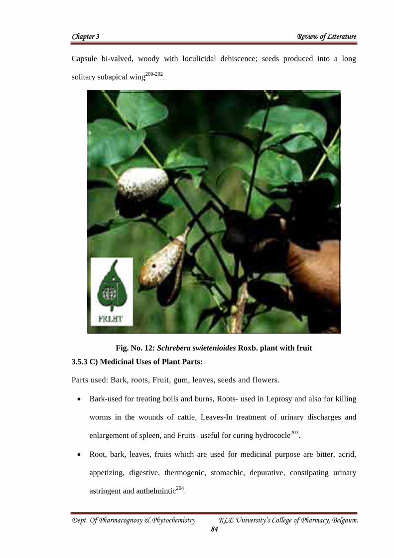
c) Metabolism of Insulin:74
Insulin is believed to be transported in the plasma bound to a specific insulin
transporting protein. Insulin is degraded primarily in the liver and kidney by the enzyme,
"Glutathione insulin transhydrogenase". The half life of plasma insulin is only 7-15
minutes.
d) Mode of action of Insulin:74
1. Muscle, adipose tissue and liver are the major sites of its action
2. It is active on the lens and leukocytes
3. It has minor action on the metabolism of renal tissue, erythrocytes and GIT
e) Extrahepatic tissues:74
It facilitates the transport of glucose across the cell membrane.
Insulin promotes metabolic pathways like glycogenesis, glycolysis and HMP
pathways.
Insulin stimulates intracellular transport of all sugars eg. arabinose, xylose and
galactose.
Insulin stimulates uptake of amino acids by the cell.
Insulin stimulates the activity of enzymes hexokinase and glycogen synthetase.
Insulin stimulates oxidative phosphorylation in mitochondria of muscle.
Insulin stimulates the entry of Na+, K+ & PO4-- into adipose tissue.
Liver: Insulin is an anabolic hormone causing increased carbohydrate metabolism,
glycogen formation, lipid synthesis, amino acid uptake and protein synthesis.
Page 9
Chapter 3 Review of Literature
Dept. Of Pharmacognosy & Phytochemistry KLE University’s College of Pharmacy, Belgaum. 24
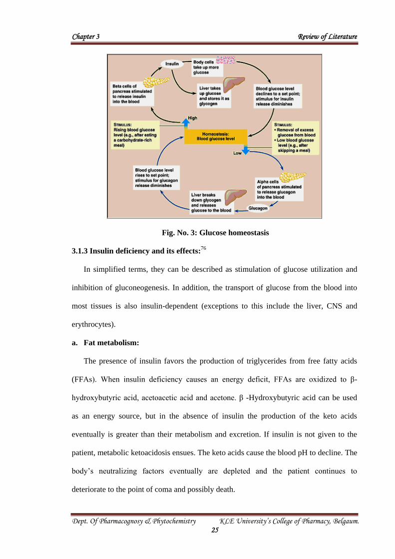
Glucose Homeostasis:75
A carbohydrate, particularly glucose, is an important source of fuel for living
organisms. It has been found that glucose homeostasis contributes to two kinds of
hormones, including insulin and anti-insulin or counter-regulatory hormones (glucagon,
growth hormones, cortisol and catecholamines). Maintenance of serum glucose
concentrations within a normal physiological range is primarily accomplished by two
pancreatic hormones, insulin and glucagon. Derangements of glucagon or insulin
regulation can result in hyperglycemia or hypoglycemia. Glucose penetrates most tissues
slowly unless, insulin is present to facilitate its uptake; however, central nervous system
(CNS) cells, capillary endothelial cells, gastrointestinal epithelial cells, pancreatic cells
and renal medullary cells are freely permeable to glucose.
The endocrine portion of the pancreas, called the islets of Langerhans, consists of
cordlike groups of cells arranged along pancreatic capillary channels. These pancreatic
cells monitor changes in the availability of small calorigenic molecules, namely glucose
and to a lesser extent amino acids, ketone bodies and fatty acids. Pancreatic β-cells
appropriately alter their rates of insulin secretion in response to fluctuations in the levels
of these calorigenic molecules, with glucose playing the dominant role in regulation of
insulin secretion. Pancreatic β-cells secrete glucagon in response to increases in amino
acid and fatty acid levels; however, glucose inhibits glucagon secretion. If blood glucose
levels fall (e.g., during hypoglycemia or fasting), glucagon secretion is augmented,
providing a counter regulatory hormonal response that stimulates gluconeogenesis in the
liver and other tissues to avoid hypoglycemia. Circulating glucose levels are determined
by the balance among absorption, storage, production and use (metabolic rate). Glucagon
and insulin are the two most important hormones that maintain glucose homeostasis
when blood concentrations are disturbed.
Page 10
Chapter 3 Review of Literature
Dept. Of Pharmacognosy & Phytochemistry KLE University’s College of Pharmacy, Belgaum. 25
Fig. No. 3: Glucose homeostasis
3.1.3 Insulin deficiency and its effects:76
In simplified terms, they can be described as stimulation of glucose utilization and
inhibition of gluconeogenesis. In addition, the transport of glucose from the blood into
most tissues is also insulin-dependent (exceptions to this include the liver, CNS and
erythrocytes).
a. Fat metabolism:
The presence of insulin favors the production of triglycerides from free fatty acids
(FFAs). When insulin deficiency causes an energy deficit, FFAs are oxidized to β-
hydroxybutyric acid, acetoacetic acid and acetone. β -Hydroxybutyric acid can be used
as an energy source, but in the absence of insulin the production of the keto acids
eventually is greater than their metabolism and excretion. If insulin is not given to the
patient, metabolic ketoacidosis ensues. The keto acids cause the blood pH to decline. The
body’s neutralizing factors eventually are depleted and the patient continues to
deteriorate to the point of coma and possibly death.
Page 11
Chapter 3 Review of Literature
Dept. Of Pharmacognosy & Phytochemistry KLE University’s College of Pharmacy, Belgaum. 26
b. Protein metabolism:
The presence of insulin favors the production of structural proteins from constituent
amino acids. When glucose is present intracellularly in sufficient quantities for needed
energy production, most structural proteins retain their integrity. In the absence of insulin,
structural protein production is not favored and intracellular glucose levels are
insufficient to match energy demands. In attempt to produce energy, skeletal muscle
converts its structural proteins to constituent amino acids. The liberated amino acids are
transported to the liver, where they are converted to glucose via gluconeogenesis. In
patients with diabetes, glucose enters the blood but is not taken up by tissues because of
a true or relative lack of insulin. Thus, hyperglycemia is escalated and structural proteins
are wasted77
.
Fig. No. 4: Insulin deficiency and its effects
Page 12
Chapter 3 Review of Literature
Dept. Of Pharmacognosy & Phytochemistry KLE University’s College of Pharmacy, Belgaum. 27
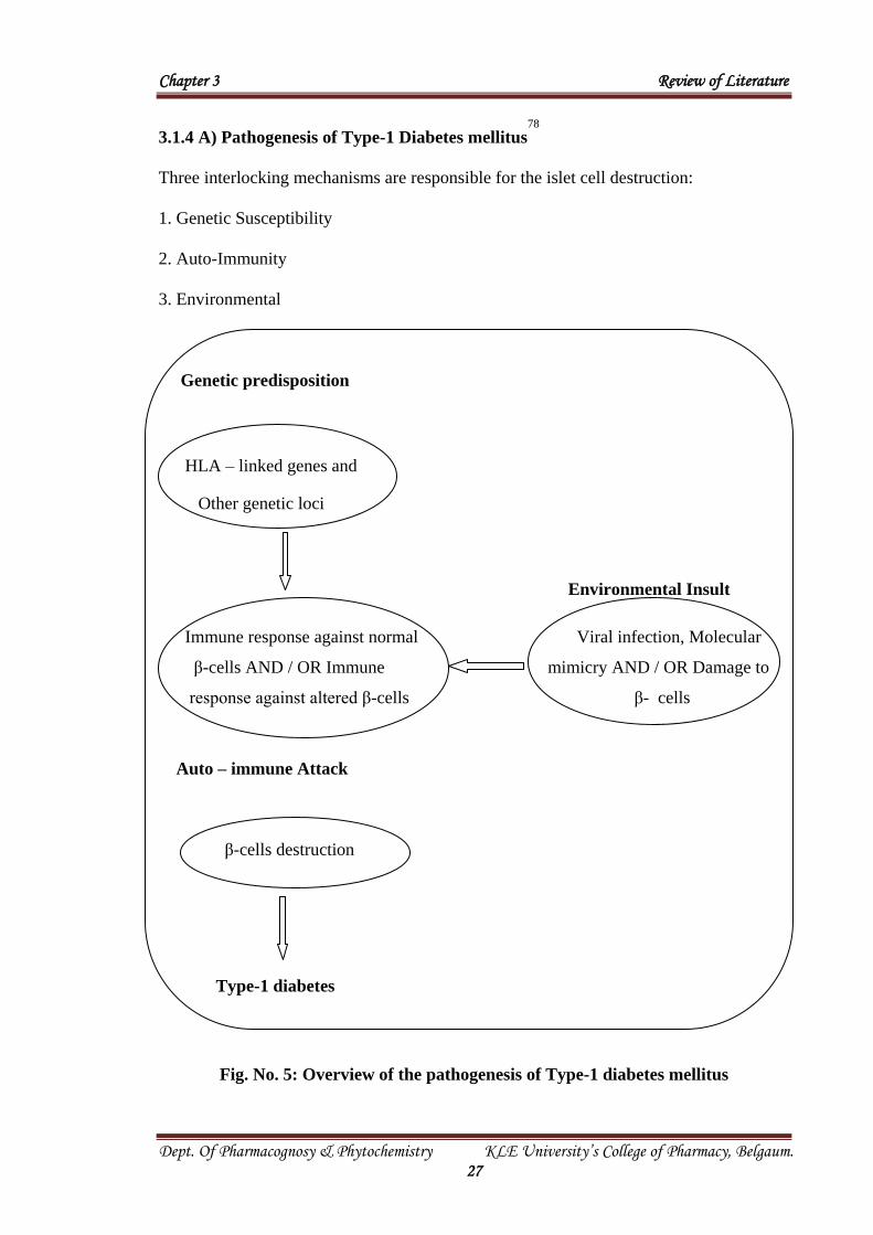
3.1.4 A) Pathogenesis of Type-1 Diabetes mellitus78
Three interlocking mechanisms are responsible for the islet cell destruction:
1. Genetic Susceptibility
2. Auto-Immunity
3. Environmental
Genetic predisposition
HLA – linked genes and
Other genetic loci
Environmental Insult
Immune response against normal Viral infection, Molecular
β-cells AND / OR Immune mimicry AND / OR Damage to
response against altered β-cells β- cells
Auto – immune Attack
β-cells destruction
Type-1 diabetes
Fig. No. 5: Overview of the pathogenesis of Type-1 diabetes mellitus
Page 13
Chapter 3 Review of Literature
Dept. Of Pharmacognosy & Phytochemistry KLE University’s College of Pharmacy, Belgaum. 28
1. Genetic Susceptibility:
At least one of the susceptibility gene for Type-1 diabetes resides in the region
that encodes the class II antigens of the Major Histocompatibility Complex (MHC) on
chromosome GP21 (HLA-D). The HLA-D region contains three classes of genes (DP,
DQ and DR). The class II molecules are highly polymorphic and each has numerous
alleles. About 95% of white patient with Type-1 diabetes have either HLA-DR3 or HLA-
DR4 alleles or both where as in the general population the prevalence of these antigens is
only 45%.
It is thought that genetic variations in the HLA class II molecules may alter
recognition by the T-cell receptor, or may modify the presentation of the antigen because
of variations in the antigen-binding cleft, thus, class II HLA gene may effect the degree
of immune responsiveness to a pancreatic β-cell autoantigen or a β-cell autoantigen may
be presented in a manner that promotes an abnormal immunologic reaction.
2. Auto-Immunity:
Clinical onset of Type-1 diabetes is abrupt; this disease in fact results from a
chronic auto-immune attack of β-cells that usually exists for many years before the
disease becomes evident79
.
A lymphocyte with rich inflammatory infiltrate (Insulitis) is observed in the islets
of patients in early diabetes. The infiltration consists mostly of CD8 T- lymphocytes.
CD4 T cell from animals with auto immune diabetes can transfer diabetes to normal
animals, thus establishing the primary of T-cell auto-immunity in Type-1 diabetes.
The Insulitis is associated with increase expression of class I MHC molecules and
aberrant expression of class II MHC molecules on the β-cells. This aberrant expression is
mediated in part by locally produced cytokines [eg. Interferon-gamma (IFN-γ) derived
from activated T-cells]. Genetic dysregulation of a cytokine that induce IFN-γ production
Page 14
Chapter 3 Review of Literature
Dept. Of Pharmacognosy & Phytochemistry KLE University’s College of Pharmacy, Belgaum. 29
promotes the development of diabetes in a mouse model. About 70%-80% of patients
with Type-1 diabetes have islet cell auto antibodies against intracellular islet cell
antigens, such as Glutamic Acid Decarboxylose (GAD) “islet auto antigen 2” (1a-2a
tyrosine phosphatases), insulin and gangliosides.
3. Environmental Factors:
Viruses
A viral infection has long been noted in the diagnosis of new cases and has the
association between coxsackie viruses of group B and pancreatic diseases including
diabetes. Other implicated viral infections include mumps, measles, cytomegalovirus,
rubella and infections mononucleosis.
It has been postulated that one of these viruses causes mild β-cells injury, which is
followed by an auto-immune reaction against previously sequestered antigens in virally
altered β-cells in persons with HLA-linked susceptibility. Another is that an immune
response develops against a viral protein that shares amino acid sequences with a β-cell
protein (molecular mimicry).
Others
Antigenic exposure may also come from other sources. Children who ingest cow’s
milk products early in life (before age of 4 months) have a 1.5 fold increase risk for
Type-1 diabetes relative to those who do not, raising the spectrum of a cross-reacting
antigen in cow’s milk.
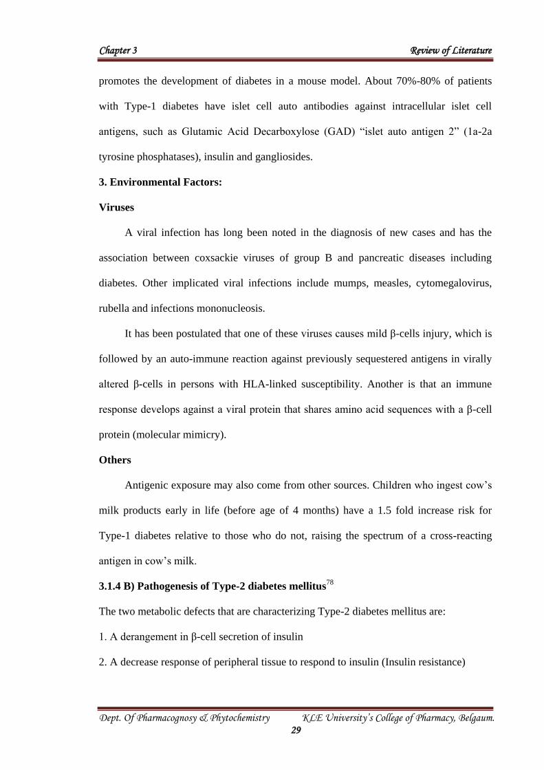
3.1.4 B) Pathogenesis of Type-2 diabetes mellitus78
The two metabolic defects that are characterizing Type-2 diabetes mellitus are:
1. A derangement in β-cell secretion of insulin
2. A decrease response of peripheral tissue to respond to insulin (Insulin resistance)
Page 15
Chapter 3 Review of Literature
Dept. Of Pharmacognosy & Phytochemistry KLE University’s College of Pharmacy, Belgaum. 30
Type-2 diabetes
Genetic Predisposition Environment
Multiple genetic defects Obesity
Primary β-cell defect Peripheral tissue
Deranged insulin secretion Insulin resistance
Hyperglycemia
β -cell exhaustion
Inadequate glucose Utilization
Type-2 diabetes mellitus
Fig. No. 6: Overview of the pathogenesis of Type-2 diabetes mellitus
Page 16
Chapter 3 Review of Literature
Dept. Of Pharmacognosy & Phytochemistry KLE University’s College of Pharmacy, Belgaum. 31
1. Deranged β-cell Secretion of Insulin:
A modest hyperinsulinemia may be observed, attributed to β-cell hyper
responsiveness to physiological elevations in blood glucose, with the development of
overt disease. The pattern of insulin secretion exhibits a subtle change. Early in the
course of Type-2 diabetes, insulin secretion appears to be normal and plasma insulin
levels are not reduced.
However, the normal pulsatile oscillating pattern of insulin secretion is lost and the
rapid first phase of insulin secretion triggered by glucose is obtunded. Collectively, these
and other observations suggest derangements in β-cell response to hyperglycemia early
in Type-2 diabetes, rather than deficiencies in insulin synthesis per se. Later in the cause
of Type-2 diabetes a mild to moderate deficiency of insulin develops which is less severe
than that of Type-1.
2. Insulin Resistance:
Insulin resistance (IR) is a common pathological state in which target cells fail to
respond to ordinary levels of circulating insulin. It results in inability of insulin to
provide normal glucose and lipid homeostasis80
. Insulin resistance is also a feature of a
number of other health disorders, including obesity, glucose intolerance, dyslipidemia
and hypertension clustering in the so-called metabolic syndrome (also commonly
referred to as syndrome X)81
.
a) Symptoms of insulin resistance:
Feeling agitated, jittery, moody, nauseated, or having a headache is common in
insulin resistance, with almost immediate relief once food is eaten.
Intestinal bloating.
Sleepiness.
Weight gain.
Page 17
Chapter 3 Review of Literature
Dept. Of Pharmacognosy & Phytochemistry KLE University’s College of Pharmacy, Belgaum. 32
Fatigue.
Increased triglycerides.
Increased blood pressure.
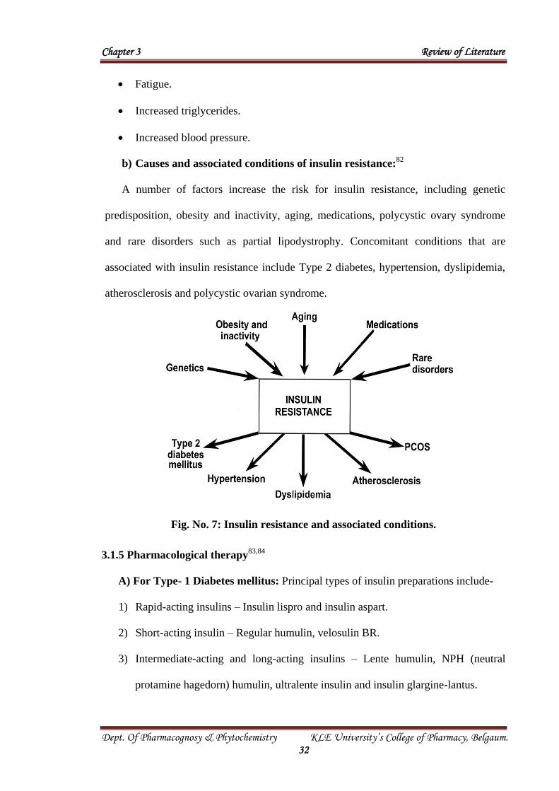
b) Causes and associated conditions of insulin resistance:82
A number of factors increase the risk for insulin resistance, including genetic
predisposition, obesity and inactivity, aging, medications, polycystic ovary syndrome
and rare disorders such as partial lipodystrophy. Concomitant conditions that are
associated with insulin resistance include Type 2 diabetes, hypertension, dyslipidemia,
atherosclerosis and polycystic ovarian syndrome.
Fig. No. 7: Insulin resistance and associated conditions.
3.1.5 Pharmacological therapy83,84
A) For Type- 1 Diabetes mellitus: Principal types of insulin preparations include-
1) Rapid-acting insulins – Insulin lispro and insulin aspart.
2) Short-acting insulin – Regular humulin, velosulin BR.
3) Intermediate-acting and long-acting insulins – Lente humulin, NPH (neutral
protamine hagedorn) humulin, ultralente insulin and insulin glargine-lantus.
Page 18
Chapter 3 Review of Literature
Dept. Of Pharmacognosy & Phytochemistry KLE University’s College of Pharmacy, Belgaum. 33
B) For Type- 2 Diabetes mellitus: Oral Hypoglycemic agents -
1) -glucosidase inhibitors (AGIs): Acarbose and miglitol.
An enzyme in the brush border of proximal small intestinal epithelium -
glucosidase serves to breakdown disaccharides and more complex carbohydrates. By
competitive inhibition of this enzyme, the AGIs delay intestinal carbohydrate absorption.
Their greatest effect is on post-prandial glucose levels and effect on fasting blood
glucose level is small.
Adverse effects: Flatulence, abdominal discomfort, diarrhea.
2) Sulfonylureas (SUs):
They have been available in United States since 1954.
First generation SUs: Chloropropamide, tolbutamide, acetohexamide and tolazamide.
Second generation SUs: Glyburide, glipizide, glimepiride, gilbenclamide.
SUs bind to the SU receptor found on the surface of pancreatic -cells. This
interaction leads to a closure of voltage-dependent KATP channels, facilitating cell
membrane depolarization, calcium entry into the cell and insulin secretion. The
possibility that such agents may also directly enhance peripheral glucose disposal (i.e.
decrease insulin resistance) has also been raised.
Adverse effects: Weight gain, hypoglycemia. They must be used cautiously in hepatic or
renal impairment.
3) Biguanides:
Over 30 years ago, biguanides like metformin, phenformin, buformin were used
for treatment of diabetes.
Metformin’s major action is to decrease hepatic glucose output primarily by
decreasing gluconeogenesis, but it may also increase glucose uptake by skeletal muscles.
Metformin activates hepatic and muscle AMPK, a cellular signal for increased energy
Page 19
Chapter 3 Review of Literature
Dept. Of Pharmacognosy & Phytochemistry KLE University’s College of Pharmacy, Belgaum. 34
requirements. Activation of hepatic AMPK results in phosphorylation and inhibition of
acetyl-coenzyme A carboxylase, which catalyzes the rate-limiting step of lipogenesis.
This block in fatty acid synthesis promotes fatty acid oxidation. In addition, activation of
hepatic AMPK decreases expression of SREBP-1, a transcription factor implicated in the
pathogenesis of insulin resistance, dyslipidemia and diabetes. Results of earlier studies
suggest disruption of coupled oxidative phosphorylation in mitochondria. Whether this
underlies increase in AMPK activity remains unclear.
Adverse effects: Gastrointestinal, lactic acidosis (rare). Contraindicated in liver, cardiac,
renal dysfunction.
4) Non-sulfonylureas: Nateglinide, repaglinide.
The mechanism of action of these drugs is similar to that of SUs (closure of KATP
channel leading to calcium-dependent insulin secretion). However they bind to the SU
receptor at a different site and with different kinetics than SUs. Their onset of action is
faster and half-life is shorter, which results in brief stimulation of insulin release.
Adverse effects: Hypoglycemia, weight gain, contraindicated in liver, kidney
dysfunction and concomitant use of repaglinide with gemfibrozil is avoided.
5) Insulin sensitizers (Thiazolidinediones):
The Currently available thiazolidinedione is pioglitazone. Troglitazone an earlier
introduced thiazolidinedione was removed from market because of risk of hepatic failure.
Thiazolidinediones function as ligands for the PPAR, which is most highly expressed in
adipocytes. These nuclear receptors, which are ligand-activated transcription factors,
play an integral part in the regulation of the expression of a variety of genes involved in
carbohydrate and lipid metabolism.
Thiazolidinediones improve insulin sensitivity, particularly in the peripheral tissues.
In the adipocyte differentiation is enhanced, lipolysis is reduced, adipokines are altered,
Page 20
Chapter 3 Review of Literature
Dept. Of Pharmacognosy & Phytochemistry KLE University’s College of Pharmacy, Belgaum. 35
namely a decrease in TNF- and free fatty acid levels and increased adiponectin levels.
These effects enhance insulin sensitivity.
Adverse effects: Weight gain, edema, anemia, pulmonary edema, congestive heart failure,
contraindicated in liver dysfunction.
6) Intestinal lipase inhibitor: Orlistat
It is an antiobesity agent that acts as a selective inhibitor of gastric and pancreatic
lipases and thereby inhibits the hydrolysis of dietary fat into absorbable free fatty acids
and monoglycerides.
Adverse effects: Flatulence, oily spotting, fecal urgency, increased frequency of
defecation and fecal incontinence. Absorption of fat-soluble vitamins can be adversely
affected. Contraindications are chronic malabsorption syndrome, cholestasis and known
hypersensitivity.
7) Herbal Drugs:
Diabetes mellitus is a common chronic endocrine disorder. Since ancient time a
number of herbal medicines were used in the treatment of DM. Many studies have been
carried out in search of a suitable plant drug that would be effective in DM.
Herbal medicines for diabetes can be classified into four categories according to
their mode of action:
i) Drugs acting like insulin
ii) Drugs acting on insulin secreting beta cells
iii) Drugs acting by modifying glucose utilisation
iv) Drugs acting by miscellaneous mechanisms85
.
3.1.6 Animal models for experimental diabetes mellitus86
There are many advantages of using animals models in research work on diabetes
as various aspects of the disease like the etiology, its multifactorial genetics,
Page 21
Chapter 3 Review of Literature
Dept. Of Pharmacognosy & Phytochemistry KLE University’s College of Pharmacy, Belgaum. 36
pathogenesis of the disease and its complication can be explicity understood. Secondly, it
also helps in the development and evaluation of newer agents for the treatment of
diabetes. However, there are some limitations in the use of animal model for studies on
diabetes.
Induction of diabetes in animals can be carried out by various ways– by using
different chemical diabetogenic agents, surgically by partial Pancreatectomy, by viral
induction and genetic manipulation by selective in breeding.
Various diabetic chemicals-
Induction of diabetes by various chemical diabetogenic agents is also dependent
on the species, the strain, sex and the diet of the animals. Variations in susceptibility
have also been observed amongst male and female mice of same strain, males being
more susceptible to insulin dependent diabetes mellitus (IDDM) than females. Types of
diabetes produced depend on the amount of diabetogenic agent used.
1. Alloxan87
Diabetogenic action of alloxan is mediated by reactive oxygen species. Alloxan
and the product of its reduction, dialuric acid, establish a redox cycle with the formation
of superoxide radicals. These radicals undergo dismutation to hydrogen peroxide.
Thereafter highly reactive hydroxyl radicals are formed by the Fenton reaction. The
action of reactive oxygen species with a simultaneous massive increase in cytosolic Ca+2
concentrations causes rapid destruction of β-cells. The action of alloxan in the pancreas
is preceded by its rapid uptake by the β-cells. Since alloxan exhibits a high affinity to the
SH-containing cellular compounds, reduced glutathione (GSH), cysteine and protein
bound sulfhydryl groups (including SH-containing enzymes) are very susceptible to its
action. The reaction between alloxan and dialuric acid is a process in which intermediate
alloxan radicals (HA•) and an unidentified “compound 305” (maximum absorption at 305
Page 22
Chapter 3 Review of Literature
Dept. Of Pharmacognosy & Phytochemistry KLE University’s College of Pharmacy, Belgaum. 37
nm) is formed. Alloxan is converted into unstable dialuric acid which is then reoxidised
back to alloxan. This reaction establishes a redox cycle for the generation of superoxide
radicals and also accompanied by reduction of oxygen to the OFR, O2 and H2O2. The
latter, through a Fanton type reaction in the presence of transition metals generates the
highly toxic OFR, OH. Increased production of OFR in the islets, together with
inadequate defense makes the β-islet cells susceptible to alloxan. In normal non fasted
animals, the blood glucose level after alloxan injection fluctuates in a triphasic pattern.
Triphasic response of alloxan
1. Early hyperglycemia of short duration (about 1-4 h) due to a sudden short lasting
decrease or cessation of insulin release and a direct glycogenolytic effect on the
liver.
2. Hypoglycemia phase lasting up to 48 h and often resulting in convulsion and death
(which may be prevented by treatment by glucose) due to uncontrolled leakage of
insulin from the damaged cells.
3. Chronic diabetes phase, consequence of insulin lack histologically only a few β-cells
if any, are detectable in animals with fully developed alloxan diabetes. Exogenous
insulin readily restores normal blood glucose level.
2. Streptozotocin87
Streptozotocin [2-deoxy-2-{3-(methyl-3-nitrosoureido)-D-glucopyranose}] is
synthesized by streptomycetes achromogenes and is used to induce both Type-1 and
Type-2. It is freely soluble in water, unstable at room temperature and has to be stored
below -200C.
Streptozotocin induces diabetes in almost all the species. Diabetes dose varies
with the species and the optimal dose required to produce diabetes in rat was found to be
(50 – 60 mg/kg i.p. or i.v.), in mice (175-200 mg/kg i.p. or i.v.) and in dogs (15 mg/kg,
Page 23
Chapter 3 Review of Literature
Dept. Of Pharmacognosy & Phytochemistry KLE University’s College of Pharmacy, Belgaum. 38
for 3 days). Due to its low stability the rapid i.v. injection appears to be the best route of
administration. STZ induces diabetes in hamster, monkey and guinea pigs. STZ diabetes
can be induced by two ways either by single injection of STZ or by multiple low dose
injection of STZ. Like alloxan, it shows triphasic fluctuation pattern in diabetes. Initial
hyperglycemia is observed by 1 h after the injection followed by hyperglycemia and
again a hyperglycemia state at 48 h, the elevated blood glucose level is observed by 48-
72 h (peak effect) and is maintained thereafter. Different mechanism of action on the β-
cells destruction by STZ has been proposed. It mainly acts through free radical
generation. Other report proposed that STZ exerts lethal damage by alkylating DNA or
its phosphate backbone as well as glycolytic or mitochondria enzyme. STZ also
influence the immune system by suppressing the T-cell function associated with atrophy
of the thymus and peripheral lymphoid tissue. Like alloxan, STZ also induces OFR
induced lipid peroxidation and DNA strand breaking in pancreatic islet cell
Streptozotocin enters the β-cell via a glucose transporter (GLUT 2) and cause alkylation
of DNA. DNA damage induces activation of poly ADP-ribosylation leads to depletion of
cellular NAD+
and ATP. Enhanced ATP dephosphorylation after streptozotocin
treatment supplies a substrate for xanthine oxidase resulting in the formation of
superoxide radicals. Consequently, hydrogen peroxide and hydroxyl radicals are also
generated. Furthermore, streptozotocin liberates toxic amounts of nitric oxide that
inhibits aconitase activity and participates in DNA damage.
3. Other diabetogenic agents84
1. Dehydroascorbic acid 650 mg/kg for three days in rat
2. Dehydroisoascorbic acid 1.5 mg/kg in rat
3. Dehydroglucoascorbic acid 3.5-3.9 gm/kg in rat
4. Methyl Alloxan 53 mg/kg in rat
Page 24
Chapter 3 Review of Literature
Dept. Of Pharmacognosy & Phytochemistry KLE University’s College of Pharmacy, Belgaum. 39
5. Ethyl Alloxan 53-130 mg/kg in rat
6. Oxime & Dithizone 53 mg/kg in rabbit
7. Sodium Diethyldithiocarbonate 0.5-1 g/kg in rabbit
8. Potassium Xanthate 200-350 mg/kg in rabbit
4. Non-insulin dependent diabetes mellitus (NIDDM) resembling animal models88
By altering the dose and the day of the STZ injection, the n-STZ models exhibit
various stages of Type-2 diabetes mellitus, such as impaired glucose tolerance, mild,
moderate and severe hyperglycemia. Neonatal STZ-induced rat (n-STZ) model of Type 2
diabetes mellitus model is generated by injecting Wistar rats on the day of their birth
(n0=birth) intravenously (sapheneous vein) or intraperitoneally with 100 mg/kg of STZ.
Also, the n-STZ rat model is developed by varying the day of the STZ injection after the
birth, such as 2nd
day or 5th
day of the birth and these are alternatively called n2-STZ and
n5-STZ models respectively. The rats treated with STZ on the day of birth, exhibit
insulin deficient acute diabetes mellitus 3-5 days after birth. They showed high plasma
glucose and about 93% decrease in plasma insulin and high plasma glucagon content. It
was found that only by 8 weeks of age and thereafter n0-STZ rats showed mild
hyperglycemia.
Sprague-Dawley pups were injected intraperitoneally on the 2nd
day after birth
with 90 mg/kg STZ and on 1.5 days after birth with 120 mg/kg STZ. By 6 weeks of age
these animals showed basal hyperglycemia and abnormal glucose tolerance. The above
two animal models are based mainly on β-cell deficiency and these models are useful for
evaluating the effect of β-cell deficiency in the development of NIDDM.
NIDDM animal models can also be prepared by neonatal alloxan induced
diabetes by injecting alloxan 200 mg/kg body weight i.p. to neonates of 6 days old.
Page 25
Chapter 3 Review of Literature
Dept. Of Pharmacognosy & Phytochemistry KLE University’s College of Pharmacy, Belgaum. 40
5. Hormone induced diabetes86
Growth hormone induced diabetes: In intact adult dogs and cats repeated administration
of growth hormone induces an intensively diabetic condition with all symptoms of
diabetes including severe ketonemia and ketonuria.
Corticosteroid induced diabetes: Hyperglycemia, glucosuria are observed in forced fed
rats treated with cortisone. In guinea pig and rabbit, experimental corticoid diabetes
could be obtained without forced feeding.
6. Insulin deficiency due to insulin antibodies
Bovine insulin (1mg) is injected subcutaneously to guinea pigs at monthly
intervals and is bleed by cardiac puncture two weeks after the second and subsequent
doses of antigen. Intravenous injection (0.25 – 1.0 ml) of guinea pig anti-insulin serum
to rats induces a dose dependent increase of blood glucose. This effect is due to
neutralization by insulin antibodies secreted by the injected animal.
7. Virus induced diabetes
Type- 1 diabetes mellitus may be due to virus infection and -cell specific
autoimmunity. The D-variant of the encephalomyocarditis virus (EMC-D) selectively
infects and destroys the -cells in the male ICR Swiss mice similar to the human insulin-
dependent diabetes.
8. Genetically diabetic animals
Several animal species, mostly rodents have been descried to exhibit spontaneous
diabetes mellitus on a hereditary basis.
E.g. * Spontaneously diabetic rats like BB rat, WBN/ KOB rat etc.
* Spontaneously diabetic mice like KK-AY mouse, NOD mouse etc.
Other prone strains to Type- 1 diabetes mellitus include New Zealand white rabbit,
Kreesbond dog, Chinese hamster and Celebes black ape.
Page 26
Chapter 3 Review of Literature
Dept. Of Pharmacognosy & Phytochemistry KLE University’s College of Pharmacy, Belgaum. 41
9. Models of diabetes accelerated atherosclerosis
Accelerated cardiovascular disease is a leading cause of both morbidity and
mortality in diabetic patients. Aggressive therapy of dyslipidemia is necessary, since the
risk of myocardial infarction is the same as in nondiabetic patients with previous
myocardial infarction. Currently, rats and mice are the most widely used models to study
diabetes and atherosclerosis.
10. Pancreatectomy
The technique of complete Pancreatectomy in the dog has been used by many
scientists as a relevant animal model for diabetes mellitus in man. Polyuria, polydipsia,
polyphagia and severe glucosuria were noted following removal of the pancreas in dogs.
Precise evaluation of consequences of reduced -cell mass in rats can be achieved
by partial Pancreatectomy. After 90% of the pancreas is removed, animals maintain
moderate hyperglycemia in fed state but show no differences in body weight and plasma
insulin concentrations as compared with sham-operated control animals. Loss of glucose-
stimulated insulin secretion was documented in the animal after oral or intravenous
glucose challenge. No glucose stimulated insulin release can be seen in perfused
pancreases in these animals. In contrast the reaction to other secretagogues is retained.
3.1.7 Diabetes and oxidative stress
It is accepted that oxidative stress results from an imbalance between the
generations of oxygen derived radicals and the organism’s antioxidant potential. Various
studies have shown that diabetes mellitus is associated with increased formation of free
radicals and decrease in antioxidant potential. Due to these events, the balance normally
present in cells between radical formation and protection against them is disturbed. This
leads to oxidative damage of cell components such as proteins, lipids and nucleic acids.
Page 27
Chapter 3 Review of Literature
Dept. Of Pharmacognosy & Phytochemistry KLE University’s College of Pharmacy, Belgaum. 42
In both insulin dependent (Type- 1) and non-insulin-dependent diabetes (Type- 2) there
is increased oxidative stress89
.
During diabetes, persistent hyperglycemia causes increased production of free
radicals especially reactive oxygen species (ROS), for all tissues from glucose auto-
oxidation and protein glycosylation. The increase in the level of ROS in diabetes could
be due to their increased production and/ or decreased destruction by nonenzymic and
enzymic catalase (CAT), glutathione peroxidase (GSH-Px) and superoxide dismutase
(SOD) antioxidants. The level of these antioxidant enzymes critically influences the
susceptibility of various tissues to oxidative stress and is associated with the
development of complications in diabetes. Also this is particularly relevant and
dangerous for the beta islet, which is among those tissues that have the lowest levels of
intrinsic antioxidant defenses90
. The peroxidation of lipoproteins is believed to play an
important role in atherosclerosis. First, aldehyde products of lipid peroxidation are
believed to react with the amino groups of low density lipoprotein (LDL), causing it to
become modified and prone to uptake by scavenger receptors. Secondly, accumulation of
oxidized phospholipids in the various fractions of lipoprotein may cause inappropriate,
pathophysiological, responses within the cell types with which they come in contact.
Precise measurement of lipid hydroperoxides would appear critical to the scrutiny of this
oxidative stress hypothesis of atherosclerosis91
.
Oxidative stress has been related to the etiopathogenesis of several chronic
diseases and plays a paramount role in the aging process. Of the many biological targets
of oxidative stress, lipids are the most involved class of biomolecules. Lipid oxidation
gives rise to a number of secondary products. These products are mainly aldehyde, with
the ability to exacerbate oxidative damage. Longevity and high reactivity allow these
molecules to act inside and outside the cells, interacting with biomolecules such as
Page 28
Chapter 3 Review of Literature
Dept. Of Pharmacognosy & Phytochemistry KLE University’s College of Pharmacy, Belgaum. 43
nucleic acids and proteins, often irreversibly damaging the delicate mechanisms involved
in cell functionality. Malondialdehyde (MDA) is the principal and most studied product
of polyunsaturated fatty acid peroxidation. Since the 1960s several methods have been
developed to assess this molecule in order to quantify the level of oxidative stress in vivo
and in vitro92
.
Various studies have shown that diabetes mellitus is associated with oxidative
stress, leading to an increased production of ROS, including superoxide radical (O2•),
hydrogen peroxide (H2O2) and hydroxyl radical (OH•) or reduction of antioxidant
defense system. Implication of oxidative stress in the pathogenesis of diabetes mellitus is
suggested not only by oxygen free radical generation but also due to non-enzymatic
protein glycosylation, auto-oxidation of glucose, impaired antioxidant enzyme, and
formation of peroxides. Lipid peroxidation (LPO) is a key marker of oxidative stress. It
is a free radical-induced process causing oxidative deterioration of polyunsaturated fatty
acids that eventually results in extensive membrane damage and dysfunction. The
significant extent of LPO products that was measured as thiobarbituric acid reactive
substances (TBARS) has been reported in diabetes93
.
Free radicals have been implicated in the causation of several diseases such as liver
cirrhosis, atherosclerosis, cancer, diabetes, etc. and compounds that can scavenge free
radicals have great potential in ameliorating these disease processes. Oxygen free radical
activity can initiate peroxidation of lipids, which in turn stimulates glycation of protein,
inactivation of enzymes and alterations in the structure and function of collagen,
basement and other membranes and play a role in the long team complications of
diabetes Oxidative stress in diabetes coexists with a reduction in the antioxidant status,
which can increase the deleterious effects of free radicals94
.
Page 29
Chapter 3 Review of Literature
Dept. Of Pharmacognosy & Phytochemistry KLE University’s College of Pharmacy, Belgaum. 44
Antioxidants have been shown to reduce the risk of diabetes onset, improve
glucose disposal and improve some of the associated complications. It is possible that a
population prone to diabetes using sources of antioxidants kept diabetes in a preclinical
state and reduced the occurrence of diabetic complications that may have arisen with
fluctuating glucose levels95
.
Diabetic patients are exposed to oxidative stress and complications of diabetes
seem to be mediated by oxidative stress. Hyperglycemia is one of the main causes of
oxidative stress in type 2 diabetes. Under hyperglycemia, the increased blood level of
various reducing sugars promotes protein glycation and advanced glycation end products
(AGEs). ROS are formed in this process and trigger tissue damage. Recently, the
progressive deterioration of β cell function in type 2 diabetes has been accounted for in
the oxidative stress-induced tissue damage. Due to a relatively low expression level of
antioxidant enzymes, b-cells are implicated to be vulnerable to oxidative stress as
compared with other tissues96
.
Many traditional plants treatments for diabetes are also used but most of the
evidence for their beneficial effects is anecdotal. Traditional antidiabetic plants might
provide new oral hypoglycemic compounds, which can counter the high cost and poor
availability of the current medicines / present day drugs for many rural populations in
developing countries. India is well known for its herbal wealth. Medicinal plants like
Trigonella foenum graecum, Allium sativum, Gymnema slyvestre and Syzigium cumini
have been studied for treatment of DM. In the indigenous Indian system of medicine
good numbers of plants were mentioned for the cure of diabetes and some of them have
been experimentally evaluated and active principle were isolated. WHO (1980) has also
recommended the evaluation of the effective of plants in conditions where there are no
safe modern drugs. The ethnobotanical information reports state that about 800 plants
Page 30
Chapter 3 Review of Literature
Dept. Of Pharmacognosy & Phytochemistry KLE University’s College of Pharmacy, Belgaum. 45
may possess antidiabetic potential. Recently the medicinal values of various plants
extracts have been studied by many scientists in the field of diabetic research97
.
3.2 HEPATOTOXICITY
3.2.1a) Anatomy of liver:98
The liver is the second largest organ of the body and is located in the right
upper quadrant (RUQ) of the abdomen, weighing 1400-1600 gm. in the males and
1200-1400 gm. in females. There are 2 main anatomical lobes – right and left, the
right being about six times the size of the left lobe. The right lobe has quadrate lobe
on its inferior surface and a caudate lobe on the posterior surface. The right and
left lobes are separated anteriorly by a fold of peritoneum called the falciform
ligament, inferiorly by the fissure for the ligamentum teres and posteriorly by the
fissure for the ligamentum venosum.
The major functional unit of the liver is the hepatic acinus, which contains the
portal vein, hepatic artery, bile duct and obviously the hepatocytes. The porta hepatis is
the region on the inferior surface of the right lobe where blood vessels, lymphatics and
common hepatic duct form the hilium of the liver. The liver has a double blood supply –
the portal vein brings the venous blood from the intestines and spleen, and the hepatic
artery coming from the coeliac axis supplies arterial blood to the liver. This dual blood
supply provides sufficient protection against infarction in the liver. The portal vein and
hepatic artery divide into branches to the right and left lobes in the porta. The right and
left hepatic ducts also join in the porta to form the common hepatic duct. The venous
drainage from the liver is into the right and left hepatic veins which enter the inferior
vena cava. Lymphatics and the nerve fibres accompany the hepatic artery into their
branchings and terminate around the porta hepatis.
Page 31
Chapter 3 Review of Literature
Dept. Of Pharmacognosy & Phytochemistry KLE University’s College of Pharmacy, Belgaum. 46
b) Liver injury:
Although drugs are usually metabolized without injury to the liver, many
fatal and near fatal drug reactions occur each year. Factors promoting the
accumulation of hepatocyte toxins include genetic alterations in enzymes that allow
the formation of the harmful metabolites, competition by another drugs and
depletion of the substrates required to detoxify the metabolites.
A few compounds produce metabolites that cause liver injury in a uniform, dose
dependent fashion. Injury to hepatocytes results in either directly from the disruption of
intracellular functions or membrane integrity or indirectly from immune-mediated
membrane damage99
.
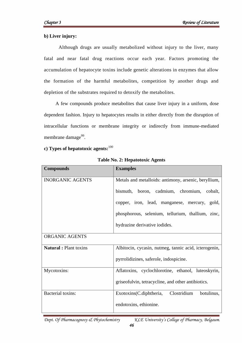
c) Types of hepatotoxic agents:100
Table No. 2: Hepatotoxic Agents
Compounds Examples
INORGANIC AGENTS Metals and metalloids: antimony, arsenic, beryllium,
bismuth, boron, cadmium, chromium, cobalt,
copper, iron, lead, manganese, mercury, gold,
phosphorous, selenium, tellurium, thallium, zinc,
hydrazine derivative iodides.
ORGANIC AGENTS
Natural : Plant toxins Albitocin, cycasin, nutmeg, tannic acid, icterogenin,
pyrrolidizines, saferole, indospicine.
Mycotoxins: Aflatoxins, cyclochlorotine, ethanol, luteoskyrin,
griseofulvin, tetracycline, and other antibiotics.
Bacterial toxins: Exotoxins(C.diphtheria, Clostridium botulinus,
endotoxins, ethionine.
Page 32
Chapter 3 Review of Literature
Dept. Of Pharmacognosy & Phytochemistry KLE University’s College of Pharmacy, Belgaum. 47
Synthetic: Non-medicinal Haloalkanes and haloolephins, Nitroalkanes,
Chloroaromatic compounds, Nitroaromatic
compound, organic amines, Azo compounds, Phenol
and derivatives various other organic compounds.
MEDICINAL AGENTS:
Category of drugs Examples
1) Neuro psychotropics Hydrazine, Tranylcypromine Anticonvulsants,
Antidepressants.
2) Anti-inflammatory and anti-
muscle spasm agents
Cinchopen, Cholchicine, Ibuprofen, Salicylates,
Indomethacin.
3) Hormonal derivatives and other
drugs used in endocrine disease
Acetohexamide, Azepinamide, Carbutamide,
Tolbutamide.
4) Antimicrobials Clindamycin, Novobiocin, Penicillin, Tetracycline,
Sulfonamide, Amodiaquine, Isoniazid, Rifampin.
5) Antineoplastic L-Asparaginase, Azacytidine, Methotrexate, 6-
Mercaptopurine, Chlorambucil, Clavicin.
d) Types of drug reactions:99
Although most hepatotoxic effects involve hepatocyte necrosis, some drugs
injure bile ducts or canaliculi, causing cholestasis without marked damage of
hepatocytes. Other therapeutic agents affect sinusoidal or endothelial cells or fat -
storing Ito cells (causing Vitamin A toxicity, which leads to fibrosis) or cause a
particular pattern of liver injury affecting multiple cell types.
Page 33
Chapter 3 Review of Literature
Dept. Of Pharmacognosy & Phytochemistry KLE University’s College of Pharmacy, Belgaum. 48
I) Direct toxic reactions:
Acetaminophen is an example of an agent that causes direct toxic reaction.
Two clinical scenarios account for most cases of acetaminophen– related hepatic
necrosis i.e. the intentional suicidal overdose and the “therapeutic misadventure”.
In the latter scenario, an alcoholic takes acetaminophen for pain relief in doses that
exceed those recommended in the package insert (4 gm per 24 hrs). The result is a
direct toxic reaction due to the enzyme-induction and glutathione depletion.
Starvation may also play a part, presumably because of glutathione depletion. This
alcohol-acetaminophen syndrome is the most common form of acute liver failure in
the United States and Australia. Extremely elevated serum alanine and aspartate
amino-transferase values (mean approx. 9000 units per liter in one study)
distinguish this condition from viral or alcoholic hepatitis.
II) Idiosyncratic reactions:
Fifteen to twenty percent of patients receiving isoniazid as a single agent for
prophylaxis against tuberculosis may have increased serum alanine and aspartate
aminotransferase levels, but only 1 percent have hepatic necrosis severe enough to
require the withdrawal of the drug. Several factors explain the relatively common toxic
reaction observed. First, the simultaneous use of alcohol or rifampin may augment the
toxicity of isoniazid. Second, elderly persons may be more likely to have toxic reactions
than younger persons. Third, genetic differences are important, since person who is
capable of rapid acetylation of isoniazid have an increased likelihood of toxic reactions
resulting from the formation of acetylhydrazine, which is then transformed by
cytochrome P-450 into a reactive metabolite. In the case of isoniazid and perhaps of
other drugs causing idiosyncratic reactions, such reactions are not truly idiosyncratic but
Page 34
Chapter 3 Review of Literature
Dept. Of Pharmacognosy & Phytochemistry KLE University’s College of Pharmacy, Belgaum. 49
occur when a series of genetic and environmental influences coincide to produce a
significant quantity of one or more toxic metabolites.
III) Combined toxic and allergic reactions:
Halothane can induce a combination of toxic and allergic reactions leading
to liver injury. Although there is usually no rash, fever and eosinophilia commonly
observed and the histological features of liver-biopsy specimens are similar to those
seen with idiosyncratic reactions. The initial elevations in serum alanine and
aspartate amino transferase levels are delayed, but the interval between the drug
administration and toxic reactions becomes shorter with each exposure. Protein
adducts formed from the initial toxic reaction provide the hapten for the formation
of antibodies, so that with subsequent exposure, antibody and cellular recognition
of the halothane-protein-adduct antigen on the hepatocyte surface leads to cell
injury.
IV) Allergic hepatitis:
Drugs such as phenytoin can cause a systemic allergic reaction characterized
by fever, rash, lymphadenopathy, eosinophillia and the presence of eosinophils or
granulomas in liver-biopsy specimens. This allergic reaction is accompanied by
both hepatocyte necrosis and cholestasis. The mechanism responsible for the
combined allergic and hepatotoxic reactions are unknown but the slow resolution of
the illness suggests that the allergen remains on the hepatocyte surface for weeks or
months. This drug-induced hypersensitivity hepatitis syndrome results in a
mononucleosis-like illness that may be confused with viral illness or streptococcal
pharyngitis, so that the agent is not withdrawn, despite signs of developing
hepatitis. The result is often a severe form of the Stevens-Johnson syndrome, with
fever lasting for weeks.
Page 35
Chapter 3 Review of Literature
Dept. Of Pharmacognosy & Phytochemistry KLE University’s College of Pharmacy, Belgaum. 50
V) Cholestatic reactions:
The drugs that mainly affect bile flow, causing cholestatic injury, include
estradiol, chlorpromazine, trimethoprim-sulphamethoxazole, rifampin, erythromycin,
nafcillin and captopril. Typically, jaundice appears early, with associated purities but
little alteration in the patient’s general well-being. A liver biopsy reveals engorgement of
the canaliculi with bile and minimal hepatocellular injury. Eosinophils may be found in
mildly inflamed portal tracts. The mechanism of cholestatic injury remains unclear.
Estradiol and other estrogens have been shown to decrease bile flow and Na+/K
+ ATPase,
change tight junctions between cells, and alter the fluidity of hepatocyte membrane.
VI) Granulomatous reactions:
Noncaseating granulomas resembling sarcoidosis in the liver are caused by
various drugs such as, Allopurinol, Isoniazid, Quinidine, Sulfonamides, Aspirin,
Diazepam, Procainamide etc. The clinical picture is the same as that of other forms
of granulomtous hepatitis i.e. low grade fever and chronic fatigue, with jaundice
only in rare cases.
VII) Drug –induced chronic hepatitis:
Methyldopa and a number of other compounds like trazodone,
nitrofurantoin, and acetaminophen have been found to cause a more indolent form
of liver damage that closely resembles autoimmune chronic active hepatitis.
Hyperglobulinemia may be present, with positive tests for antinuclear antibodies.
The classic agent producing this reaction is oxyphenisatin, a laxative that has been
withdrawn from the market. Early identification of drug-related chronic hepatitis is
not easy, cirrhosis may develop before the hepatitis is diagnosed. Multiple
prescription renewals may be a problem in the case of nitrofurantoin, which is used
to control recurrent urinary tract infections.
Page 36
Chapter 3 Review of Literature
Dept. Of Pharmacognosy & Phytochemistry KLE University’s College of Pharmacy, Belgaum. 51
VIII) Fatty liver and alcoholic hepatitis – like reactions:
Although fatty liver is most commonly related to obesity, diabetes, alcoholism
or corticosteroid therapy, amiodarone and several other drugs can cause a disorder
similar to alcoholic hepatitis, termed non-alcoholic steatohepatitis. This drug and
some related compounds have been shown to cause severe liver toxicity, in an acute
or chronic form, as a part of a multisystem syndrome. Patients typically have
moderately elevated serum alanine and aspartate aminotransferase levels, with a
characteristic lesion of steatohepatitis and cirrhosis can develop in just a few
months. The presence of microvesicular fat within hepatocytes has a different
meaning from that of the macro vesicular steatosis. Fine vesicles are associated
with considerable cellular dysfunction but without cell death. This is the
characteristic lesion of fatty liver caused by pregnancy, high doses of tetracyclines
and Reye’s syndrome associated with aspirin.
IX) Indolent cirrhosis:
Of the several agents capable of causing a gradual progression to cirrhosis without
any manifestation of clinical illness, methotrexate is the most frequently cited example.
This agent is used in patient with severe psoriasis or rheumatoid arthritis, and toxicity
may develop over a period of several years without any symptoms or evidence of
hepatitis or other biochemical abnormalities. A liver biopsy is the only sure way to
establish the diagnosis of indolent cirrhosis caused by a drug reaction. Methyldopa and
Vitamin A have been reported to cause a similar syndrome.
3.2.2 Mechanism of hepatotoxicity:101
There are numerous ways in which the structure and/or function of the liver
can be altered. In view of this, the pathogenesis of hepatic injury requires
consideration of at least several factors.
Page 37
Chapter 3 Review of Literature
Dept. Of Pharmacognosy & Phytochemistry KLE University’s College of Pharmacy, Belgaum. 52
A. Pathogenesis of fatty liver:
The accumulation of abnormal amounts of fat within the liver may be due to
either by extra hepatic causes that provoke a higher input of triglyceride (TG)
precursors into the liver or as a consequence of changes in the function of the liver
itself. In general, the mechanisms that can account for accumulation of TG include;
(i) the rate of synthesis of hepatic TG is normal, but the liver cells are unable to
secrete the TG into the plasma (ii) the secretion of hepatic TG is normal, but the
rate of synthesis is increased (iii) there is both an increase in the rate of synthesis
and a block in the secretion of the synthesized TG and (iv) the TG synthesis takes
place in a compartment of the cell other than the endoplasmic reticulum and thus
this pool is not accessible to the normal secretory pathway.
Impairment of lipid release:
The movement of fat from the liver may be blocked by either interference
with the formation of VLDL or by defective movement of the VLDL across a
damaged plasma membrane. Defective formation of the VLDL may be due to
impairment of synthesis of apoprotein moiety or of the mechanism for assembly of
its three components, such as destruction of cellular site of protein synthesis like,
rough endoplasmic reticulum (RER) and its ribosome. For example, CCl4 acts
through destruction of RER, puromycin inhibits protein synthesis by attaching itself
to the ribosome as the “P” site, tetracycline and its other congeners inhibit protein
synthesis, by binding to t-RNA, ethionine inhibit protein synthesis by ATP
depletion or interfering with other steps of the synthetic pathway. However, there
are several agents which inhibit protein synthesis without producing fatty liver (e.g.
cyclohexamide and actinomycin D) and others which produce fatty liver without
affecting protein synthesis e.g. orotic acid.
Page 38
Chapter 3 Review of Literature
Dept. Of Pharmacognosy & Phytochemistry KLE University’s College of Pharmacy, Belgaum. 53
Increased mobilisation of depot lipid:
The studies have clearly shown that increased mobilization of lipid from depot
can provoke toxin induced catecholamine release which is responsible for the
increased mobilization of fat from depots and thereby contribute to fatty liver.
B. Pathogenesis of necrosis:
Although many hepatotoxic substances that produce necrosis have been shown
to cause similar morphological changes, the exact mechanism by which these agents
lead to necrosis remains to be understood. Several studies in the past have focused
attention on the organelles of hepatocyte as the probable sites of injury responsible
for the necrosis in animals, exposed to hepatotoxins. Toxic damage of
mitochondria, lysosomes, smooth (SER) and rough endoplasmic reticulum (RER)
and the plasma membrane may be responsible for necrosis. It was suggested that
injury to mitochondria might lead to loss of bioenergetics required to maintain
cellular integrity and thus results in necrosis. The possibility that it plays a
subsidiary role in the necrogenic process continues to be the subject of study. The
lysosomes seem to play a little role in necrosis but the main role of lysosomes in
injury seems to be that of scavenger of the debris.
An injury to the RER or to the synthesis of a protein for maintenance of cell
integrity or inability to particular protein that might be essential for maintenance of
cell integrity of inability to synthesize protein destroyed by the toxic agent might
contribute to necrogenesis but damage to the SER may not contribute much in the
development of necrosis.
Recently, much attention has been given on the plasma membrane and the
molecular basis for the membrane injury and their role in the pathogenesis of the
necrosis. On the basis of several studies it has been concluded that the offending
Page 39
Chapter 3 Review of Literature
Dept. Of Pharmacognosy & Phytochemistry KLE University’s College of Pharmacy, Belgaum. 54
agent leads to injury to the plasma membrane which permits intra hepatic
accumulation of calcium ion. The high concentration of calcium ions in turn
enhances plasma membrane injury permitting even higher intracellular content of
the ion which leads to necrosis. In addition, several other molecular mechanisms
lead to necrosis through membrane injury and physico-chemical changes in
hepatocyte mainly by peroxidation of lipids, by trapping and depletion of cellular
uridine triphosphate (UTP), and by alkylation or arylation of key macromolecules.
For example, dimethylnitrosamine alkylates purines, pyrimidines and proteins.
Bromobenzene and large overdoses of acetaminophen lead to arylation of cell
macromolecules.
C. Cholestatic reactions:
The interference with the bile flow induced by the hepatic injury can result
from (i) damage to the bile ducts and ductules (ii) damage to the canalicular
membrane of the hepatocyte (iii) injury to its ATPase activity (iv) interference with
the energy source required for the active transport of constituents of bile into the
canaliculus (v) defects in the synthesis and transport of bile acids into bile and (vi)
defects in the metabolic conversion of substances into the molecular form required
for excretion. Besides these several physico-chemical changes produced in the
micelles of the bile could result into cholestasis.
Some chemicals may block the transport of bile constituents (bilirubin and
bile acids) from the sinusoidal blood into the hepatocyte or their conjugation in the
hepatocyte or transportation of bile into the canaliculus for excretion. For example,
saramycetin, mirex, kepone and rifampicin inhibit the transport of bile from
sinusoidal blood into the hepatocyte perhaps by competing for binding proteins of
the hepatocyte or by affecting changes in the plasma membrane or both. Novobiocin
Page 40
Chapter 3 Review of Literature
Dept. Of Pharmacognosy & Phytochemistry KLE University’s College of Pharmacy, Belgaum. 55
inhibits glucuronyl transferase enzyme responsible for conjugation of bilirubin and
thereby decrease the transport into the canaliculus for excretion. This in turn
results decrease clearance of bilirubin from blood.
Chemicals like C-17 alkylated steroids cause damage to fibrillar network of
the canaliculi and thus produce anatomic obstruction of the extra hepatic biliary tree
which may prove to be an important factor in the production of cholestasis.
Similarly, chemicals like manganese sulphate and norethindrolone lead to definite
manifestations of intrahepatic cholestasis, such as hyperbilirubinemia of
intrahepatic cholestasis and canalicular bile plugs.
D. Pathogenesis of cirrhosis:
The hepatoxins which produce necrosis in experimental animals can produce
cirrhosis but little is known about the mechanism responsible for cirrhotogenesis.
Several clinical cases which show steatosis do not lead to cirrhosis but so far the
evidence is less than conclusive. The exact mechanism by which necrosis triggers
cirrhosis or the difference between the steatosis that does not lead to cirrhosis and
that which appears to do so is obscure. However, it is certain that factors liberated
from injured tissue provoke the fibrogenesis which in turn lead to cirrhosis. The
hepatotoxic chemicals which lead to chronic inflammatory response or injury to
hepatocytes may contribute to fibrogenesis.
E. Hepato-carcinogenesis:
In most instances, induction of hepatic carcinoma requires prolonged
administration of the carcinogens. Most hepatocarcinogens are also hepatotoxic, but
all hepatotoxins are not carcinogens. The carcinogens are electrophilic reactants in
their own right or must be converted so in vivo by metabolism and/or by chemical
breakdown. The reactions so formed bind with cellular macromolecules and so
Page 41
Chapter 3 Review of Literature
Dept. Of Pharmacognosy & Phytochemistry KLE University’s College of Pharmacy, Belgaum. 56
initiate a chain of events that lead to cancer. Thus, the hepatoma inducing action of
aromatic amines resides in their ability to produce electrophilic radicals, formed
during the course of the metabolism by the N-hydroxylation pathway. The crucial
significance of these radicals in malignancy induction is increased by observations
indicating a failure of non-carcinogenic analogues (e.g. several amines or
anthracene) to generate free radicals in liver hepatocytes and mitochondria. The
initial strong generation of radicals subsides to fluctuating changes during tumor
genesis. Treatments with the noncarcinogenic counterparts do not lead to similar
variation in free radical content of the liver.
The new precursor cell population produced, have distinctive biochemical
properties, including the acquisition of one or more new antigens that appear early
and persist in the different cell populations in the ultimate cancer.
F. Hepatic injury due to host idiosyncrasy:
Some drugs can produce hepatic injury unpredictably in a small proportion of
recipients. The injury produced is an expression of unique, individual susceptibility
instead of intrinsic toxicity of the offending agents. The mechanism is presumed to
be that of drug allergy. Several other chemicals produce hepatic injury probably
through a different mechanism, may be through an aberrant metabolic pathway of
the drugs.
G. Hypersensitivity:
Indeed, no firm evidence for the role of hypersensitivity in chemical induced
hepatic injury is available. The evidence available so far indicates that chemical -
induced allergy as the cause of hepatic injury is incomplete because the antigen
responsible for the presumed allergic state might be an unknown metabolite of the
chemical. Despite lack of concrete evidence, chemical-induced allergy is probably
Page 42
Chapter 3 Review of Literature
Dept. Of Pharmacognosy & Phytochemistry KLE University’s College of Pharmacy, Belgaum. 57
responsible for many instances of hepatic injury. For example, sulfonamides, p-
amino-salicylic acid, arsenicals and halothane, all produce hepatic injury under
circumstances which suggest that chemical allergy plays an important role.
H. Biochemical mechanism of hepatic injury:
As discussed previously, changes produced by hepatotoxins are preceded or
succeeded by functional or metabolic changes in the liver. Some biochemical
changes which disturb the liver function are summarized as under:
i) Depletion of coenzyme:
Some chemicals disturb the liver function by depleting an essential metaboli te
or coenzyme followed by morphological changes in the particular cell concerned.
For example, ethionine depletes ATP in rat liver which results changes in protein
synthesis. Another chemical CCl4 causes rapid depletion of NADPH, and antibiotic
azaserine depletes the liver of NAD+ + NADH.
ii) Activation of insulting agents:
Now, a variety of hepatotoxic agents are known which require preliminary
metabolism through an interaction with the NADPH-cytochrome P-450 chain before
their toxic potential can become fully expressed. The process whereby a material is
metabolized to a biologically more active and toxic form is called activation.
Chemicals in this category include CCl4, halothane, dimethyl-nitrosamine,
trichlorethylene, vinyl chloride, paracetamol, aflatoxins etc.
iii) Lipid peroxidation:
During the last few years much evidence has accumulated showing that
lipoperoxidation occurs in living tissues and is of importance in some pathological
phenomena. CCl4 was metabolized to chloroform and concluded that this
transformation was caused by homolytic cleavage, yielding free radicals that could
Page 43
Chapter 3 Review of Literature
Dept. Of Pharmacognosy & Phytochemistry KLE University’s College of Pharmacy, Belgaum. 58
alkylate sulfhydryl groups of enzyme. It was reported that free radicals arising from
the homolytic cleavage of CCl4 could attack the methylene bridges of unsaturated
fatty acid side chains of microsomal lipids, resulting in morphological alteration of
the endoplasmic reticulum, loss of drug metabolizing enzyme activity, loss of
protein synthesis, loss of the capacity of liver to form and excrete VLDL.
There is evidence in support of the role of lipid peroxidation as the cause of
hepatic injury and against the importance of alkylation or oxidation of thiol group
or other direct attacks on proteins, nucleic acid polymers, or nucleotides. According
to them the free radical leads to peroxidation of the unsaturated lipids of the ER
resulting in destruction of the membranes, and to the generation of secondary free
radicals derived from the lipids of the membrane-a form of chain reaction, and
damage to other organelles of the hepatocyte following exposure to these secondary
free radicals derived from the initial effect of the CCl4 on the lipids of the ER.
3.2.3 Biochemical and functional manifestations of injury:
In toxicity studies much attention is devoted to the effect of drugs or chemicals
on liver function. Since the liver is an organ with such diverse functional activities,
no single parameter can be selected to be representative of “Liver Function”. The
development of the so-called liver function tests has largely followed the
development of new knowledge about the biochemistry of the liver. So, tests
indirectly become a measure of hepatic functions.
a. Transaminases:
The two transaminase enzymes, which are sensitive indicators of parenchymal
cell integrity, are:
Aspartate aminotransferase (AST), formerly known as serum glutamate
oxaloacetate transaminase (SGOT).
Page 44
Chapter 3 Review of Literature
Dept. Of Pharmacognosy & Phytochemistry KLE University’s College of Pharmacy, Belgaum. 59
Alanine aminotransferase (ALT), formerly known as serum glutamate pyruvate
transaminase (SGPT).
There is little to choose better ALT and AST in terms of sensitivity and,
though ALT has the greater specificity for liver damage, the tendency for AST to be
elevated in primary skeletal muscle disease as well as in myocardial infarction
seldom causes problems of interpretation. In alcoholic hepatitis, the increase in
AST is usually more pronounced than that of ALT.
Clinical significance:
i) Hepatitis: AST and ALT both show their greatest elevations in acute hepatitis, in
which the rise in enzyme activity begins in the prodromal phase, preceding the
onset of jaundice and occurring also in those patients who remain anicteric. Levels
may reach more than 20 times, or even 50 times, the upper reference limit, with
AST and ALT reaching comparable levels. Declining values accompany clinical
improvement, though falling enzyme levels accompanying clinical deterioration
may result from widespread necrosis and signal impending liver failure.
ii) Other infections involving the liver: Infections such as infectious mononucleosis,
cause elevated transaminase levels.
iii) Poisoning and drugs: AST and ALT levels similar to those seen in acute
hepatitis may follow poisoning with chemicals or plant toxins (e.g. from
mushrooms). Hepatotoxic or hepatitis-inducing drugs usually result in smaller
enzyme increases.
iv) Chronic parenchymal disease: Transaminase levels may reach over five times
the upper reference limit in chronic active or persistent hepatitis, but are generally
much lower than those typical of acute hepatitis. In cirrhosis or fatty change, levels
are usually only slightly or moderately elevated and in cirrhosis may fluctuate. A
Page 45
Chapter 3 Review of Literature
Dept. Of Pharmacognosy & Phytochemistry KLE University’s College of Pharmacy, Belgaum. 60
mild, long-standing elevation of AST may be observed in the presymptomatic phase
of Wilson’s disease.
v) Malignancy: Moderate increases of transaminase levels with occasional
exacerbations, which presumably also reflect episodes of cell destruction, are seen
in malignant disease of the liver. AST is more markedly increased than ALT.
vi) Reduced liver perfusion: The sensitivity of the transaminase is such that even
minor degrees of parenchymal cell damage result in elevated transaminase levels in
serum. Reduced perfusion of the liver in congestive cardiac failure leads to
hypoxia, a potent cause of enzyme leakage from cells, and hence to the escape of
transaminases and other enzymes from the centrilobular regions. Similarly,
haemodynamic changes, such as the loss of fluid due to diarrhea and vomiting can
result in small, transient elevations.
vii) Cholestasis: Transaminase levels are often normal in extrahepatic cholestasis,
but may rise slightly or moderately in prolonged obstruction. Levels are also
generally low in intrahepatic cholestasis. However, because of the frequent
concurrence of cholestasis and hepatitis, many patients do not fall neatly into these
classes.
b. Bilirubin and bile pigments:102
The liver disposes of considerable quantities of bilirubin each day. This
process involves several stages and malfunction at any stage can give rise to useful
biochemical signs.
Bilirubin entering the plasma is normally derived mainly from the senescence
of circulating erythrocytes, with a smaller contribution from the degradation of
erythropoietic elements in the bone marrow. It is almost completely bound to
Page 46
Chapter 3 Review of Literature
Dept. Of Pharmacognosy & Phytochemistry KLE University’s College of Pharmacy, Belgaum. 61
albumin and transported to the liver, normally the only organ that removes bilirubin
from the circulation.
Uptake by hepatocyte is mediated by a carrier. The carrier also takes up
bilirubin glucuronide from plasma as well as the exogenous dyes, such as
bromsulphalein, sometimes used to test excretory capacity; such tests are useless,
therefore, when plasma bilirubin is already raised.
Conjugation: The formation of bilirubin monoglucuronides and diglucuronides
takes place in the hepatocyte. It is affected by the enzyme bilirubin uridine
diphosphate glucuronyltransferase and converts bilirubin from a lipid-soluble to a
water-soluble form.
Bile excretion of conjugated bilirubin: Some of the bilirubin glucuronide refluxes
back into the plasma, but most is excreted in the bile by mechanisms that are still
incompletely understood, but which limit the overall rate of transport of bilirubin.
Conjugated bilirubin in the plasma: Impairment of biliary excretion of conjugated
bilirubin in intrahepatic or extrahepatic cholestasis increases its regurgitation into
the plasma, raising both total and ‘direct’ bilirubin concentrations.
Conjugated bilirubin in the urine: Conjugated bilirubin is water-soluble and so can
pass into the urine. Bilirubinuria can be detected by ‘stick-tests’ before the level of
plasmabilirubin begins to rise; therefore, the fractionation of plasma bilirubin into
its conjugated and unconjugated components is unnecessary in most cases. Bilirubin
bound to albumin in this way cannot pass the glomerulus, and therefore may persist
in plasma after bilirubinuria has disappeared.
Fate of bilirubin in the bile (urobilinogen formation): Bilirubin glucuronides
excreted into the bile undergo hydrolysis by the glucuronidases of the intestinal
flora followed by oxidation and reduction of bilirubin to the colorless urobilinogen.
Page 47
Chapter 3 Review of Literature
Dept. Of Pharmacognosy & Phytochemistry KLE University’s College of Pharmacy, Belgaum. 62
Most urobilinogen is excreted in the faeces; oxidation of urobilinogen
produces the faecal pigment, stercobilin. Some urobilinogen is reabsorbed from the
intestine into the plasma, from which most is re-excreted by the liver into bile.
However, re-excretion is not complete and some urobilinogen passes into the urine.
3.2.4 Histological assessment of the liver:103
a. Normal liver
The normal portal tract contains a hepatic artery, portal vein and a bile duct,
with a few inflammatory cells present. The interface between the hepatocytes and
the portal tracts is known as the limiting plate. The hepatocytes are arranged as cell
plates radially distributed down to hepatic venules. Blood flows from the hepatic
artery and portal vein through the hepatic sinusoids to the venules. These sinusoids
are lined by fenestrated endothelial cells and Kupffer cells. The space between the
endothelium and the surface of hepatocytes contains a matrix of fibrillar and non-
fibrillar collagens with proteoglycans, along with perisinusoidal cells. In contrast to
the flow of blood, bile flows from the hepatocytes into canaliculi and then via the
canalicular system to the bile ductules and ducts in the portal tracts.
b. Chronic active hepatitis
Two histological types (chronic active hepatitis and chronic persistent hepatitis)
are defined, though there is a large, grey area between these two. In chronic
persistent hepatitis, the inflammatory infiltrate is limited to the portal tract.
Hepatocyte necrosis is absent. In chronic active hepatitis, the chronic inflammatory
infiltrate crosses the limiting plate to cause piecemeal hepatocyte necrosis.
c. Granulomatous Inflammation
A granuloma is a small nodular collection of modified macrophages, which
are often termed epithelioid macrophages. In this biopsy, there were numerous
Page 48
Chapter 3 Review of Literature
Dept. Of Pharmacognosy & Phytochemistry KLE University’s College of Pharmacy, Belgaum. 63
granulomas scattered through the liver parenchyma and the portal tracts. The
clinical history was suggestive of drug reaction (carbamazepine). Granulomatous
inflammation is merely a descriptive term, with many factors causing such
inflammation, viz:
Sarcoidosis
Infection
Drugs
Primary biliary cirrhosis
Foreign body reactions (e.g. in drug addicts)
d. Alcoholic Hepatitis
Alcoholic hepatitis is shown, with fatty change in the liver. Fatty change in
readily reversible if alcohol consumption stops. Alcoholic hepatitis is recognized by
liver cell necrosis with polymorph infiltration around hepatocytes (satellitosis) and
the presence of Mallory hyaline bodies (aggregates of ubiquitin filaments) in the
cytoplasm. It is almost always accompanied by some new collagen formation in
perivenular sinusoids. Cirrhosis may result if alcohol ingestion is heavy and
prolonged.
e. Cholestasis
Cholestasis is defined as an accumulation of bile pigments within the liver. In
this case, brown bile can be seen within canaliculi, and also within the cytoplasm of
hepatocytes. Cholestasis can be caused by many different diseases.
f. Cirrhosis
The liver parenchyma is composed of proliferating parenchymal nodules
divided by interconnecting fibrous septa. Classifying cirrhosis into macro nodular
or micro nodular forms is of little histological value. The causes of cirrhosis include
Page 49
Chapter 3 Review of Literature
Dept. Of Pharmacognosy & Phytochemistry KLE University’s College of Pharmacy, Belgaum. 64
alcohol, chronic viral hepatitis, chronic active hepatitis, biliary cirrhosis and
haemochromatosis. A significant proportion may be cryptogenic (idiopathic).
Complications include portal hypertension and hepatocellular carcinoma.
g. Hepatocellular carcinoma
Hepatocellular carcinoma is also known as hepatoma; this is a misnomer – it
is not a benign tumour and the term should be dropped. This lesion is composed of
cords and trabeculae of pleomorphic hepatocytes, with no normal architectural
features (e.g. portal tracts). The histological appearances can vary from solid to
acinar, and sclerosing to papillary, with some lesions being well differentiated and
others anaplastic.
h. Metastatic adenocarcinoma
The most common form of carcinoma in the liver is a metastasis. This patient
presented with a single lesion in the right lobe of the liver, which was biopsied
under ultrasound control. Histology revealed irregular gland structures lined by
pleomorphic epithelial cells, reminiscent of bowel epithelium.
I. Carbon tetrachloride induced hepatotoxicity:
It is useful to divide the mechanism of CCl4 into the following sequence.
- Initial events
- Secondary evoked mechanism
- End stage pathological consequences
The initial event involves carbon-halogen bond cleavage, probably by a one-
electron reduction of CCl4, by a particular ferrous cytochrome P-450, to form
chloride anion and trichloromethyl radical (CCl3). Trichloromethyl peroxy radical
(OOCCl3) is probably generated and small quantity of CO may appear, mostly
through dichlorocarbene intermediate.
Page 50
Chapter 3 Review of Literature
Dept. Of Pharmacognosy & Phytochemistry KLE University’s College of Pharmacy, Belgaum. 65
In next stage, CCl4-carbon is covalently bound to microsomal lipids and
proteins. This placed CCl4 into a general class of xenobiotics, the toxicity of which
appears to depend on their metabolism and subsequent covalent bindings to cellular
macromolecules. However within the first hour, there is inhibition of movement of
liver triglycerides to the plasma as VLDL, polyribosomal desegregation and
findings of protein synthesis also set in well. Protein synthesis could not take place
as the specific binding site is already occupied by cytochrome P-450 induced free
radicals.
The peroxidative decomposition of lipids of the ER is initiated by CCl4
metabolism. Lipid peroxidation generates a wide variety of more or less toxic
products, not organic radicals, which presumably could migrate from membrane
sites near cytochrome P-450 to the other parts of the cell. This states that CCl4
hepatotoxicity is primarily a matter of lipid peroxidation rather than covalent
binding of CCl4 cleavage products104
.
Free radical reactions are implicated in the progression of cancer, inflammation,
atherosclerosis, hepatocellular damage and the biological process of aging. The
hepatoprotective action combined with antioxidant activity has a synergistic effect to
prevent the process of initiation and progress of hepatocellular diseases105
.
3.2.5 Liver damage due to free radicals:105
The pioneering studies on the role of free radical reactions in the genesis and the
expression of cellular and tissue damage have been carried out mainly in the liver. Most
recently, the hepatotoxicity of several free radical-generating compounds like
paracetamol, halothane and iron overload has been reported.
It is now generally accepted that reactive free radicals can exert cellular damage
through a variety of mechanisms e.g. lipid peroxidation, covalent bonding, depletion of
Page 51
Chapter 3 Review of Literature
Dept. Of Pharmacognosy & Phytochemistry KLE University’s College of Pharmacy, Belgaum. 66
glutathione and protein thiols, derangement of intracellular free calcium homeostasis,
DNA fragmentation etc with different relevance in the various conditions. An essential
involvement of lipid peroxidation in the events leading to hepatocyte death has been
proved in the in-vitro and in-vivo acute intoxication.
Plant drugs are known to play a vital role in the management of liver diseases.
There are numerous plants and polyherbal formulations claimed to have hepatoprotective
action. However, numerous medicinal preparations have been advocated a traditional
system of medicine, especially in Ayurvedic, for treating liver disorders. Only a small
portion of the hepatoprotective plants as well as formulations used in traditional
medicine are pharmacologically evaluated for their efficiency.
Plant derived natural products such as flavonoids, terpenoids and steroids etc.
have received considerable attention in recent years due to their diverse pharmacological
properties including hepatoprotective and antioxidant activity. There has been growing
interest in the analysis of certain flavonoids, triterpenoids and steroids stimulated by
intense research in to their potential benefits to human health. Anti-oxidant plays an
important role in inhibiting and scavenging radicals, thus providing protection to humans
against infection and degenerative diseases106
.
3.3 FREE RADICALS IN BIOLOGICAL SYSTEM
A free radical is any atom or group of atoms capable of independent existence
that contains one or more unpaired valence electrons. The unpaired electrons do not
contribute to intramolecular bonding. That unpaired electron/s controls the properties of
radicals107
. They are produced by oxidation/reduction reactions, in which there is a
transfer of only one electron at a time, or when a covalent bond is broken and one
electron from each pair remains with each group. Free radicals can be either highly
Page 52
Chapter 3 Review of Literature
Dept. Of Pharmacognosy & Phytochemistry KLE University’s College of Pharmacy, Belgaum. 67
reactive species like hydrogen atom, hydroxyl radical, or they can be stable entities like
nitric oxide, DPPH radical. In biological systems free radicals have a range of transitory
existences depending upon their reactivity. Some are stable, e.g. melanin can have a long
lifetime, moderately stable ones such as nitric oxide can have lifetimes of ~5 seconds and
highly unstable ones such as hydroxyl radicals exist for only a hundredth of a
microsecond. The importance of free radicals and reactive oxygen species (ROS) has
attracted increasing attention over the past decade. ROS, which include free radicals such
as hydroxyl radicals (.OH), superoxide anion radicals (O2
.) and non free radical species
such as H2O2 and singlet oxygen (.O2), are various forms of activated oxygen. These
molecules exacerbate cellular injury and aging process108
. In living organisms, various
ROS can be formed in different ways. Normal aerobic respiration and the stimulation of
polymorphonuclear leukocytes, macrophages and peroxisomes constitute prominent
sources of ROS. These are major endogenous sources of cellular oxidants. Exogenous
sources of ROS include tobacco smoke, certain pollutants, organic solvents and
pesticides109
.
Many present day diseases are reported to be due to the shift in the balance of the
pro-oxidant and the antioxidant homeostatic phenomenon in the body109
. Pro-oxidant
conditions dominate either on account of increased generation of free radicals caused by
excessive oxidative stress, or due to poor scavenging in the body caused by depletion of
the dietary anti-oxidants. ROS differ significantly in their interactions and can cause
extensive cellular damage such as nucleic acid strand scission modification of
polypeptides, lipid peroxidation etc110, 111
.
Free radicals are assumed to play an important role in aging, cancer, radiation
injury, inflammation, atherosclerosis, ischemia of the heart, brain, small intestine, kidney
and liver; neurodegenerative diseases, diabetes mellitus and disorders of prematurity.
Page 53
Chapter 3 Review of Literature
Dept. Of Pharmacognosy & Phytochemistry KLE University’s College of Pharmacy, Belgaum. 68
Free radicals seem to be one of the final common pathways of cell damage and affect the
cell membrane and the nuclear DNA. The cell membrane damage is by cross-linking of
proteins and by critical alterations of lipids112
.
3.4 ANTIOXIDANT
Antioxidant can be defined as “Any substance, present at low concentrations
compare to those of an oxidizable substrate, significantly delays or prevents oxidation of
the oxidizable substrate” 113
. Antioxidants are the first line of defense against free radical
damage, and are critical for maintaining optimum health. The need for antioxidants
becomes even more critical with increased exposure to free radicals. As part of a healthy
lifestyle and a well-balanced, wholesome diet, antioxidant supplementation is now being
recognized as an important means of improving free radical protection.
The human body employs many antioxidant systems. The exact activity of an
antioxidant depends on the reactive species involved, the area of the body affected by
reactive species and the exact molecular target.
In general, an antioxidant in the body may work in one of the following five ways.
(i) The removal of or decrease in the local O2 concentrations
(ii) The removal of catalytic metal ions
(iii) The removal of ROS such as O2• and H2O2
•
(iv) Scavenging initiating radicals such as •OH, RO
• and RO2
•
(v) Breaking the chain of an initiated sequence.
3.4.1 Mode of action of antioxidant
Antioxidant means "against oxidation." Under normal conditions the damaging
actions of ROS and RNS are minimized by abundant protective and repair mechanisms
that cells possess, including many enzymes and redox active molecules. The human body
Page 54
Chapter 3 Review of Literature
Dept. Of Pharmacognosy & Phytochemistry KLE University’s College of Pharmacy, Belgaum. 69
has an elaborate antioxidant defense system. Antioxidants are effective because they are
willing to give up their own electrons to free radicals. When a free radical gains the
electron from an antioxidant it no longer needs to attack the cell and the chain reaction of
oxidation is broken. After donating an electron an antioxidant becomes a free radical.
Antioxidants in this state are not harmful because they have the ability to accommodate
the change in electrons without becoming reactive. Steric and electronic factors are also
responsible for a chain breaking antioxidant. Antioxidants are manufactured within the
body and can also be extracted from the food humans eat such as fruits, vegetables, seeds,
nuts, meats, and oil. There are two lines of antioxidant defence within the cell. The first
line, found in the fat-soluble cellular membrane consists of vitamin E, beta-carotene etc
Of these, vitamin E is considered the most potent chain breaking antioxidant within the
membrane of the cell. Inside the cell water soluble antioxidant scavengers are present.
These include vitamin C, glutathione peroxidase, superoxide dismutase, and catalase107
.
3.4.2 Classification of antioxidants
The antioxidant systems are classified into two major groups, enzymatic antioxidants and
non enzymatic antioxidants.
Enzymatic antioxidants
Enzyme antioxidants are produced in the body and they act as body’s first line of
defense against free radicals. They convert reactive free radicals into less reactive or inert
species. Enzymatic antioxidant present in the body includes superoxide dismutase (SOD),
catalase (CAT), and glutathione peroxidase (GPx)114-116
.
Catalase
Catalase is an enzyme, which can function either in the catabolism of H2O2 or in the
peroxidase oxidation of small substrates such as ethanol, methanol, and quinine117
. Most
of the aerobic cells have catalyse activity. It was first crystallized from beef liver by
Page 55
Chapter 3 Review of Literature
Dept. Of Pharmacognosy & Phytochemistry KLE University’s College of Pharmacy, Belgaum. 70
Sumner and Dounce118
. Catalase is present in all major body organs, especially in liver.
The catalyse activity of animal and plant is largely located in sub cellular organelles
known as peroxisome119
. Peroxisomes in animal cells are involved in the oxidation of
fatty acids, synthesis of cholesterol and bile acids. Hydrogen peroxide is a byproduct of
fatty acid oxidation. Catalase promotes the conversion of hydrogen peroxide into
molecular oxygen and water without the production of free radicals120
.
Peroxisomes in plant cells are involved in photorespiration (the use of oxygen and
production of carbon dioxide) and symbiotic nitrogen fixation (the breaking apart of the
nitrogen molecule N2 to reactive nitrogen atoms). Hydrogen peroxide is produced as an
intermediate during these chemical processes removed by catalase to prevent damage to
cellular machinery. White blood cells produce hydrogen peroxide to kill bacteria during
which excess of hydrogen peroxide is removed by the catalase. Catalase is composed of
four identical subunits, each containing a protoporphyrin ring and a central iron (Fe)
atom that are very much like the familiar hemoglobins, cytochromes, chlorophylls and
nitrogen-fixing enzymes in legumes121
.
Non-enzymatic antioxidants
Endogenous non-enzymatic antioxidants such as GSH and total thiol were playing
an important role in scavenging ROS. Low molecular weight non-enzymatic antioxidants
such as carotenoids, and dietary phenolic compounds are not manufactured by cell itself
so they are required to supplement through food and diet. In the absence of effective and
affordable interventions for both types of diabetes, the frequency of the disease is
expected to escalate worldwide, with a major impact on the population of developing
countries122
. The preventing activity of allopathic drugs against progressive nature of
diabetes and its complications has been modest and sub-optimal. Insulin therapy affords
effective glycemic control, yet its drawbacks such as ineffectiveness on oral
Page 56
Chapter 3 Review of Literature
Dept. Of Pharmacognosy & Phytochemistry KLE University’s College of Pharmacy, Belgaum. 71
administration, short shelf life, requirement of constant refrigeration, and in the event of
excess dosage leads to hypoglycemia etc, limits its usage123
. Treatment with
sulfonylureas, bigaunides and thiazolidenediones is also associated with side effects124
.
For such various reasons in recent years, the popularity of complementary medicine has
increased. WHO (1980) has also recommended the evaluation of the plants in conditions
where we lack safe modern drugs125
. This leads to increasing demand for herbal products
with anti-diabetic activity with fewer side effects. Further, the selection of herbal
products is easier because of supporting folklore claims and evidence gathered from
traditional usage126
.
There has been growing interest in the analysis of certain flavonoids, triterpenoids,
quinones and steroids, stimulated by intense research in to their potential benefits to
human health127
. Plants provide a rich source of antioxidants, which include tochopherols,
Vit.C, phenolic compounds, carotenoids128
, flavonoids, terpenoids, anthraquinones,
steroids, strychnine and eugenol alkaloids129
etc.
Under normal circumstances, reactive oxygen species (ROS) such as O2•-, •OH,
and H2O2 are detoxified by an efficient antioxidant system that includes enzymes such as
superoxide dismutase, catalase and glutathione peroxidases. In case this defense system
is inefficient, the cells experiences an oxidative stress which contributes in a variety of
chronic inflammatory diseases such as arthritis and atherosclerosis as well as other
ailments viz. cancer, diabetes, hepatitis, neurodegeneration and early aging. Likewise in
liver injury, free radicals and lipid peroxidative metabolites also cause damages to
hepatocytes leading to severe necrosis, sepsis or endotoxemia. Carbon tetrachloride
(CCl4) is a widely used hepatotoxin in rodents and its trichloromethyl radical (•CCl3)-
induced toxicity in rat liver closely resembles to human cirrhosis and hence is an
acceptable animal model for analyzing hepatoprotective agents130
.
Page 57
Chapter 3 Review of Literature
Dept. Of Pharmacognosy & Phytochemistry KLE University’s College of Pharmacy, Belgaum. 72
3.5.1 REVIEW OF BAUHINIA VARIEGATA PLANT
Botanical name : Bauhinia variegata
Family : Caesalpiniaceae
3.5.1 A) Description:
Bauhinia variegata Linn is a medium-sized, deciduous tree, found throughout
India, ascending to an altitude up to 1800 meter in the Himalayas. The Hong Kong
Orchid Tree, botanically known as genus Bauhinia. The origin of the Hong Kong
Orchid Tree is China. The name Bauhinia was named after the Bauhin brothers who
were sixteenth century herbalists131
.
The plant is known by various names in different languages as under.
English : Mountain Ebony
Marathi : Rakta kanchan
Kannada : Kempu mandara
Hindi : Kachnar
Tamil : Shemmandarai
Telgu : Daevakanchanamu
3.5.1 B) Morphology:
Bark is grey with longitudinal cracks, pale pink inside. Leaves are rather broader than
deep, rigidly sub-coriaceous, deeply cordate with two leaflets, connate for about two-
thirds up, leaflets are ovate, rounded at apex, 10-15 cm long, pubescent beneath when
young.
Its young stem Flowers are variously coloured, in few-flowered, lateral, sessile or
short peduncled corymbs, the uppermost petal darker and variegated usually appearing
before the leaves in short axillary or terminal racemes, stamens 5, staminodes absent,
fruits flat; hard glabrous dehiscent pods, 10-15 seeded131, 132
.
Page 58
Chapter 3 Review of Literature
Dept. Of Pharmacognosy & Phytochemistry KLE University’s College of Pharmacy, Belgaum. 73
Fig. No. 8: Bauhinia variegata Linn. Plant
Fig. No. 9: Roots of Bauhinia variegata Linn. Plant
Page 59
Chapter 3 Review of Literature
Dept. Of Pharmacognosy & Phytochemistry KLE University’s College of Pharmacy, Belgaum. 74
3.5.1 C) Medicinal Uses of Plant Parts:
Parts used: Bark, roots, buds, gum, leaves, seeds and flowers.
The bark is astringent, tonic and anthelmintic and is used for ulcers and leprosy.
A decoction of the bark is taken for dysentery. It is used to give tone and vitality
to body. It is used against tuberculosis and skin ailments131,132
.
The leaves contain Vitamin C (146mg %). They are rich in reducing sugars and
have good nutritive value. The infusion of the leaves is used as a laxative and for
cure of diarrhoea, dysentery and piles132
.
The dried buds are used for the treatment of diarrhoea, dysentery, worms, piles
and tumours133
.
A decoction of the buds is given in cough, piles, haematuria and menorrhagia.
The flowers are laxative. Flower buds are pickled131-133
.
An aqueous extract of the plant was found to be effective in induced goiter in rats.
A gargle made from the bark with the addition of extract of acacia pods and
pomegranate flowers is a remedy in salivation and sore throat133
.
Bark rubbed into an emulsion with rice water and administered with the addition
of ginger in scrofulous enlargement of the glands of the neck. A paste made of
the bark together with dried ginger is also applied to scrofulous tumours134
.
This plant is used in malaria and is also an antidote to snake poison135
.
Both roots and bark are astringent, acrid, constipating and anthelmintic. They are
useful in diarrhea, dysentery, cough, leprosy and diabetes135
.
Bark is tonic to the liver136
.
Plant is described as astringent to bowels, tonic to the liver and useful in
treatment of leucoderma, leprosy, menorrhagia, asthma, wounds and ulcers137
.
Page 60
Chapter 3 Review of Literature
Dept. Of Pharmacognosy & Phytochemistry KLE University’s College of Pharmacy, Belgaum. 75
3.5.1 D) Constituents Present in Various Part of B.variegata Linn:
The stem--sitosterol, lupeol, kaempferol-3-glucoside and 5,7-dehydroxy and
5,7-dimethoxy flavanone-4-O--L-rhamnopyranosyl--D-glucopyranosides131
.
The pale violet flowers- Cyaniding-3-glucoside, maluidin-3-glucoside, maluidin-
3-diglucoside, peonidin 3-diglucoside132
.
White flowers- Kaempferol-3-galactoside and kaempferol-3-rhamnoglucoside132
.
The bark yields a fibre133
.
The tree yields a gum similar to cherry gum134
.
Root and bark - Flavanone, (2S)-5,7-dimethoxy-3’,4’-methylenedioxyflavanone
and a new dihydrodibenzoxepin, 5,6-dihydro-1,7-dihydroxy-3,4-dimethoxy-2-
methyldibenzoxepin138
.
Root- Flavonol glycoside 5,7,3',4'-tetrahydroxy-3-methoxy-7-O-α-
Lrhamnopyranosyl (1--)3)-O-β galactopyranoside139
.
Stem bark- hentriacontane, octacosanol, stigmasterol140
and sterols, glycosides,
reducing sugars and nitrogenous substances141
.
Stem- Flavonone glycoside-5, 7- dihydroxyflavonone-4–O–α-L–rhanmo-
pyranosyl- β–D– glucopyranoside142
.
Stem-β-sitosterol, lupeol, kaempferol-3-glucoside and a 5, 7-dimethoxy-
flavonone-4–O–α–L– rhanmopyranosyl- β-D-glucopyranoside143,144
.
Stem-Flavonol glycoside-Kaempferol-3-glucoside144
.
Plant-Phenanthraquinone-bauhinone-2, 7-dimethoxy-3-methyl-9,10-dihydro-
phenanthrene-1, 4-dione145
.
Leaves-Two new long chain compounds- heptatriacontan-12, 13-diol and
dotetracont-15-en-9-ol146,147
.
Page 61
Chapter 3 Review of Literature
Dept. Of Pharmacognosy & Phytochemistry KLE University’s College of Pharmacy, Belgaum. 76
Leaves-Saponins, steroids, flavonoids, alkaloids, tannins and sugars148
.
Volatile oil of leaves-Sesquiterpenes, -caryophyllene, germacrene D and
spathulenol along with - - cadinene149
.
3.5.1 E) Reported activity:
Antitumor activity: Ethanolic extract of stem was evaluated against Dalton's
Ascytic lymphoma in swiss albino mice. A significant enhancement of survival
time of tumor bearing mice was found with respect to control group. Extract was
able to reverse changes in haematological parameters protein and PCV
consequent to tumor inoculation150
.
Methanolic extract of leaves was tested for antimicrobial activity. Antifungal
activity was shown against Aspergillus fumogalus, A.niger. The activity maxima
was displaced by A.fumigatus, Bacillus anthracis, S.agalcties151
.
Kanchanar (B. variegata) along with Manjishtha was given orally in non-healing
diabetic foot ulcers and gangrene. 80% of patients showed improvement with
10% partial amputation152
.
The Kanchanar guggulu is an ethical preparation advocated for the management
of various glandular swellings like galgand, gandmala, granthi and arbuda
etc153
.
Oral administration of Kanchanara (B .variegata) bark and Ghanastava of
Manjishtha (Rubia cordifolia) root to the patients of diabetic microangiopathy
gave satisfactory results in newly formed ulcers154
.
Solid extract of B.variegata in a polyherbal formulation has been found to be
useful as a thyrocap in treatment of simple diffuse goiter with physical and
biochemical improvement155
.
Page 62
Chapter 3 Review of Literature
Dept. Of Pharmacognosy & Phytochemistry KLE University’s College of Pharmacy, Belgaum. 77
Anti-inflamatory activity: Six flavonoids together with one triterpene caffeate were
evaluated as inhibitors of some macrophage functions involved in the inflammatory
process. These experimental findings suggest, use of the plant B.variegata in the
management of inflammatory conditions156
.
Hepatoprotective activity: Alcoholic stem bark extract exhibited hepatoprotective
activity in carbon tetrachloride (CCl4) intoxicated Sprague-Dawley rats157
.
Anti-arthritic activity: Ethanol extract has significant antiarthritic effect
158.
Immunomodulatory activity: Ethenolic extract of the stem bark possesses
immunomodulatory property159
.
Antibacterial activity: Aqueous and methanolic extract of plant shown remarkable
antibacterial activity160
.
Antihyperglycemic and anti-hyperlipidemic activity: Aqueous and ethenolic
extract of leaves has shown antihyperglycemic and anti-hyperlipidemic activity in
normal and STZ induced diabetic rats161
.
Antioxidant and antihyperlipidemic activity: Alcoholic and aqueous extract of
stem bark and root showed significant antioxidant and antihyperlipidemic activity in
rats162
.
Antiobesity effect: Methanolic extract of bark has shown antiobesity action on
female rats163
.
Antinociceptive and anti-inflammatory activity of Triterpene Saponin was found
in leaves164
.
Analgesic activity: Aqueous and ethenolic extract of root has shown dose dependent
analgesic activity165
.
Anticarcinogenic and antimutagenic potential of ethanolic and aqueous extract in
Swiss Albino mice166
.
Page 63
Chapter 3 Review of Literature
Dept. Of Pharmacognosy & Phytochemistry KLE University’s College of Pharmacy, Belgaum. 78
3.5.2 REVIEW OF TECTONA GRANDIS PLANT
Botanical name : Tectona grandis
Family : Verbenaceae
3.5.2 A) Description:
Tectona grandis Linn. (Verbenaceae) is a large deciduous tree. Branchlets are
quadrangular, channeled and stellately tomentose. The tree is growing in higher
situations, native to central India, Konkan, Western Deccan peninsula, South India and
Burma167
. Teak is a hardwood species of worldwide reputation168
.
The plant is known by various names in different languages as under168,169
English : Teak
Marathi : Sag
Kannada : Tega, Jadi
Hindi : Sagvan
Sanskrit : Sakah
Telgu : Peddateku
3.5.2 B) Morphology:
This is an erect, large, deciduous tree growing up to 20 meters or more in height. It
grows best in warm, moist tropical climates with 1,250 to 3,000 mm of mean annual
precipitation and a marked dry season of 3 to 6 months. The branchlets are 4-angled. The
leaves are large, elliptic or obovate, 20 to 30 centimeters in length, pointed at both ends,
usually wedge-shaped at the base, and entire at the margins; the upper surface is rough,
but without hairs, and the lower is densely covered with gray or yellowish hairs. The
calyx is small, board, bell-shaped, and covered with stellate hairs, with subequal and
spreading lobes. The corolla is white, and smooth, and less than 1 centimeter across, with
subequal and spreading lobes. The fruit is somewhat rounded, about 1.3 centimeters in
Page 64
Chapter 3 Review of Literature
Dept. Of Pharmacognosy & Phytochemistry KLE University’s College of Pharmacy, Belgaum. 79
diameter, and somewhat 4-lobed, the soft pericarp densely clothed with felted, stellate
hairs167,170
.
Fig. No. 10: Tectona grandis Linn. Plant
Fig. No. 11: Bark of Tectona grandis Linn.
Page 65
Chapter 3 Review of Literature
Dept. Of Pharmacognosy & Phytochemistry KLE University’s College of Pharmacy, Belgaum. 80
3.5.2 C) Medicinal Uses of Plant Parts:
Parts used: Bark, roots, leaves and flowers.
The roots are useful in anuria167,171
.
The bark is astringent, acrid, sweet, cooling, constipating, anthelmintic and
depurative. It is useful in bronchitis, hyperacidity, diabetes, leprosy and skin
diseases168
.
Leaves are useful in inflammation, leprosy and in skin diseases171
.
The flowers are acrid, bitter, refrigerant, diuretic and anti-inflammatory and
are useful in leprosy, skin diseases, burning sensation and diabetes171
.
3.5.2 D) Constituents Present in Various Part of T. grandis Linn:
Wood- Resin, silica, calcium, ammonium and magnesium phosphate169
Anthraquinone-2-carboxylic acid, anthraquinone-2-carboxaldehyde172
Triterpenic and hemiterpenic compound173
9, 10-dimethoxy-2-methyl-1, 4-anthraquinone, 5-hydroxy-2-methyl-9, 10-
anthraquinone, 1-hydroxy-5-methoxy-2-methyl-9, 10-anthraquinone,1, 5-
dihydroxy-2-methyl-9,10-anthraquinone,tecomaquinone-I, tectoquinone,
dehydro-a-lapachone174,175
.
lapachol, 5-hydroxy-lapachol, methlyquinizarin, squalene176
Dehydro-a-isodunnione177
Lignins178
Root- Lapachol, tectol, dehydrotectol, tectoquinone, b-lapachone, dehydro-a-
lapachone,b-sitosterol, new diterpene, tectograndinol172,179
Non-structural carbohydrates180
hydroxy-2-methyl anthraquinone, obtusifolina, betulinic acid179
Leaves- Tectoleafquinone173
Page 66
Chapter 3 Review of Literature
Dept. Of Pharmacognosy & Phytochemistry KLE University’s College of Pharmacy, Belgaum. 81
Tannins,dye-Tectoionols-B,tectoionols-A,monoterpene, apocarotenoids181
Protein (7.1%), crude fiber (22.3%), calcium (3%), phosphorous (0.46%)
Steroidal compound squalene, polyisoprene-a-tolylmethyl ether and
betulinic acid, a anthraquinonenapthaquinone pigment174,182
Seed - Seed oil contain fatty acids as caprylic (1.45%) myristic acid (2.86%),
palmitic acid (12.12%), stearic acid (9.52%), oleic acid (23.33%) and
linoleic acid (43.22%)(5).Xanthene183
Bark - Tannin (7.14%), quinone173
5-hydroxy-1,4-napthalenedione (juglone), sterols184
Obtusifolina, Desidro-A-lapachona184
.
3.5.2. E) Reported activity:
Antifungal activity: Teak (T. grandis) sawdust extract inhibited the growth of
Aspergillus niger185
.
Antiulcer activity: Lapachol, a naphthaquinone isolated from the roots was found to
have an anti-ulcerogenic effect on subsequently induced experimental gastric and
duodenal ulcers in rats and guinea-pigs186
.
Anti-anaemic activity: Teak ethanolic extract increases significantly the
concentration of haemoglobin, osmotic resistance of red blood cells and the number
of reticulocytes after 7 days of Phenyl hydrazine administration. This study supports
the use of Tectona grandis in the treatment of anaemia187
.
Nitric oxide scavenging activity: The plant extract exhibited a dose-dependent NO
scavenging activity188
.
Wound Healing activity: Leaf extract when applied topically (5% and 10% gel
formulation) or given orally (250mg and 500 mg/kg body weight), promoted the
Page 67
Chapter 3 Review of Literature
Dept. Of Pharmacognosy & Phytochemistry KLE University’s College of Pharmacy, Belgaum. 82
breaking strength, wound contraction and period of epithelization so it is used to
promote wound healing189
.
According Ayurveda, wood is acrid, cooling laxative sedative to gravid uterus and
useful in treatment of piles, leucoderma and dysentery. It allays thirst and possess
anthelmintic and expectorant properties. Tectona grandis leaf extract are widely
used in the folklore for the treatment of various kinds of wound, especially burn
wound190
.
Antiasthmatic activity: Ethyl acetate extract of bark showed significant
antiasthmatic activity191
.
Antihyperglycemic activity:. Bark extract exerted antihyperglycemic activity in
alloxan induced diabetic rats192
.
Antibacterial, cytotoxic and antioxidant activity: Different extracts from leaves,
leaf, bark and wood showed Antibacterial, cytotoxic and antioxidant activity193
.
Tocolytic effect: Stem extract possess tocolytic effect on uterus of female albino
wistar rats194
.
Wound healing activity: Aqueous and methanolic extracts of leaves have significant
wound healing activity195
.
Methanolic extract (root)196
, ethenolic extract (bark)197
and water juice extract
(leaves)198
showed antioxidant activity.
Page 68
Chapter 3 Review of Literature
Dept. Of Pharmacognosy & Phytochemistry KLE University’s College of Pharmacy, Belgaum. 83
3.5.3 REVIEW OF SCHREBERA SWIETENIOIDES PLANT
Botanical name : Schrebera swietenioides Roxb.
Family : Oleaceae
3.5.3 A) Description:
Schrebera swietenioides Roxb. (Weaver's Beam tree) belonging to family
Oleaceae, is a moderate sized tree of 20 m height with thick grey bark growing in
deciduous forests, to an altitude of 1200 m, throughout India. Mostly found in tropical
and sub-tropical Himalayas, south and Central India, from Rajasthan to west Bengal199
.
The plant is known by various names in different languages as under.
English : Weaver’s beam tree
Kannada : Bula,Gante, Nagganti, Mogalingamara
Hindi : Moka, Banpalas
Tamil : Mogalingam
Telgu : Mogalinga,Tondamukkudi
Sanskrit : Muskakah
3.5.3 B) Morphology:
Leaves opposite, simple or imparipinnate; rachis usually winged. Infloresence a
paniculate cyme. Flowers heterostylous and bisexual. Calyx campanulate, loosely
enveloping the corolla, truncate or irregularly and obscurely lobed. Corolla salver-
shaped, white, sometimes tinged with pink or puce; tube well developed, cylindrical;
segments 6 or more, spreading to reflexed, each with a group of swollen brown to
purplish hairs at the base. Stamens 2, inserted on the corolla; filaments short, anthers
large, introrse. Ovary bilocular, small, truncate or obscurely bi-lobed at apex; ovules 4 in
each loculus; style filiform; stigma included or excerted, subcapitate or oblong in outline.
Page 69
Chapter 3 Review of Literature
Dept. Of Pharmacognosy & Phytochemistry KLE University’s College of Pharmacy, Belgaum. 84
Capsule bi-valved, woody with loculicidal dehiscence; seeds produced into a long
solitary subapical wing200-202
.
Fig. No. 12: Schrebera swietenioides Roxb. plant with fruit
3.5.3 C) Medicinal Uses of Plant Parts:
Parts used: Bark, roots, Fruit, gum, leaves, seeds and flowers.
Bark-used for treating boils and burns, Roots- used in Leprosy and also for killing
worms in the wounds of cattle, Leaves-In treatment of urinary discharges and
enlargement of spleen, and Fruits- useful for curing hydrococle203
.
Root, bark, leaves, fruits which are used for medicinal purpose are bitter, acrid,
appetizing, digestive, thermogenic, stomachic, depurative, constipating urinary
astringent and anthelmintic204
.
Page 70
Chapter 3 Review of Literature
Dept. Of Pharmacognosy & Phytochemistry KLE University’s College of Pharmacy, Belgaum. 85
The roots, bark and leaves are bitter, acrid, appetizing, digestive, constipating and
anthelmintic. They are useful in flatulence, skin diseases, leprosy, diarrhea, anemia
and rectal disorders. The fruit is digestive, purgative and stomachic, and is useful in
flatulence, anorexia, colic and diabetes205
.
3.5.3 D) Constituents Present in Various Part of S. swietenioides Roxb:
The tree exudes a grey gum which is sweet in taste. It consists of mannitol,
fructose and a digalactoside named swietenose203, 206
.
Fruit reported the presence of triterpenoids- Oleanolic acid, Betulinic acid207
.
3.5.3 E) Reported activity:
Antioxidant, Anti-Inflammatory and Antipyretic Activity: Ethenolic extract of
roots showed significant antioxidant, anti-inflammatory and antipyretic activities208
.
Its stem bark is used in joint and body pains, headache, itching209
.
Its boiled fruit with ginger and sugar is used for haemorrhoids by tribals of
Saurashtra, Gujrat210
.
Powdered leaf material methanol, ethanol and aqueous extracts were found
significant antibacterial activity211
.