Page 1
44
CHAPTER 3
THE EFFECT OF METAL IONS ON LIPID OXIDATION, COLOR AND CHEMICAL
PROPERTIES OF CUTTLEFISH (SEPIA PHARAONIS) SUBJECTED TO MULTIPLE
FREEZE-THAW CYCLES
3.1 Abstract
The effect of different metal ions (Fe, Cu and Cd) at various concentrations (0, 5
and 25 ppm) on the lipid oxidation, discoloration and physicochemical properties of muscle
protein in cuttlefish (Sepia pharoanis) subjected to multiple freeze-thaw cycles, were
investigated. Lipid oxidation of all treatments increased as the freeze-thaw cycle increased.
However, the rate of TBARS increases varied, depending on concentration, type and valency of
metal ion. Fe(II) induced the lipid oxidation mostly effectively and its prooxidative effect was in a
concentration dependent manner. The increased lipid oxidation of cuttlefish added with iron was
coincidental with the increase in b*-value (yellowness), especially with increasing freeze-thaw
cycles. Cu(I) and Cu(II) altered cuttlefish protein sulfhydryl content and the protein solubility
decreased with the concomitant increased disulfide bond content. The oxidative changes of
protein were observed only when a concentration of metal ions of 25 ppm was used. Those
changes were more intense with increasing freeze-thaw cycles. Ca2+-, Mg
2+- and Mg
2+-Ca
2+-
ATPase activity of cuttlefish natural actomyosin decreased markedly in the presence of copper,
whereas Mg2+-EGTA-ATPase activity was increased. SDS-PAGE revealed that Cu(I) and Cu(II)
induced the polymerization of muscle proteins stabilized by disulfide bond formation. However,
Fe(II), Fe(III) and Cd(II) exhibited no pronounced effect on the oxidation of cuttlefish muscle
proteins. Therefore, copper mainly caused the oxidation of protein, while iron induced the lipid
oxidation and the formation of yellow color from cuttlefish muscle, particularly with multiple
freeze-thaw cycles.
Page 2
45
3.2 Introduction
Cephalopods, including cuttlefish, squid and octopus, are an important marine
resource for human consumption (Sikorski and Kolodziejska, 1986). Lipid oxidation in muscle
foods including cephalopods is one of the major deteriorative reactions causing a loss in quality
during frozen storage. The oxidation leads to the formation of free radicals and hydroperoxides.
Such intermediary compounds are unstable and cause oxidation of pigments, flavors, and
vitamins. Other compounds such as ketones, aldehydes, alcohols, hydrocarbons, acids and
epoxides are formed during the oxidation of unsaturated fatty acids, produce off-flavor and can
react with protein to produce off-color (Khayat and Schwall, 1983). Oxidized unsaturated fatty
acids bind to protein and form insoluble lipid-protein complexes. Thus lipid oxidation processes
lead to discoloration, drip losses, and off-flavor development (Decker and Hultin, 1992) and
production of potentially toxic compounds (Xiong, 2000). Protein and lipid oxidation can
therefore account for the toughened texture, poor flavor and/or unappealing odor of poorly stored
frozen seafood (Khayat and Schwall, 1983).
Transition metals and heme protein are the major prooxidants in muscle foods
(Decker et al., 1992). Both iron (Fe) and copper (Cu) are an essential trace element in
cephalopods, since they are required as a cofactor for a number of enzymes and other cellular
activities. For instance, cephalopods use Cu-containing protein, haemocyanin, as a respiratory
pigment (Decleir et al., 1978). Both Fe and Cu are known to promote oxidative reaction through
the pathways such as Fenton reaction (Walling, 1975).
H2O2 + Fe(II)/Cu(I) 0OH + OH
+ Fe(III)/Cu(II)
The highly reactive hydroxyl radical causes oxidative damage to lipid
membrances (Decker et al., 1989; Lauritzsen and Martinsen, 1999; Lauridsen et al., 2000).
Proteins are also major targets of reactive oxygen species in cells, either through oxidation of
their amino acid side chains to hydroxyl or carbonyl derivatives, or by a splitting of their peptide
bonds (Srinivasan and Hultin, 1997). Degradation and polymerization of myofibrillar proteins
Page 3
46
occurs in different model oxidation systems that closely resembling meat or processed meat
conditions (Decker et al., 1993; Martinard et al., 1997; Srinivasan and Hultin, 1997; Liu et al.,
2000; Liu and Xing, 2000). Liu and Xiong (2000) found marked changes in the electrophoretic
pattern change of myosin treated with FeCl3/H2O2/ascorbate system. Oxidation caused a disulfide
cross-linkage of myosin to form a polymer. Decker et al. (1993) reported that hydroxyl radicals
promoted the deterioration in functional properties of myofibrillar proteins (solubility, water
holding capacity and gel strength). Liu et al. (2000) found that myofibrils oxidized by
FeCl3/H2O2/ascorbate showed a decrease in gel-forming ability and alteration in the functional
groups of amino acids in the myofibrillar proteins. Site-specific metal-catalyzed protein oxidation
occurs via hydroxyl free radicals, which are produced from H2O2 at specific iron-biding site on
proteins.
Although metal ions in cephalopods have been intensively studied, the influence
of metal ions on the quality changes of cephalopods during processing or storage is poorly
understood. The objective of this work was to study the effects of metal ions on lipid oxidation,
discoloration and physicochemical changes of cuttlefish muscle subjected to multiple freeze-thaw
cycles.
3.3 Materials and Methods
Chemicals
Ammonium molybdate, 5-5A-dithio-bis (2-nitrobenzoic acid) (DTNB), adenosine
5A-triphosphate (ATP), β-mercaptoethanol (βME), cysteine, sodium bisulfite, iron (II) chloride,
iron (III) chloride, copper (I) chloride, copper (II) chloride and cadmium (II) chloride were
obtained from Sigma (St. Louise, MO, USA).
Materials
Cuttlefish (Sepia pharaonis) caught by cast net from Songkhla coast along the
Gulf of Thailand and offloaded about 24 h after capture, were purchased. The cuttlefish with the
Page 4
47
size of 8-10 cuttlefish/kg was placed in ice with a cuttlefish/ice ratio of 1:2 (w/w) and transported
to the Department of Food Technology, Prince of Songkla University within 1 h. The cuttlefish
were cleaned and deskinned, eviscerated and the eyes were removed. The cuttlefish was kept in
ice during preparation.
Sample preparation
Minced cuttlefish were frozen in liquid nitrogen and then powdered in a blender.
The cuttlefish powder was mixed with 8 ml of water or stock solution containing different ions
including iron, copper and cadmium to obtain the final concentration of 5 or 25 ppm. The
cuttlefish paste was frozen at -18 °C using an air blast-freezer for 48 h, followed by thawing
using running tap water (25-27 °C) until the core temperature reached 0-2 °C. The mixtures were
subjected to different freeze-thaw cycles (0, 1, 2, 3, 5 and 7 cycles).
Preparation of natural actomyosin
Natural actomyosin (NAM) was prepared according to the methods of Benjakul
et al. (1997). Cuttlefish paste was homogenized (IKA Labortechnik, Salangor, Malaysia) in 10
volumes of 0.6 M KCl (4 °C) for a total of 4 min on ice. Homogenization was performed in 20
sec bursts, followed by 20 sec rest periods to avoid over heating. The homogenate was
centrifuged at 5000 xg for 30 min at 4 °C and the supernatant, to which 3 volumes of chilled
water were added, was collected. The diluted supernatant was centrifuged at 5000 xg for 30 min
at 4 °C to precipitate the NAM. NAM was solubilized in 0.6 M KCl (1:1) and then recentrifuged
at 5000 xg for 30 min at 4 °C to precipitate insoluble protein. The supernatant was collected
and the protein content was adjusted to 4 mg/mL. The NAM solution was used for analysis of
surface hydrophobicity, sulfhydryl contents, disulfide bond contents and enzyme activity.
Thiobarbituric acid reactive substances (TBARS)
Page 5
48
Thiobarbituric acid reactive substances (TBARS) were determined as described
by Buege and Aust (1978). Cuttlefish paste, containing different types and concentrations of ions
(2 g), was dispersed in 10 mL of thiobarbituric acid solution (0.375% thiobarbituric acid, 15%
trichloroacetic acid and 0.25 N HCl). The mixture was heated in boiling water for 10 min,
followed by cooling in running tap water. The mixture was centrifuged at 3600 xg for 20 min at
room temperature. The absorbance of the supernatant was measured at 532 nm. The standard
curve was prepared using malonaldehyde, and TBARS were expressed as mg malonaldehyde/kg
sample.
Color
The color of the cuttlefish paste was measured using a colorimeter (Hunter Lab,
Model Color Flex, Virginia, USA) and reported in the CIE color profile system as L*, a* and b*-
values.
Surface hydrophobicity
Surface hydrophobicity (S0ANS) was determined as described by Benjakul et al.
(1997) using 1-anilinonaphthalene-8-sulphonic acid (ANS) as a probe. NAM solution (4 mg/mL)
was diluted in 10 mM phosphate buffer, pH 6.0, containing 0.6 M NaCl to produce protein
concentrations of 0.1%, 0.2%, 0.3% and 0.5%, followed by incubation at room temperature for 10
min. The diluted protein solution (2 mL) was mixed with 20 µL of 8mM ANS in 0.1 M
phosphate buffer, pH 7.0 and the fluorescence intensity of ANS-conjugates was immediately
measured at an excitation wavelength of 374 nm and an emission wavelength of 485 nm. The
initial slope of the plot of fluorescence intensity versus protein concentration was referred to as
S0ANS.
Total sulfhydryl content
Page 6
49
The total sulfhydryl content was determined using 5,5A-dithio-bis(2-nitrobenzoic
acid) (DTNB) according to the method of Ellman (1959) as modified by Benjakul et al. (1997).
One mL of NAM solution (4 mg/mL) was mixed with 9 mL of 0.2 M Tris-HCl buffer, pH 6.8,
containing 8 M urea, 2% SDS and 10 mM EDTA. Four mL of the mixture were mixed with 0.4
mL of 0.1% DTNB and incubated at 40 °C for 25 min. The absorbance at 412 nm was measured
using a 0.6 M KCl solution as a blank. The sulfhydryl content was calculated using the extinction
coefficient of 13500 M-1 cm
-1.
Disulfide bond content
The disulfide bond content was determined using the 2-nitro-5-thiosulfobenzoate
(NTSB) assay according to the method of Thannhauser et al. (1987). To 0.5 mL of NAM solution
(4 mg/mL), 3.0 mL of freshly prepared NTSB assay solution were added. The mixture was mixed
thoroughly and incubated in the dark at room temperature for 25 min. The absorbance at 412 nm
was measured. The disulfide bond content was calculated using the extinction coefficient of
13900 M-1 cm
-1.
Sodium dodecyl sulfate-polyacrylamide gel electrophoresis
Sodium dodecyl sulfate-polyacrylamide gel electrophoresis (SDS-PAGE) was
used to monitor the polymerization of the modified proteins. The cuttlefish paste was solubilized
in 5% SDS (1:9, w/v) and dissolved in sample buffer with and without β-mercaptoethanol. SDS-
PAGE was performed using 4% stacking gels and 10% running gels (Laemmli, 1970).
ATPase activity
ATPase activity was determined according to the method of Benjakul et al.
(1997). NAM solution was diluted to 3.0 mg/mL with 0.6 M KCl, pH 7.0. Diluted protein
Page 7
50
solution (1 mL) was added with one of the following solutions for each ATPase activity assay to a
total volume of 9.5 mL: 10 mM CaCl2 for Ca2+-ATPase; 2 mM MgCl2 for Mg
2+-ATPase; 0.1 mM
EGTA for Mg2+-EGTA-ATPase, 10 mM CaCl2 and 2 mM MgCl2 for Ca
2+-Mg
2+-ATPase. To each
assay solution, 0.5 mL of 20 mM ATP was added to initiate the reaction. The reaction was
conducted for 8 min at 25 °C and terminated by adding 5 ml of chilled 15% (w/v) trichloroacetic
acid. The reaction mixture was centrifuged at 3500 xg for 5 min and the inorganic phosphate
liberated in the supernatant was measured by the method of Fiske and Subbarow (1925). Specific
activity was expressed as µmoles inorganic phosphate released/mg protein/min. A blank solution
was prepared by adding chilled trichoroacetic acid prior to addition of ATP. Ca2+-sensitivity was
calculated according to Benjakul et al. (1997) as follows:
Ca2+-sensitivity = [ 1- Mg
2+-EGTA-ATPase activity ] x 100
Mg2+-Ca
2+-ATPase activity
Protein solubility
Solubility was determined according to Benjakul and Bauer (2000). To 1 g cuttlefish
paste, 20 mL of 0.6 M KCl were added and the mixture was homogenized for 1 min at a speed of
12000 rpm, using an IKA homogenizer (Selangor, Malaysia). The homogenate was stirred at 4 °
C for 4 h, followed by centrifuging at 8500 xg for 30 min at 4 °C. To 10 mL of supernatant, cold
50% (w/v) trichloroacetic acid was added to obtain a final concentration of 10%. The precipitate
was washed with 10% trichloroacetic acid and solubilized in 0.5 M NaOH. The cuttlefish paste
was also directly solubilized by 0.5 M NaOH to determine total protein. Protein content was
determined using the Biuret method (Robinson and Hodgen, 1940).
Statistical analysis
A factorial design (5 metal ions × 2 concentrations × 6 storage times) was used.
Data were subjected to analysis of variance (ANOVA) and mean comparison were carried out
using DuncanAs multiple rang test (DMRT) (Steel and Torrie, 1980). Statistical analyses were
Page 8
51
performed using the Statistical Package for Social Science (SPSS 10.0 for windows, SPSS Inc.,
Chicago, IL).
3.3 Results and Discussion
Lipid oxidation
Lipid oxidation in muscle foods is one of the major deteriorative reactions
causing losses in quality during frozen storage (Sahoo and Verma, 1999). The levels of TBARS
in cuttlefish paste with added metal-ions at 5 and 25 ppm during different freeze-thaw cycles are
presented in Table 9. TBARS, in all treatments, increased as the number of freeze-thaw cycles
increased (p<0.05). The rate of lipid oxidation varied, depending on concentration and type of
metal ion. TBARS formation increased with increasing metal ion concentrations. The TBARS
values of Fe(II)- and Fe(III)-treated samples increased more rapidly, than did other samples. Of
the two iron-containing samples, Fe(II) induced lipid oxidation in cuttlefish paste most
effectively, as evidenced by the greatest TBARS values after 3-5 and 1-5 freeze-thaw cycles for 5
and 25 ppm iron, respectively. Samples containing Cu or Cd had TBARS levels similar to the
control. Mizushima et al. (1977) reported the catalytic effect of copper, iron, and hemin on lipid
oxidation of Alaska Pollack and mackerel oil at 40 °C in the order: Fe(II)>hemin>Cu(II)Fe>
III). Freeze-thaw cycling produces the repeated ice crystal formation that could disrupt cellular
structure (Benjakul and Bauer, 2000). Lipid oxidation in muscle foods is thought to occur at the
cellular membrane level and thus oxidative deterioration is a problem in both lean and fatty fish
muscle. Therefore, freeze-thawing could result in changes in the physical organization of
membrane lipids which could impact lipid oxidation pathways.
Discoloration
L*, a*, and b*-values of cuttlefish paste, with and without metal ions, as affected
by multiple freeze-thaw cycles are shown in Figure 10. In samples with added metals, only those
with Fe(II) and Fe(III) had an increase in a*-value and b*-value as the number of freeze-thaw
Page 9
52
cycles increased. In addition, a decrease in L*-value was only observed in the samples containing
Fe(II) and Fe(III). No changes in L*-value or b*-value were observed in samples containing Cu
or Cd. Cu(II) caused a decrease in a*-value during the freeze-thawing process. A positive
correlation between lipid oxidation (TBARS) and yellow color (b*-value) formation was seen in
the samples containing Fe. Free radicals and carbonyl compounds, produced from oxidation of
the highly unsaturated fatty acids in the cell membranes, can react with free amino groups in
proteins with a subsequent condensation to polymeric brown pigments (Pokorny, 1981).
Both soluble and insoluble brown pigments and flavor substances resembling
fishy odor were produced by interaction of hydroperoxide decomposition products with primary
and secondary amino groups of protein (Pokorny et al., 1974). The pigments are probably formed
by ionic condensation of primary amino groups of protein with conjugated unsaturated aldehydes
or similar reactive lipid-oxidation products that are produced by cleavage of unsaturated
hydroperoxides. Frozen herring fillets became discolored because the lipid oxidation induced the
formation of yellow fluorescent pigments in the fillet (Hamre et al., 2003).
Surface hydrophobicity
Changes in surface hydrophobicity (S0ANS) indicate conformational changes in
protein structure. The changes in S0ANS in cuttlefish muscle protein, with and without different
metal ions (25 ppm), as influenced by freeze-thaw cycles are shown in Figure 11. Samples with 5
ppm of the metal ions showed no differences in S0ANS from the control (data not shown). After 1
cycle of freeze-thawing, S0ANS of all cuttlefish samples increased (p<0.05), except for the
sample treated with Cd. The S0ANS gradually increased as the number of freeze-thaw cycles
increased. Cu was the only metal ion that was able to increase S0ANS more than the control.
S0ANS in samples containing Cu(II) decreased from 5 to 7 freeze-thaw cycles. The decrease in
S0ANS of sample with Cu(II) after seven freeze-thaw cycles suggested that the protein molecules
might aggregate with each other via hydrophobic interaction, leading to fewer hydrophobic
groups being available to react with ANS. Wang et al. (1997) reported an increase in S0ANS of
beef heart surimi protein stored at either -15 or -29 °C for more than 3 months. The enhanced
hydrophobicity was coincidental with the increased oxidation of lipids, suggesting that protein
Page 10
53
structural changes might result from modifications of amino acid residue side chains by lipid free
radicals or fatty acid decomposition products. However in these experiments, an increase in
S0ANS was only seen in the Cu-containing samples, where lipid oxidation was not different from
the control. In general, proteins can bind copper ions more effectively than iron, a factor that can
induce site-specific oxidation of proteins in the presence of hydroperoxides (Uchida and
Kawakishi, 1990; Decker et al., 1992; Uchida et al., 1992).
Table 9. Changes in TBARS of cuttlefish paste with different metal ions at concentration 5 ppm
(*) and 25 ppm (**) during multiple freeze-thaw cycles
Freeze-thaw TBARS values (mg malonaldehyde/kg sample) *
Cycles
Control Fe (II) Fe (III) Cu (I) Cu (II) Cd (II)
0 5.30+1.74c 7.53+0.32
f 4.05+0.35
e 3.85+0.19
c 3.30+2.86
c
4.16+0.17d
1 5.23+0.78c 17.73+0.23
e 9.41+0.71
e 4.66+0.14b
c 4.67+0.67
c
4.60+0.40cd
2 4.99+0.18c 27.91+3.71
d 23.02+1.11
d 6.60+1.83
b 4.79+1.83
c
5.04+0.10cd
3 6.03+0.33c 60.85+2.88
c 44.26+2.20
c 4.92+0.90b
c 5.24+0.10
c
5.66+0.13cd
5 8.37+1.93b 85.47+1.10
b 71.58+5.67
b 5.66+0.66b
c 6.27+1.21
b
7.24+1.25b
Page 11
54
7 10.44+0.42a 119.52+0.44
a 107.81+4.90
a 5.67+0.45
a 13.05+1.36
a
11.75+0.09a
Freeze-thaw TBARS values (mg malonaldehyde /kg sample) **
Cycles
Control Fe (II) Fe (III) Cu (I) Cu (II) Cd (II)
0 5.30+1.74c 21.77+1.23
d 15.83+0.55
d 5.28+0.23
bc 4.49+0.08
d
4.66+0.08c
1 5.23+0.78c 87.40+5.81
c 63.56+4.46
c 4.72+0.10
d 4.85+0.36
d
4.50+0.39c
2 4.99+0.18c 145.44+0.17
b 111.15+9.88
b 4.95+0.23
cd 5.29+0.12
bc
5.30+0.64bc
3 6.03+0.33c 141.77+7.03
b 112.67+13.40
b 4.76+0.26
d 5.07+0.62
bc
5.32+0.71bc
5 8.37+1.93b 136.83+16.73
b 100.41+11.40
b 5.67+0.45
b 5.07+0.62
b
6.54+0.69b
7 10.44+0.42a 146.19+3.95
a 145.03+3.94
a 11.83+0.17
a 13.57+1.10
a
9.52+1.05a
Values are given as mean + SD from three replication. Values in the same column with different
superscripts are significantly different (p<0.05).
Page 12
55
5 0
5 5
6 0
6 5
7 0
7 5
8 0
8 5
0 1 2 3 4 5 6 7
F re e z e - th a w c y c le s
L*
C o n tro l F e ( I I ) F e ( I I I ) C u (I ) C u ( I I ) C d ( I I )
A
-6 .0
-5 .0
-4 .0
-3 .0
-2 .0
-1 .0
0 .0
0 1 2 3 4 5 6 7
Freeze-thaw cycles
a*
C ontrol Fe (II) Fe (III) Cu (I) Cu (II) C d (II)
B
0
3
6
9
1 2
1 5
1 8
2 1
0 1 2 3 4 5 6 7F reeze -th aw cy c les
b*
C o n tro l
F e (II )
F e (II I)
C u (I)
C u (II)
C d (II)
C
Page 13
56
Figure 10. Changes in L* (A), a* (B) and *b (C)-values of cuttlefish paste containing different
metal ions (25 ppm) and subjected to multiple freeze-thaw cycles. Error bars
indicate the standard deviations from the mean of triplicate determinations.
Figure 11. Changes in surface hydrophobicity (S0ANS) of cuttlefish proteins exposed to
different metal ions (25 ppm) and subjected to multiple freeze-thaw cycles. Error
bars indicate the standard deviations from the mean of triplicate determinations.
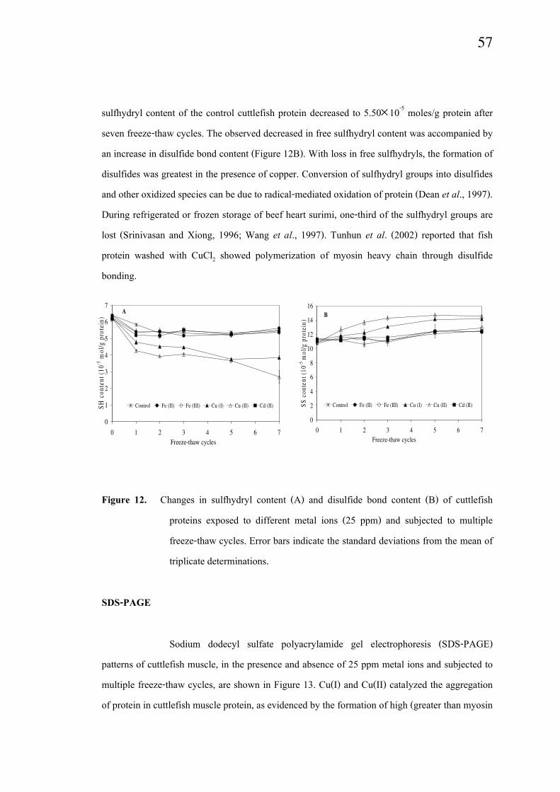
Sulfhydryl and disulfide bond content
The impacts of metal ions and freeze-thaw cycling on free sulfhydryl and
disulfide bond contents in cuttlefish protein are shown in Figure 12. Neither free sulfhydryl nor
disulfide bond concentrations in samples containing 5 ppm of the metal ions were different from
the control (data not shown). In the presence of 25 ppm metals, the free sulfhydryl content of
cuttlefish muscle protein decreased after one freeze-thaw cycle (Figure 12A). The extent of
decrease was dependent on the metal ion and copper caused the greatest loss of free sulfhydryls.
Free sulfhydryl contents in the control, Fe(II)-, Fe(III)- and Cd(II)-containing samples were
constant after one cycle of freeze thawing. After seven freeze-thaw cycles, Cu(I)- and Cu(II)-
treated samples had 3.84 and 2.68×10⁻⁵ moles free sulfhydryls/g protein, respectively. The free
0
20
40
60
80
100
120
140
0 1 2 3 4 5 6 7Freeze-thaw cycles
So
AN
S
Control Fe (II) Fe (III) Cu (I) Cu (II) Cd (II)
Page 14
57
sulfhydryl content of the control cuttlefish protein decreased to 5.50×10⁻⁵ moles/g protein after
seven freeze-thaw cycles. The observed decreased in free sulfhydryl content was accompanied by
an increase in disulfide bond content (Figure 12B). With loss in free sulfhydryls, the formation of
disulfides was greatest in the presence of copper. Conversion of sulfhydryl groups into disulfides
and other oxidized species can be due to radical-mediated oxidation of protein (Dean et al., 1997).
During refrigerated or frozen storage of beef heart surimi, one-third of the sulfhydryl groups are
lost (Srinivasan and Xiong, 1996; Wang et al., 1997). Tunhun et al. (2002) reported that fish
protein washed with CuCl2 showed polymerization of myosin heavy chain through disulfide
bonding.
Figure 12. Changes in sulfhydryl content (A) and disulfide bond content (B) of cuttlefish
proteins exposed to different metal ions (25 ppm) and subjected to multiple
freeze-thaw cycles. Error bars indicate the standard deviations from the mean of
triplicate determinations.
SDS-PAGE
Sodium dodecyl sulfate polyacrylamide gel electrophoresis (SDS-PAGE)
patterns of cuttlefish muscle, in the presence and absence of 25 ppm metal ions and subjected to
multiple freeze-thaw cycles, are shown in Figure 13. Cu(I) and Cu(II) catalyzed the aggregation
of protein in cuttlefish muscle protein, as evidenced by the formation of high (greater than myosin
0
1
2
3
4
5
6
7
0 1 2 3 4 5 6 7
Freeze-thaw cycles
SH
co
nte
nt
(10
-5 m
ol/
g p
rote
in)
Control Fe (II) Fe (III) Cu (I) Cu (II) Cd (II)
A
0
2
4
6
8
10
12
14
16
0 1 2 3 4 5 6 7
Freeze-thaw cycles
SS
co
nte
nt
(10
-5 m
ol/
g p
rote
in)
Control Fe (II) Fe (III) Cu (I) Cu (II) Cd (II)
B
Page 15
58
heavy chain, MHC) and intermediate (between actin and paramyosin) molecular weight polymers
with concomitant decreases in myosin, actin and troponin T band intensities (Figure 13A). The
disappearance of polymers and the reappearance of myosin, actin and troponin T in the presence
of reducing agents suggested that the polymers were formed via disulfide linkages between these
proteins (Figure 13B). Similar changes in electrophoretic patterns were also reported (Srinivasan
and Hultin, 1997) in oxidized cod muscle. No fragmentation could be observed in any of the
cuttlefish protein samples. Srinivasan and Hultin (1997) also did not observe fragmentation in
oxidized cod protein.
1 2 3 4 5 6 7
MHC
Paramyosin
Actin
MHC
Paramyosin
Actin
A
B
Troponin TTropomyosin
Troponin TTropomyosin
Figure 13. SDS-PAGE pattern of cuttlefish mince with subjected to different metal-ions (25
ppm) and subjected three freeze-thaw cycles, lane 1: non frozen cuttlefish, 2:
Control (non added metal ion), lane 3: Fe(II), lane 4: Fe(III), 5: Cu(I), 6: Cu(II)
and 7: Cd(II); A= Non-reducing condition, B= Reducing condition.
ATPase activity
Page 16
59
Ca2+-ATPase, Mg
2+-ATPase, Mg
2+-Ca
2+-ATPase and Mg
2+-EGTA-ATPase
activities can be used as indicators for chemical or structural changes in myosin, actin, actin-
myosin and troponin-tropomyosin, respectively (Azuma and Konno, 1998; Benjakul et al., 1997;
Roura and Crupin, 1995). A decrease in ATPase activity can be due to conformational changes in
the myofibrillar proteins (Okada et al., 1986) as well as protein-protein polymerization (Benjakul
and Bauer, 2000). ATPase activity in cuttlefish proteins subjected to freeze-thaw cycling, in the
presence and absence of metal ion, are depicted in Figure 14. As seen with the other
measurements of protein oxidation, Cu(II) and Cu(I) caused the greatest loss of activity in all the
different measurements of ATPase activities. Copper was able to decrease all the ATPase
activities measured, suggesting that myosin, actin, actin-myosin and troponin-tropomyosin were
all susceptible to oxidation. The oxidation of these myofibrillar proteins, as measured by loss of
ATPase activity, was in good agreement with the SDS-electrophoresis data (Figure 13) which
also showed that actin, myosin and troponin T were susceptible to copper-induced oxidation. Of
all the ATPase activities, copper was able to decrease Ca2+-ATPase activity most effectively,
suggesting that myosin was the most susceptible to Cu-induced oxidation. Mg2+-ATPase activity,
in actomyosin extracted from mantles of squid, decreased during 3 months of frozen storage
(Joseph et al., 1997; Paredi and Crupkin, 1997). Igochi et al. (1981) reported a slight decrease in
the Ca2+-ATPase of squid actomyosin caused by freezing. A decrease in Ca
2+-sensitivity indicates
the loss in Ca2+ regulation of tropomyosin (Ebashi et al., 1968; Benjakul et al., 1997). The Ca
2+-
sensitivity of all samples decreased with increasing numbers of freeze-thaw cycles (Figure 15).
Cu-treated samples showed the most dramatic decrease in Ca2+-sensitivity, especially, with
repeated freeze-thaw cycling. These results suggested that copper was able to alter the biological
activity of tropomyosin, even though no changes in its molecular weight could be observed by
electrophoresis (Figure 13).
Solubility
Decrease in protein solubility is often used as a marker of oxidative deterioration
of muscle protein quality (Decker et al., 1993; Xiong and Decker, 1995; Srinivasan and Hultin,
1997). Solubilities of cuttlefish proteins in 0.6 M KCl in the presence of different metal ions
Page 17
60
during multiple freeze-thaw cycles are depicted in Figure 16. Protein solubility decreased only
slightly during freeze-thaw cycling with the exception of samples treated with copper. Unlike
other measurements of protein oxidation, a large difference between the activities of Cu(I) and Cu
(II) was observed with Cu(II) causing a larger decrease in protein solubility. Thermodynamically,
a decrease in protein solubility is the result of a shift from a tendency of proteins to interact with
water towards a situation where proteins interact with each other (Vojdani, 1996). The decrease in
salt-soluble protein concentrations in copper-treated samples was in agreement with other protein
oxidation data, which showed that copper induced an increase in surface hydrophobicity (Figure
11), disulfide bond formation (Figure 12B) and protein polymerization (Figure 13). Moral et al.
(1983) reported a 60% or greater decrease in protein solubility in gutted muscle of squid (Loligo
valgaris) after 13.5 months of frozen storage. A gradual decrease in protein extractability during
frozen storage of squid (Loligo duvauceli) and voladur (Illex coindentii) was also reported
(Joseph et al., 1997; Ruiz-Capillas et al., 2002).
0.00
0.05
0.10
0.15
0.20
0.25
0.30
0 1 2 3 4 5 6 7
Freeze-thaw cycles
Act
ivit
y (
um
ol
Pi/
mg
pro
tein
/min
)
Control Fe (II) Fe (III) Cu (I) Cu (II) Cd (II)
A
0.00
0.05
0.10
0.15
0.20
0.25
0.30
0 1 2 3 4 5 6 7Freeze-thaw cycles
Act
ivit
y (
um
ol
Pi/
mg
pro
tein
/min
)
Control Fe (II) Fe (III) Cu (I) Cu (II) Cd (II)
B
0.00
0.05
0.10
0.15
0.20
0.25
0.30
0 1 2 3 4 5 6 7
Freeze-thaw cycles
Act
ivit
y (
um
ol
Pi/
mg
pro
tein
/min
)
Control Fe (II) Fe (III) Cu (I) Cu (II) Cd (II)
C
0.00
0.02
0.04
0.06
0.08
0.10
0.12
0.14
0 1 2 3 4 5 6 7
Freeze-thaw cycles
Act
ivit
y (
um
ol
Pi/
mg
pro
tein
/min
)
Control Fe (II) Fe (III) Cu (I) Cu (II) Cd (II)
D
Page 18
61
Figure 14. Changes in ATPase activity of cuttlefish proteins exposed to different metal ions (25
ppm) and subjected to multiple freeze-thaw cycles, A: Ca2+-ATPase Activity, B:
Mg2+-ATPase Activity, C: Mg
2+-Ca
2+-ATPase Activity, D: Mg
2+-EGTA-ATPase
Activity. Error bars indicate the standard deviations from the mean of triplicate
determinations.
Figure 15. Changes in Ca2+-sensitivity of cuttlefish proteins exposed to different metal ions
(25 ppm) and subjected to multiple freeze-thaw cycles. Error bars indicate the
standard deviations from the mean of triplicate determinations.
0
20
40
60
80
100
0 1 2 3 4 5 6 7
Freeze-thaw cycles
Ca2
+se
nsi
tiv
ity
(%
)
Control Fe (II) Fe (III) Cu (I) Cu (II) Cd (II)
0
20
40
60
80
100
0 1 2 3 4 5 6 7
Freeze-thaw cycles
% S
olu
bil
ity
Control Fe (II) Fe (III) Cu (I) Cu (II) Cd (II)
Page 19
62
Figure 16. Changes in protein solubility of cuttlefish paste exposed to different metal ions (25
ppm) and subjected to multiple freeze-thaw cycles. Error bars indicate the
standard deviations from the mean of triplicate determinations.
3.5 Conclusion
Cuttlefish are susceptible to lipid oxidation, discoloration and loss of protein
functionality during frozen storage. The addition of metal ions to cuttlefish paste accelerated lipid
oxidation, discoloration and loss of protein functionality during freeze-thaw cycling, although the
components in the cuttlefish that were oxidized were highly dependent on the metal ion type. Iron
showed the highest prooxidant effect on lipid oxidation and discoloration, while copper mainly
caused alterations in the physical and chemical properties of cuttlefish muscle proteins. Binding
of copper to proteins could explain why copper promoted protein oxidation while subsequently
being unable to promote lipid oxidation since the protein binding could prevent copper-lipid
interactions. Only iron caused the formation of yellow pigments (increase in b*-value) in
cuttlefish paste, suggesting that lipid oxidation is more closely related to discoloration than is
protein oxidation. Since cuttlefish are susceptible to oxidation of both its lipids and proteins, this
suggests that both metals could be active prooxidants during storage.