19
Chapter 3 Isolation and Identification of Different Parasites of Freshwater Carp
Chapter 3
Isolation and Identification of
Different Parasites of
Freshwater Carp

Chapter 3
3.1 Introduction
The major carp farming mainly dominates fresh water aquaculture of West
Bengal. The most important prerequisite of fish production is availability of
healthy fish fingerlings of carps. It is evident from the available literature
that parasitic diseases caused significant damage in fish industry mostly
affecting the fry and fingerlings.
Some groups of parasites possess unique features that enable a diagnosis of
whole organisms to be made, many of these features may not be easily
observed or identified and the degree of taxonomic detail available may vary
between the major groups. Generally, identification of parasites based on
recognizing morphological detail and determining the life cycle of the
parasites in the fish. The host specificity of particular parasite, its habitat
along with water temperature and other environmental conditions, can also
provide additional clues to parasite’s identity. The location of the parasite in
or on the host is also important, as some parasites are only found in certain
organs or tissues.
3.2 Materials and Methods
3.2.1 Examination of host fishes and collection of parasites
Live host specimens namely, Labeo bata and Labeo calbasu were randomly
sampled and collected from the fish farms, ponds and near by markets of
Howrah, Hooghly, Purba and Paschim Medinipur, South and North 24
parganas and Nadia district. The hosts were examined immediately after
collection. The parasites were collected from infected areas for gross
observation and identification. At first, the external surface of the host body
including scales, fins, skin, fin- base etc. were examined by collecting the
mucus for ectoparasites. Gills were removed from the branchial cavity and
placed on a glass slide for microscopic examination. After examination of the
external surface, the organs were dissected to search out the internal
parasites. To investigate the body cavity and general viscera, the body of the
31

Chapter 3
host fish were dissected out for examination. The parietal peritoneal lining of
the body cavity, outer surface of the visceral organs and serous membranes
were examined for encysted larva.
3.2.3 Preparation of slide for myxozoan parasites
i. Myxosporidian cysts when found attached on the tissues like gills, fins,
scales, and body surface, were isolated carefully.
ii. Free floating cysts found in the gill were isolated with the help of dropper.
iii. For detailed study and taxonomic identification, fresh cysts were first
taken on clean grease free glass slides.
iv. The cysts were slightly ruptured on one end with a needle. The spores
released from the cyst were mounted with cover glass and examined.
v. When cysts were not found gill, body, and fin smears were prepared on
grease free clean slides with a drop of 0.5% NaCl solution and air-dried for
delection of parasites.
vi. For detection of iodinophyllic vacoules in the sporoplasm, fresh spores
sealed with cover slips, were treated with Lugol’s Iodine solution and
examined under oil immersion microscope.
vii. To detect any external shell envelopes surroundig the spores, Indian ink
method as described by Lom and Vavra (1963) were followed.
viii. Some of the fresh spores were treated with 5% potassium hydroxide
(KOH) solution or saturated aqueous solution of urea [CO(NH2)2] for the
extrusion of polar filament.
ix. Permanent mounting of myxosporen parasites were done by staining with
Giemsa. Airdried smears on grease free clean slides were treated with
acetone free absolute methyl alcohol for about eight minutes to fix the
parasites and again dried.
x. Now the stock solution of Giemsa was diluted with water in the ratio of 1:
2 and buffered at pH 7.2.
xi. The slides were then placed in the staining rack and covered with dilute
stain for forty to fifty minutes.
32

Chapter 3
xii. Finally, the slides were washed by pouring neutral distilled water or
buffer solutions until the colour did not turn to a noticeable extent and the
slides were dried in air.
xiii. The slides are now ready for examination under microscope and
photographs were taken with the help of Olympus phase contrast
microscope fitted with digital camera.
3.2.4 Preparation of slides for ciliophoran parasites
i. The gill arches were removed and rubbed against the surface of a clean,
grease free dry microscopic slide.
ii. The smear thus produced, was allowed to dry in air for three to five
minutes. Precautionary measures were taken for a minimum loss of water
from the gill tissues.
iii. Mucus collected from body surface was also treated the same way to
prepare body smear.
iv. The preparations were then subjected to silver impregnation after the
method of Klein (1958). The slides were stained with 2% silver nitrate
(AgNO3) for seven to ten minutes and rinsed three times with distilled water
to remove excess stain by holding the slides in slanting position.
v. The stained slides were then transferred to petri dishes filled with distilled
water up to the brim so as to immerse the slides completely.
vi. The petridishes were placed over white filter papers and kept into a small
UV sterilization chamber containing UV tube and irradiated for twenty five
to thirty minutes.
vii. The slides were finally air dried completely and mounted in D.P.X.
viii. During exposure to UV light, the smears turned brown as the silver in
argentophilic structures was reduced. The darker their appearance, the
better was the impregnation.
ix. To stain the trophonts of Ichthyophthirius, Giemsa stain was used.
Trophonts were collected by scrapping the gill and body surface of infected
fishes and smeared on grease free slides.
x. Dried smears were fixed in acetone free absolute methyl alcohol for ten
minutes and again dried.
33

Chapter 3
xi. Now the stock solution of Giemsa was diluted with water in the ratio of 1:
2 and buffered at pH 7.2.
xii. The slides were then placed on a staining rack and covered with dilute
stain for forty to fifty minutes.
xiii. Finally, the slides were washed by pouring neutral distilled water or
buffer solutions until the colour did not turn to a noticeable extent and the
slides were dried in air.
3.2.5 Preparation of monogenean parasites
i. Parasites in the cyst form were released from the cyst and preserved.
ii. Mucus and other dirt particles attached to the parasite body were
removed by vigorous irrigation with a narrow-mouthed dropper before
fixation.
iii. Digeneans and monogeneans were press fixed and stored in 5% NBF and
stained in Gowers carmine or Alum carmine.
iv. Properly stained specimens were dehydrated in alcohol series, cleared in
creosote and mounted in Canada balsam.
3.2.6 Preparation of crustacean parasites
Crustacean parasites were fixed in 7% NBF and later on washed properly
and transferred to 70% alcohol for long term preservation. For detailed
study, the parasites were washed and dissected in 50% aqueous lactic acid
using wooden slides (Humes and Gooding, 1994).
3.2.7 Measurement and figure
The measurements of the parasites were taken using a calibrated ocular
micrometer. Photomicrographs were taken with an Olympus phase contrast
microscope fitted with Olympus camera.
34

Chapter 3
3.3 Results
3.3.1 Isolation and identification of myxozoan parasites
Myxosporidians constitute typical fish parasites known to produce cysts on
different regions of the body and internal tissues and organs. Myxozoans are
an extremely abundant and diverse group of organisms, speciated by spore
shape and size. The common myxosporidians genera are Myxoobolus,
Henneguya and Thelohanellus etc. Symptoms of this infestation include
weakness, emaciation, raising of the scales etc.
3.3.1.1 CLASSIFICATION OF THE PHYLUM MYXOZOA
The system of classification presented here is based on that of Kent et al.
(2000b) with the addition of the Class Malacosporea.
Phylum: Myxozoa Grasse, 1970
Class: Myxosporea Butschli, 1881.
Order: Bivalvulida Schulman, 1959.
Sub-order: Sphaeromyxina Lom and Noble, 1984 with one family and one genus Sphaeromyxa.
Sub-order: Variisporina Lom and Noble, 1984 with ten familie and thirty three genera e.g. Myxidium, Ceratomyxa, Sphaerospora.
Sub-order: Platysporina Kudo, 1919 with one family and thirteen genera e.g. Myxobolus, Henneguya, Thelohanellus.
Order: Multivalvulida Schulman, 1959 with six families and seven genera.
Class: Malacosporea Canning,Curry, Feist, Longshaw and
Okamura, 2000.
Order: Malacovalvulida Canning, Curry, Feist, Longshaw and Okamura, 2000, with one family and two genera Buddenbrockia and Tetracapsuloides.
35

Chapter 3
3.3.1.2 Myxosporidian belonging to the genus Myxobolus Butschli,
1882.
Description of Myxobolus sp.I
The species has been described from Labeo calbasu collected collected from
Naihati, North 24 Parganas, West Bengal. These were isolated from the gills
of the infected fish. Plasmodia were creamy white in colour and rounded in
shape (2-3 mm in diameter). It contains both late developmental stages and
mature spores. Mature spores are slightly rounded to oval shaped and
anterior and posterior ends blunt semicircular. The two polar capsules are
unequal in size. The polar filaments are not extruded out. Inside the polar
capsules there are 8-9 coils in case of larger polar capsules and 4-5 coils
were found inside the smaller polar capsules (Fig 3.1A and 3.1B).
Table3.1. Statistical analysis of measurement of the spores of Myxobolus sp.I
Spore Index:
LS: BS = 1: 0.728 LLPC: BLPC = 1:0.642 LSPC: BSPC = 1: 0.696 LLPC: LSPC = 1: 0.599 BLPC: BSPC = 1: 0.651
36

Chapter 3
Fig 3.1A and 3.1B: Myxobolus sp.I
Description of Myxobolus potaili Lalitha Kumari, 1969
The species has been observed from the freshwater carp Labeo calbasu
collected from Kalyani, Nadia, West Bengal. White rounded plasmodia are
observed attached with the gills of the host body. Mature spores are pear
shaped measuring [7.13±0.14 (6.9-7.1) X 4.69±0.44 (4.1-5.1)] with rounded
posterior and blunt anterior end (Fig 3.2A and 3.2B). The shape and size of
the specimen is similar to that of Myxobolus potaili described from Labeo
potail skyes. The shell valves are devoid of any partietal fold or marking (Fig
b). Two equal sized pyriform polar capsules measuring [3.48±0.4 (3-3.9) X
1.97±0.11 (1.8-2.1)] with greatly rounded posterior and sharply pointed
anterior end converge closely. There is no mucous envelop around the spore
as well as iodinophilous vacuole in the sporoplasm.
Fig 3.2A and 3.2B: Myxobolus potaili
37
Chapter 3
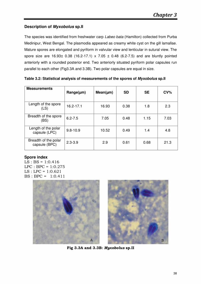
Description of Myxobolus sp.II
The species was identified from freshwater carp Labeo bata (Hamilton) collected from Purba
Medinipur, West Bengal. The plasmodia appeared as creamy white cyst on the gill lamallae.
Mature spores are elongated and pyriform in valvular view and lenticular in sutural view. The
spore size are 16.93± 0.38 (16.2-17.1) x 7.05 ± 0.48 (6.2-7.5) and are bluntly pointed
anteriorly with a rounded posterior end. Two anteriorly situated pyriform polar capsules run
parallel to each other (Fig3.3A and 3.3B). Two polar capsules are equal in size.
Table 3.2: Statistical analysis of measurements of the spores of Myxobolus sp.II
Measurements
Range(µm) Mean(µm) SD SE CV%
Length of the spore (LS)
16.2-17.1 16.93 0.38 1.8 2.3
Breadth of the spore (BS)
6.2-7.5 7.05 0.48 1.15 7.03
Length of the polar capsule (LPC)
9.8-10.9 10.52 0.49 1.4 4.8
Breadth of the polar capsule (BPC)
2.3-3.9 2.9 0.61 0.68 21.3
Spore index LS : BS = 1:0.416 LPC : BPC = 1:0.275 LS : LPC = 1:0.621 BS : BPC = 1:0.411
Fig 3.3A and 3.3B: Myxobolus sp.II
38
Chapter 3
Description of Thelohanellus sp.
The species has been identified from the freshwater carp Labeo calbasu
(Hamilton, 1822) collected from Naihati, West Bengal. The spores are
pyriform in shape and strikingly elongate with rounded posterior and a
blunt anterior proximity. There are no markings or folds on the valves.
Mature spores measure 13.68±0.69μm in length and 4.32±0.36 μm in
breadth.
Large sized pyriform polar capsule measuring 8.75±0.44 μm in length and
3.876±0.24 μm in breadth occupy a major portion of the spore cavity. There
are about seventeen to nineteen coils of the polar filament. When
completely extruded, anterior end of the long thread like polar filament
measuring 95.5±0.5 (92-95.8) μm appeared to be straight line (Fig 3.4A and
3.4B).
Table 3.3: Statistical analysis of measurements of the spores of Thelohanellus sp.
Measurements Range(µm) Mean(µm)
SD SE CV%
Length of the spore (LS)
12.8-14.2 13.68 0.69 1.6 5.1
Breadth of the spore (BS)
3.8-4.8 4.32 0.36 0.89 8.6
Length of the polar capsule
(LPC) 7.8-8.9 8.75 0.44 1.2 5.17
Breadth of the polar capsule
(BPC) 3.5-4.1 3.876 0.24 0.82 6.44
Length of the polar filament
(LPF) 92-95.8 95.5 0.5 1.3
39

Chapter 3
Fig 3.4A and 3.4B: Thelohanellus sp.
3.3.2 Isolation and identification of ciliophoran parasites
Most of the ciliate protozoans have tiny hair like structures called cilia that
are used for locomotion and/or feeding. Ciliates have a direct life cycle and
many are common inhabitants of pond-reared fish.
During the present study, members of three genera, viz. Trichodina, and
Trichodinella, Tripartiella, have been identified from fresh water carp fishes.
Ichthyophtheirius multifilis causing ich or white spot disease in fishes was
also observed.
Trichodiniasis disease is caused by trichodinid ciliophorans. Clinically, fish
usually exhibit flashing and become lethargic. The skin may develop ulcers
and increase of mucus production. Though the masses of organisms are
attached by the adhesive discs and denticles of exoskeleton, there is
secondary hyperplasia and hypertrophy of the gill epithelium and underlying
epithelial cells undergo necrosis. Ichthyophthiriasis disease is caused in
carps by protozoan ciliate, Ichthyophthirius multifilis, which infect different
regions of the body externally. Whitish cysts appear on the skin, gill and
fins.
40

Chapter 3
3.3.2.1 CLASSIFICATION OF THE PHYLUM CILIOPHORA
The system of classification adopted in this review is based on that of Lynn,
2003. The phylum is divided into two subphyla: Postciliodesmatophora with
characteristic microtubular ribbons linking all kinetosomes in a kinety,
comprising two classes; and Intramacronucleata, in which macronuclear
division involves microtubules that lie inside it. This subphylum comprises
nine classes with a total of nineteen subclasses. The freshwater fish
inhibiting symbiotic or parasitic ciliates trichodinids are grouped under the
subclass Peritrichia of the class Oligohymenophorea, one of the major taxon
of the subphylum Intramacronucleata. Another important ciliate,
Ichthyophtherius is included under the class Prostomatea.
Phylum: Ciliophora Doflein, 1901
Subphylum: Intramacronucleata Lynn, 1996
Class: Oligohymenophorea de Puytorac et al, 1974
Subclass: Peritrichia Stein, 1859
Order: Mobilina Kahl, 1933 (e.g. Trichodina sp., Tripertiella sp., Trichodinella sp.)
Class: Prostomatea Schewiakoff, 1896 (e.g. Ichthyophtherius sp.)
3.3.2.2 Trichodinid parasites
The genus Trichodina Ehrenberg, 1830 is identified with its adoral ciliary
spiral making a turn of 330°-540°. The denticulate ring composed of
denticles with straight or curved blades, distinct rays of various shapes and
lengths, and central parts lack and anteriorly directed projections. The
parasites are found in gills and skins of the host fish.
Description of Trichodina nandusi
The species has been identified from Labeo bata collected from Kalyani,
Nadia, West Bengal. These are medium sized trichodinids measuring 42.1-
41

Chapter 3
53.0(47.1±3.4) μm in diameter and are disc shaped. Denticulate ring
consists of 20.2-28.5(24.1±1.1) μm large sized denticles measuring
12.5±0.6μm in span and 5.2±0.2μm in length. There are 5-9 (6.7±0.8μm)
radial pins per denticle (Fig 3.5). The species was identified by the presence
of central clear area which is subdivided into many small granular
structures and spatulate rays.
Fig 3.5: Trichodina nandusi
Description of Tripertiella bulbosa
The species is identified by the elongated blade with parallel lateral margins,
which are constricted at either end, and is joined at the centre by a
prominent constriction at the mid length of the blade. It has been observed
from Labeo bata. These are free moving disc shaped trichodinid with a
diameter of 15.5-20.2(17.7±1.8). Around the adhesive disc measuring 12.5-
18.1(15.2 ± 2.2), there is a finely starited border membrane. The central area
of the disc is finely granular with a diameter of 4.1-6.5(5.5±0.8). Denticulate
ring consists of 22-25(23±1.8) denticles and 3-5 radial pins per denticle
(Fig3.6). The species is identified by the presence of parallel lateral margins
of the spherical blade which is attached to the anterior projection through
its stem-like narrow basal part.
42

Chapter 3
Fig 3.6: Tripertiella bulbosa
Description of Ichthyophthirius multifilis Fouquet, 1876
The spherical to ovoid trophonts may reach 1mm or more in diameter, have
short cilia covering the entire surface and have a single horseshoe-shaped
macronucleus and a single round or oval micronucleus which is sometimes
visible under 100 x magnification (Fig 3.7) and a smaller rounded
micronuclus which is visible in stained preparation.The adult parasite
moves slowly in a tumbling manner. The immature forms (tomites) are
smaller, translucent, and move quickly.
Fig 3.7: Ichthyophthirius multifilis
43

Chapter 3
3.3.3 Isolation and identification of monogenean parasites
Monogeneans are a class of parasitic flateworms or flukes commonly invade
the gills, skin and fins of fish. Monogeneans have direct life cycles (no
intermediate host) and are host and site-specific. These parasites are still
widespread in freshwater wildlife, farm fishes and marine habitats.
Gyrodactylus and Dactylogyrus are the two most common genera of
monogeneans that infect freshwater fish. They differ in their reproductive
strategies and their method of attachment to the host fish.
Classification of Dactylogyrous sp. (According to Diesing, 1850)
Kingdom: Animalia
Phylum: Platyhelminthes
Class: Monogenea
Order: Monopisthocotylea
Family: Dactylogyridae
Genus: Dactylogyrus
Classification of Gyrodactylus sp. (According to Malmberg, 1957)
Kingdom: Animalia
Phylum: Platyhelminthes
Class: Monogenea
Order: Monopisthocotylea
Family: Gyrodactylidae
Genus: Gyrodactylus
44
Chapter 3
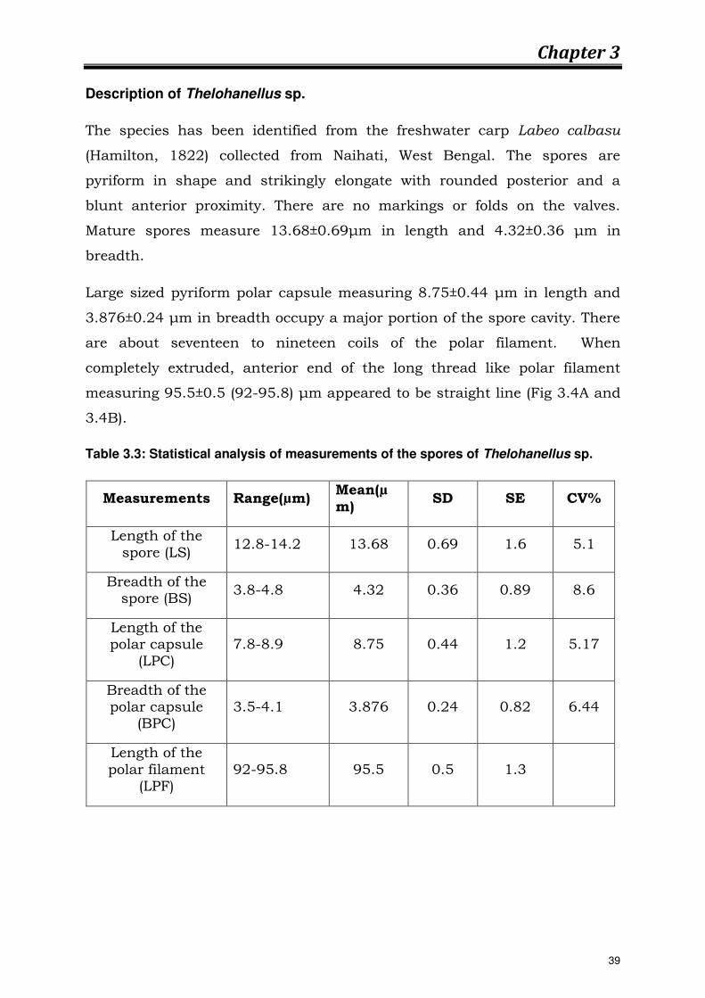
Description of Dactylogyrous sp.
The genus Dactylogyrus is found on the gills of mostly cyprinid fishes.
Dactylogyrus is recognized by a four-lobed head with four eye spots. The
average length of this species is 1.2 mm and width 0.33 mm. Body is short
and flattened, with uniform width throughout, but narrowing towards both
anterior and posterior ends. Haptors are slightly separated. Anchors are
bifid with well developed outer and inner roots and strongly recurved
pointed tips. Both dorsal and ventral connecting bars present (Fig 3.8).
When the worm is present in large numbers, gill hyperplasia and necrosis
can result.
Fig 3.8: Dactylogyrous sp.
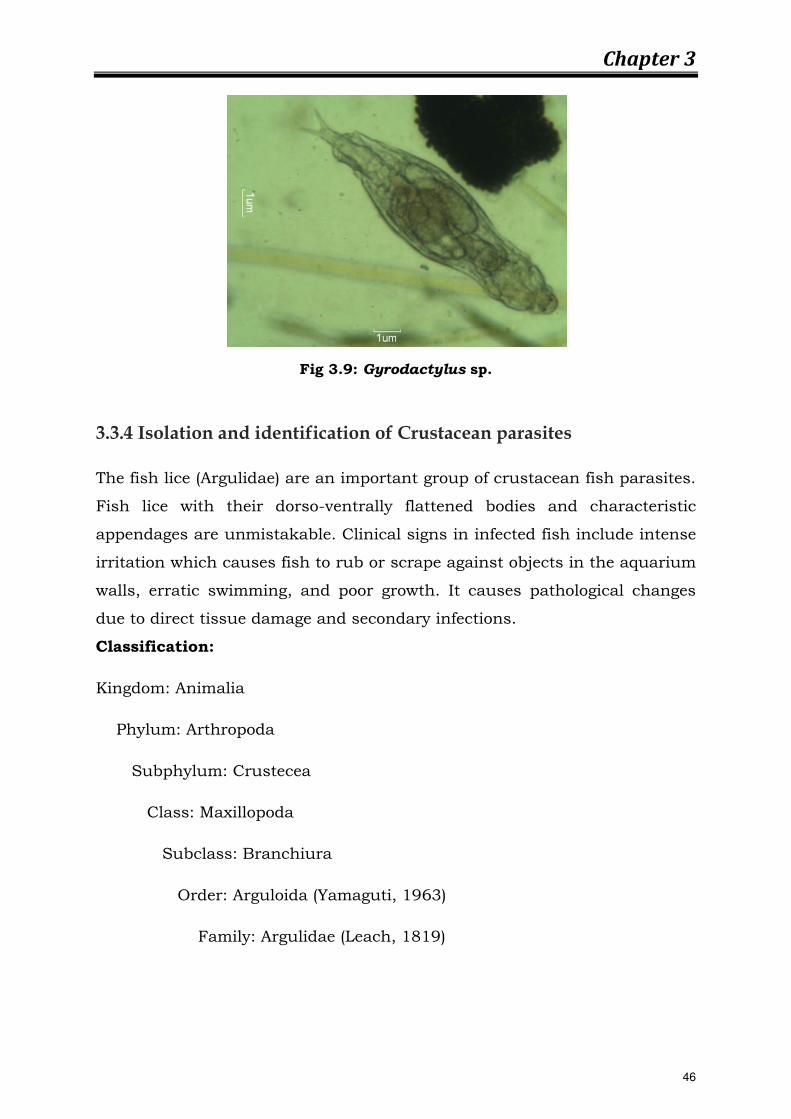
Description of Gyrodactylus sp.
The genus Gyrodactylus is a small monogenetic fluke attaches to gills, fins
and skin epithelium using an attachment organ known as an opisthohaptor
armed with a pair of large hooks and sixteen marginal hooklets. The average
length of this species is 0.75 mm. The head of the worm is bilobed, lacks
eyespots (3.9). Heavy infestations by the parasite can result in destruction of
the gills or skin epithelium due to mechanical damage caused by the
attachment organ.
45
Chapter 3
Fig 3.9: Gyrodactylus sp.
3.3.4 Isolation and identification of Crustacean parasites
The fish lice (Argulidae) are an important group of crustacean fish parasites.
Fish lice with their dorso-ventrally flattened bodies and characteristic
appendages are unmistakable. Clinical signs in infected fish include intense
irritation which causes fish to rub or scrape against objects in the aquarium
walls, erratic swimming, and poor growth. It causes pathological changes
due to direct tissue damage and secondary infections.
Classification:
Kingdom: Animalia
Phylum: Arthropoda
Subphylum: Crustecea
Class: Maxillopoda
Subclass: Branchiura
Order: Arguloida (Yamaguti, 1963)
Family: Argulidae (Leach, 1819)
46
Chapter 3
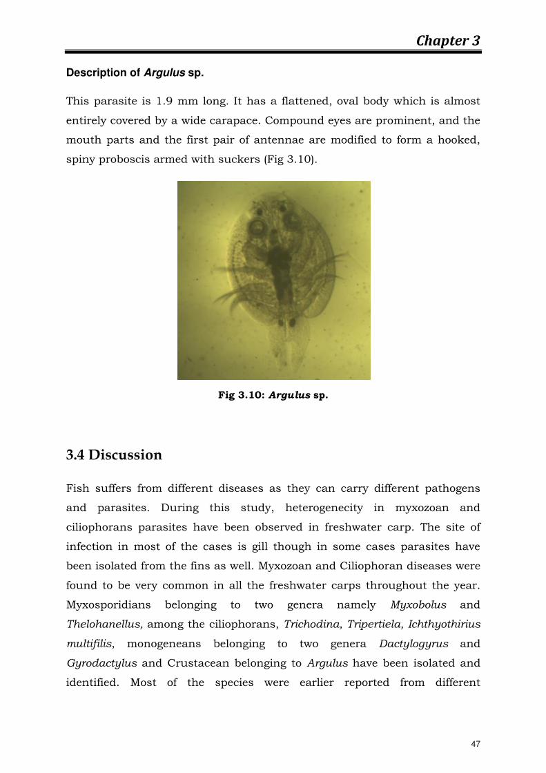
Description of Argulus sp.
This parasite is 1.9 mm long. It has a flattened, oval body which is almost
entirely covered by a wide carapace. Compound eyes are prominent, and the
mouth parts and the first pair of antennae are modified to form a hooked,
spiny proboscis armed with suckers (Fig 3.10).
Fig 3.10: Argulus sp.
3.4 Discussion
Fish suffers from different diseases as they can carry different pathogens
and parasites. During this study, heterogenecity in myxozoan and
ciliophorans parasites have been observed in freshwater carp. The site of
infection in most of the cases is gill though in some cases parasites have
been isolated from the fins as well. Myxozoan and Ciliophoran diseases were
found to be very common in all the freshwater carps throughout the year.
Myxosporidians belonging to two genera namely Myxobolus and
Thelohanellus, among the ciliophorans, Trichodina, Tripertiela, Ichthyothirius
multifilis, monogeneans belonging to two genera Dactylogyrus and
Gyrodactylus and Crustacean belonging to Argulus have been isolated and
identified. Most of the species were earlier reported from different
47

Chapter 3
geographical areas of India. Although they showed morphometric variations,
the overall characters were similar and could easily be identified. Some new
species with distinguished characteristics have also been observed. The
abundance and diversity in Myxobolus sp. was much more than the other
genus. Only a single speceis of Thelohanellus sp. have been observed.
Numerous members of ciliates Trichodina, and Tripartiella have been
observed and identified by their characteristic features during the course of
study. All the species was previously described by different authors from
India as well as abroad. A majority of freshwater fishes carry heavy infection
of parasites which cause deterioration in the food value of fish and may even
result in their mortality. These parasites use the fish for their shelter and
food and destruct more or less each and every organ resulting in pathogenic
effects (Lilley et al., 1992). Parasites interfere with the nutrition, metabolism
and secretary function of alimentary canal, damage nervous system and
even upset the normal reproduction of the hosts (Rahman et al., 1998a, b).
The distribution of these parasites of the same host and their incidence and
intensity of infestation varies from one place to another. Fish diseases are
the great threat in our fish culture system. Many fish species affects by
various types of diseases every year and as a result, production of fishes
decreases significantly. Proper steps should be taken to prevent fish
diseases and to protect these important fish species from extinction. From
the study it was observed that the parasites were most important pathogen
for disease outbreak.
48


