17
Chapter 4. Effect of ‘trans-acting elements’ Page 89 Chapter 4 Effect of ‘trans-acting elements’ on substrate half-life

Chapter 4. Effect of ‘trans-acting elements’ Page 89
Chapter 4
Effect of ‘trans-acting elements’ on substrate half-life

Chapter 4. Effect of ‘trans-acting elements’ Page 90
4.1. INTRODUCTION
Depending on their function, the half-life of proteins in a living cell ranges from few
seconds to several days. This controlled turnover of a protein is tightly regulated by the
proteasome machinery. However, the mechanism by which the half-life of a protein is
decided is still not very clear. Some of the short lived proteins carry a signature known
as degradation signals or degrons. In bacterial and archebacterial system, a degron
called SsrA is popular. SsrA is an 11 residue (AANDENYALAA) sequence, that when
fused to proteins with very long half-life in trans, shortens their half- life (Benaroudj
and Goldberg, 2000). For example green fluorescence protein (GFP) is very stable in
the cell while, GFP SsrA is relatively unstable (Benaroudj and Goldberg, 2000;
Benaroudj et al., 2003). In eukaryotic system SsrA like sequences are not well
characterized. C-terminus of ornithine decarboxylase (ODC) may be considered as the
eukaryotic equivalent. ODC gets degraded by proteasome in an Ub-independent
manner. The C-terminal sequences of ODC and antizyme has been shown to be
important for this process (Chattopadhyay et al., 2001; Li and Coffino, 1993). The C-
terminal sequence of ODC has been shown to compete with Ub for proteasomal
interaction (Zhang et al., 2003). This C-terminal sequence was found to be rich in
proline (P), glutamic acid (E), serine (S), and threonine (T) called PEST. Apart from
ODC, other proteins with similar PEST rich sequences were found to exhibit less than 2
h of intracellular half-lives (Rogers et al., 1986). In addition to such signals, N-terminal
residues have also been shown to dictate half-life of proteins and these signals are
termed as ‘N-end rule’ (Bachmair et al., 1986; Varshavsky, 1996). However, how
exactly these sequence help in shortening the half-life is still unanswered. There are
several hypothesis, for instance, the ‘degrons’ might help in substrate recognition and/or
act as initiators of degradation. The length, sequence, complexity and location of fusion

Chapter 4. Effect of ‘trans-acting elements’ Page 91
of these ‘degrons’ are other important factors. Most probably these ‘degrons’ are
context dependent, it is likely it may or may not help in degradation of all the test
proteins.
ApoMb was found self-sufficient to interact tightly with proteasome but degradation
was a long process. We therefore asked whether fusion of ‘degrons’ in ‘trans’ will affect
the half-life of apoMb. Some of the known PEST sequences as well as sequences
known to interact with proteasome were tested to address this.
4.2 MATERIALS and METHODS
4.2.1 PEST fusion to the C-terminus of Mb: The PEST sequences from ABCA1
(ATP dependent cassette transporter1), GCN4 (Yeast transactivator) and Hac1
(transcription factor) were fused to the C terminus of Mb cDNA, purified by ion-
exchange chromatography and tested for proteasomal degradation assay.
4.2.1A Insertion of PEST sequences in Mb: Synthetic sperm whale Mb cDNA was
cloned between PstI and KpnI site of pUC19. PEST sequences from GCN4 (apoMb
PEST 1), Hac1 (apoMb PEST 2) and ABCA1 (apoMb PEST 3) were converted to
nucleotide and codon preferred by bacterial system were used for the same. This was
then annealed to form a cassette. The cassette was prepared in a such way that it
contained nucleotide corresponding to PEST along with two stop codons and for
directional cloning, sticky end of BsteII (in Mb cDNA) and KnpI (vector) at the ends.
Material: Restriction enzymes (Fermentas) PstI, KpnI and BsteII, T4 DNA ligation kit
(Fermentas).
Single stranded oligonucleotides containing PEST sequence were synthesized in
such a way which when annealed to each other would generate a double stranded

Chapter 4. Effect of ‘trans-acting elements’ Page 92
cassette comprising of sticky ends corresponding to restriction enzymes BsteII and KpnI
(Table 4.1). 10 M of both the single stranded oligonucleotides were diluted in Tris
EDTA buffer (10mM Tris pH 8, 1mM EDTA), denatured at 95ºC for 5 min and
incubated at 72ºC for 10 min in a water bath. Following this, the water bath was turned
off to allow slow reduction of temperature to room temperature, ensuring proper
annealing of two strands. This double stranded cassette was annealed to BsteII and KpnI
digested pUC19 plasmid using T4 DNA ligase. The annealed product was transformed
in DH5α and colonies were screened for fusion product by restriction digestion using
PstI and KpnI enzymes. Positive clones were confirmed by gel shift as compared to
wild type (wt Mb =500 bp). The fusion of all the PEST sequences in Mb cDNA was
further verified by Sanger sequencing.
Table 4.1: Oligonucleotides used for PEST fusion in Mb
Name Primer sequence
PEST
1Mb F
GTTACCAGGGTAGCAGCAGCACCGATAGCACCCCGTAATGAGGTAC
PEST
1Mb R
CTCATTACGGGGTGCTATCGGTGCTGCTGCTACCCTG
PEST
2Mb F
GTTACCAGGGTCATAGCAGCAGCGATACCTTCACCCCGAGCCCGCTGAA
CTGCACCATGGAACCGGCGACCCTGAGCCCGTAATGAGGTAC
PEST
2Mb R
CTCATTACGGGCTCAGGGTCGCCGGTTCCATGGTGCAGTTCAGCGGGCT
CGGGGTGAAGGTATCGCTGCTGCTATGACCCTG
PEST
3Mb F
GTTACCAGGGTCGCCCGTTTACCGAAGATGATGCGGCGGATCCGAACGA
TAGCG ATATTGATCCGGAAAGCCGCGAAACCGATTAATGAGGTAC
PEST
3Mb R
CTCATTAATCGGTTTCGCGGCTTTCCGGATCAATATCGCTATCGTTCGGA
TCCGCCGCATCATCTTCGGTAAACGGGCGACCCTG
4.2.1B Expression and purification of PEST fused Mb: PEST fusion proteins were
expressed in DH5α as discussed above. Due to the fusion of PEST sequences, the pI of
the fusion protein might change. Therefore, depending on the calculated pI, the
purification strategy was designed. The calculated pI of PEST1 Mb was similar to wt.
Hence, it was purified by cation exchange chromatography. Since, the expression level
of PEST 2 Mb was suboptimal, several attempts to purify PEST2 Mb in optimum

Chapter 4. Effect of ‘trans-acting elements’ Page 93
amount failed. PEST3 Mb was purified by anion-exchange followed by gel-filtration
chromatography.
Material: DEAE cellulose (Sigma), 25 ml column (Bio-rad), Amicon ultra centrifugal
filter units (Millipore).
Essential buffer and reagents:
For DEAE cellulose regeneration: 0.1N NaOH containing 0.5 M NaCl, 0.5M NaCl, 0.1
M HCl containing 0.5 M NaCl and 1M NaCl pH 7-8 ( with NaOH).
Purification buffer: 10 mM sodium phosphate buffer pH 6.8
Elution buffer: 30 mM sodium phosphate buffer pH 6.8 supplemented with 25 mM or 50
mM NaCl.
Regeneration of DEAE resin: DEAE resin was suspended in MQ water (5 ml/g
bead) and poured in column. Resin was washed twice with 2 column volume (CV) 0.1
M NaOH containing 0.5 M NaCl followed by 0.5 M NaCl and 0.1 M HCl containing
0.5 M NaCl. Resin was washed with MQ until the effluent pH was 5 or greater. At this
stage resin can be stored in 1M NaCl pH 7-8 at 4°C (with preservative).
Note: A. If in the final stage, pH of effluent is not ~5, wash several times with MQ and
repeat the regeneration all over again.
B. Resin should not be left in NaOH or HCl for more than 30 min.
Partial purification PEST3 Mb by DEAE cellulose: DEAE cellulose resin was
equilibrated with 2 CV of 10X purification buffer followed by 5 CV of 1X purification
buffer. PEST3 Mb expressing cells were lysed by sonication, cleared lysate was loaded
on DEAE cellulose column, bound protein was washed with 10-15 CV of purification

Chapter 4. Effect of ‘trans-acting elements’ Page 94
buffer. PEST3 Mb was eluted in holo form (reddish brown in color) with the help of 30
mM sodium phosphate buffer supplemented with 25 or 50 mM NaCl.
PSET3 Mb purification by gel-filtration chromatography: Purity of Mb PEST3
eluted from DEAE cellulose column was analyzed on 15% SDS PAGE. To get rid of
contaminating proteins, eluted proteins were concentrated using Amicon filter units (3.5
kDa cut-off, Millipore) and further purified by gel-filtration chromatography. Gel-
filtration fractions were resolved on 15% SDS PAGE and pure protein fractions were
pooled together.
Mb PEST1 and Mb PEST3 were converted to apo form; heme removal was confirmed
by UV-Visible spectrometry and proteasomal degradation of PEST1 and PEST3 Mb
was done as described previously.
4.2.2 Fusion of probable ‘degron’ to C-terminus of Mb: We chose to test the effect
of the following trans-acting elements on apoMb half-life.
The C-terminus of ODC was shown to be essential for its ubiquitin independent
degradation. It was able to shorten the intracellular half-life of GFP when fused to its C-
terminus (Corish and Tyler-Smith, 1999).
Short sequences derived from N and C-terminus of proteins found to interact with 26S
proteasome by indigenous screening (Mb P8 and Mb P13).
Due to the fusion of sequences of varying length and character, the
physicochemical properties of Mb might change and interfere with purification. Hence,
we decided to switch to affinity purification of Mb using 6 X- histidine tags. The
cloning and purification strategy of Mb fusion is summarized in the following
schematic (Fig. 4.1).

Chapter 4. Effect of ‘trans-acting elements’ Page 95
Figure 4.1: Overview of cloning, expression and purification of Mb C-terminal fusion
proteins.
4.2.2A. Generating pRSTEV Vector: The pRSETA vector (Invitrogen) has 6X-
histidin tag the T7 gene, 10 leader sequences for high level of protein expression,
Xpress epitope and enterokinase (EK) recognition and cleavage sequence. All together
these components contribute to ~3 kDa extra sequences. In order to get tag free protein
(if needed) we replaced Xpress epitope, ER recognition and cleavage site with available
protease site (TEV or thrombin) using single step PCR without any cloning in single
shot, deletion of Xpress epitope and EK as well as insertion of TEV site was achieved
without affecting the MCS.
Material: pfu PCR kit (Fermentas), 25 mM dNTPs, DMSO, DpnI (Fermentas).
Bacterial strain: XL1 blue
A 50 l PCR reaction containing 50-60 ng template plasmid and control reaction
(without pfu) was set-up. The proofreading activity of pfu enzyme provided error free
amplification. The following PCR condition was used: initial denaturation- 95ºC for 5
min, denaturation- 95ºC for 1 min, annealing- 50ºC for 1 min, extension- 72ºC for

Chapter 4. Effect of ‘trans-acting elements’ Page 96
1min/kbp and number of cycle 19. The 10 l PCR product and control reaction (every
component except pfu enzyme) were resolved on 0.8% agarose gel. After confirming
amplification, DpnI digestion was setup in a 25 l reaction using 20 l of PCR product,
2 l of 10X Tango buffer and 10 units of DpnI for at least 8 h at 37ºC. DpnI would
digest the parental plasmid (cleave adenomethylated dam sites). The digested product
was then transformed in XL1 blue cells. The colonies were screened, plasmids isolated
and finally sequence verified for insertion of TEV recognition sequence and deletion of
EK site.
4.2.2B. Cloning wtMb in pRSTEV: The cDNA of Wt Mb was amplified from pUC
19 Mb and cloned in pRSTEV vector via PCR cloning vector pJET.
Material: pfu PCR kit, pJET 1.2/blunt PCR cloning kit, BamHI and KpnI from
Fermentas. Gene gel elute kit (Sigma).
Bacterial strain: DH5α
Mb was amplified using pfu enzyme and Mb cDNA cloned in pUC19 as
template. The following PCR condition was used: initial denaturation - 95ºC for 5 min,
denaturation- 95ºC for 1 min, annealing- 55ºC for 1 min, extension- 72ºC for 1 min/kbp
and number of cycle 29. Pfu polymerase does not have terminal transferase activity,
hence the blunt end PCR product was gel extracted using gene gel elute kit, ligated with
50 ng of linearized pJET vector and transformed in DH5α cells. pJET PCR cloning kit
utilizes insertional inactivation of the lethal eco47IR gene to obtain a positive clones.
Clones were screened for insert using BamHI and KnpI digestion and the insert was gel
extracted. The pRSTEV vector was serially digested with BamHI and KpnI, resolved on
0.8% agarose gel and gel extracted. The insert and vector were ligated with the help of
T4 DNA ligase. The ligated product was transformed in DH5α, colonies were screened
Chapter 4. Effect of ‘trans-acting elements’ Page 97
using BamHI and KpnI digestion and one of them was sequence verified for wtMb
sequence.
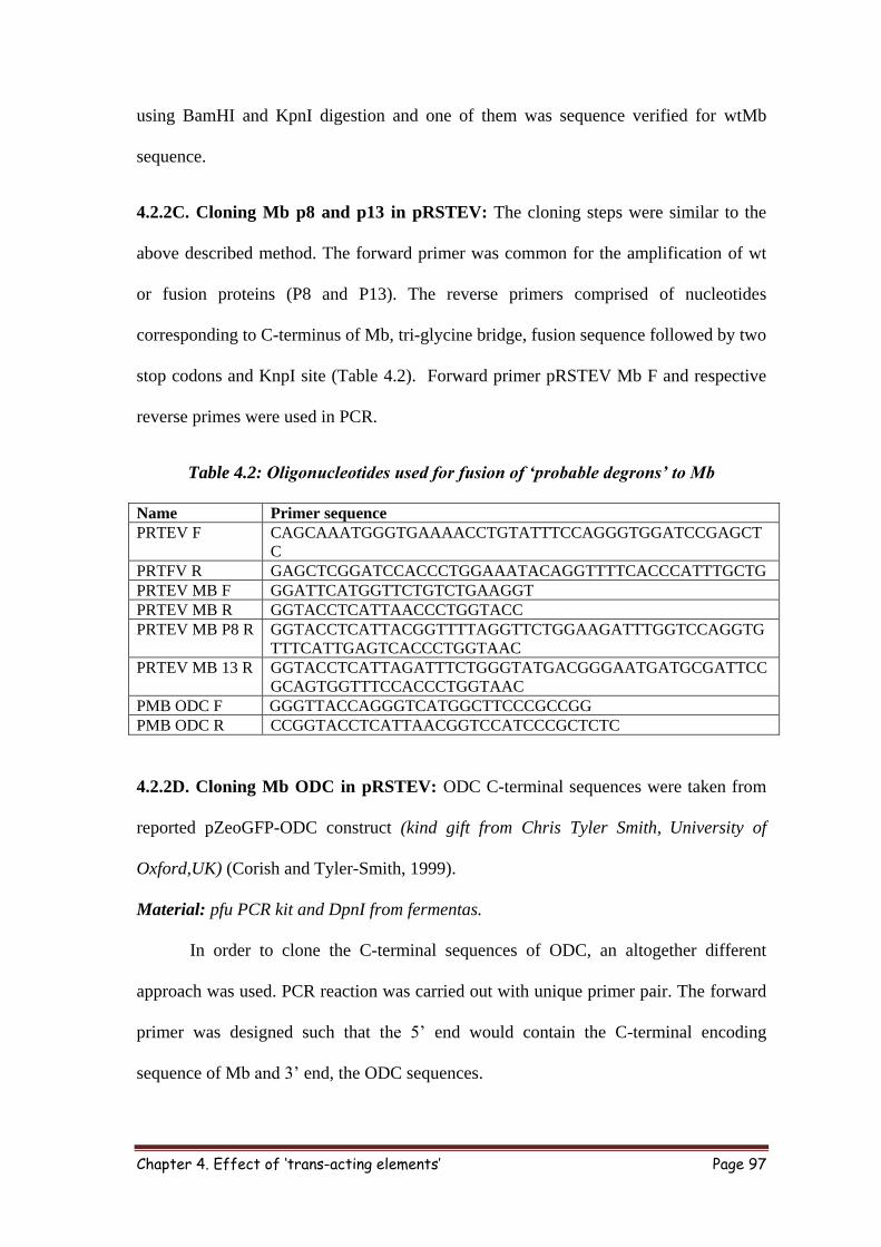
4.2.2C. Cloning Mb p8 and p13 in pRSTEV: The cloning steps were similar to the
above described method. The forward primer was common for the amplification of wt
or fusion proteins (P8 and P13). The reverse primers comprised of nucleotides
corresponding to C-terminus of Mb, tri-glycine bridge, fusion sequence followed by two
stop codons and KnpI site (Table 4.2). Forward primer pRSTEV Mb F and respective
reverse primes were used in PCR.
Table 4.2: Oligonucleotides used for fusion of ‘probable degrons’ to Mb
Name Primer sequence
PRTEV F CAGCAAATGGGTGAAAACCTGTATTTCCAGGGTGGATCCGAGCT
C
PRTFV R GAGCTCGGATCCACCCTGGAAATACAGGTTTTCACCCATTTGCTG
PRTEV MB F GGATTCATGGTTCTGTCTGAAGGT
PRTEV MB R GGTACCTCATTAACCCTGGTACC
PRTEV MB P8 R GGTACCTCATTACGGTTTTAGGTTCTGGAAGATTTGGTCCAGGTG
TTTCATTGAGTCACCCTGGTAAC
PRTEV MB 13 R GGTACCTCATTAGATTTCTGGGTATGACGGGAATGATGCGATTCC
GCAGTGGTTTCCACCCTGGTAAC
PMB ODC F GGGTTACCAGGGTCATGGCTTCCCGCCGG
PMB ODC R CCGGTACCTCATTAACGGTCCATCCCGCTCTC
4.2.2D. Cloning Mb ODC in pRSTEV: ODC C-terminal sequences were taken from
reported pZeoGFP-ODC construct (kind gift from Chris Tyler Smith, University of
Oxford,UK) (Corish and Tyler-Smith, 1999).
Material: pfu PCR kit and DpnI from fermentas.
In order to clone the C-terminal sequences of ODC, an altogether different
approach was used. PCR reaction was carried out with unique primer pair. The forward
primer was designed such that the 5’ end would contain the C-terminal encoding
sequence of Mb and 3’ end, the ODC sequences.

Chapter 4. Effect of ‘trans-acting elements’ Page 98
Figure 4.2: Cloning strategy of MbODC. With the help of specially designed primers
set comprising of ODC, C-terminal Mb (forward) and ODC, stop codon and vector
sequences (reverse), ODC sequence was amplified from pZeoGFP-ODC (A). This PCR
product was treated with DpnI and used as a primer for second step of PCR using
pRSTEV MB as template; this PCR step will allow the ODC sequence to fuse to Mb.
After DpnI digestion pRSTEV Mb ODC was used for fusion protein production (B).
The 3’end of the reverse primer on the other hand comprised of ODC sequences,
two stop codons and pRSTEV complimentary sequences (Fig. 4.2). Using this primer
set and pZeoGEP-ODC as template, PCR was done using following conditions: initial
denaturation - 95ºC for 5 min, denaturation- 95ºC for 1 min, annealing - 55ºC for 30
sec, extension- 72ºC for 1 min/kbp and number of cycle 29. The template (pZEO GFP-
ODC) was next digested by DpnI, leaving the unmethylated amplified product intact.
The amplified product harbored the ODC C-terminal sequence in between Mb (last few
residues) and pRSTEV vector sequences (few nucleotides after KpnI site). For second
round of PCR, 2l of this product was used as mega primers and pRSTEV Mb as
template. Following condition was used for PCR initial denaturation - 95ºC for 5 min,

Chapter 4. Effect of ‘trans-acting elements’ Page 99
denaturation- 95ºC for 1 min, annealing - 55ºC for 1 min, extension- 72ºC for 1 min/kbp
and number of cycle 19. After confirming amplification on 0.8% agarose gel, as
described above unmodified parental plasmid was digested using DpnI and transformed
in XL1 blue cells. The colonies were picked, plasmid DNA was extracted and digested
with BamHI and KpnI. One of the positive clones was verified by Sanger sequencing.
4.2.3 Expression and purification of wt and fusion proteins: Wt and mutant Mb were
transformed in BL21 DE3 cells. Growth condition and IPTG induction was optimized
for apoMb production.
Material: IPTG (Sigma), Ni-NTA-agarose (Invitrogen), 25 ml column (Bio-Rad), 10X
protease inhibitor cocktail (Sigma) and lysozyme (Sigma).
Bacterial strain: BL21 DE3.
Essential buffers and reagents:
5X Native purification buffer (100 ml): 250 mM Sodium phosphate monobasic
(NaH2PO4), 2.5 M NaCl, Add 90 ml of Milli Q water, adjust pH to 8 with NaOH. Make
volume to 100 ml.
1X Lysis buffer (20ml): 5X Native binding buffer (4 ml), 2 mM β-Mercaptoethanol,
0.1% Triton X -100, 10% Glycerol, and 1 X protease inhibitor (pH 8.0).
Binding buffer (50 ml): 1X Native binding buffer, 2 mM β-Mercaptoethanol, 0.1%
Triton X -100, 10% Glycerol and 10 mM imidazole, pH 8.0.
Wash buffer (50 ml): 1X Native binding buffer, 2 mM β-Mercaptoethanol, 0.1% Triton
X -100, 10% Glycerol and 20 mM imidazole, pH 8.0.
Elution buffer (50 ml): 1X Native binding buffer, 2mM β-Mercaptoethanol, 0.1% Triton
X -100, 10% Glycerol and 250 mM imidazole, pH 8.0.
Instrument: Ultrasonic homogenizer (300VT, BioLogics, Inc), Sorval RC 5C (for
centrifugation), Rotospin (Tarson).

Chapter 4. Effect of ‘trans-acting elements’ Page 100
The BL21 DE3 cells were transformed with pRSTEV Mb or mutant plasmid.
Single colony was inoculated in 10 ml of LB broth and incubated at 37ºC for 5 h, 2 ml
of this starter culture was inoculated in 1 l LB broth. Cells were grown up to A600nm 0.4-
0.6 at 37ºC then 100 μM IPTG was added to induce the recombinant protein expression.
The cells were further incubated for 16 h at 18°C and harvested by centrifugation.
Pellets were either stored at -80ºC freezer or used for purification, whereby the pellet
was suspended in 1X lysis buffer and incubated on ice for 30 min followed by cell lysis
using 5 mm sonication probe for 6 to 8 cycle of 1 min each (70% pulsar). Cell debris
was removed by centrifugation for 1 h at 18K and cleared lysate was stored on ice. The
Ni-NTA beads (2 to 3ml) were taken in 25 ml column washed several times with MQ
water and equilibrated with 15 ml of native binding buffer. The cleared cells lysate was
incubated with Ni-NTA column on rotospin at 15 rpm for 1 h at ~4ºC. The flow through
was collected to test the protein binding. The column was washed with 100 ml of 1X
wash buffer to remove impurity and protein was then eluted using imidazole. The
purification was checked on 15% SDS PAGE.
Note: A. Incubation on ice up to 2h after addition of lysis buffer require less sonication
cycle.
B. Used Ni-NTA beads can be regenerated by incubating it with 0.5 N NaOH for 30 min
and washing thoroughly with MQ water.
Protein purified by Ni-NTA affinity column were concentrated to about 2 mg/ml
and further purified by gel filtration chromatography as described earlier. The protein
fractions were collected and analyzed on 15% SDS PAGE. Pure protein fractions were
pooled together, concentrated and dialyzed overnight with 50 mM Tris pH 7.5.
4.2.4 Proteasome degradation assay: Apo form of wt and different Mb fusion protein
were tested for proteasomal degradation as described earlier.

Chapter 4. Effect of ‘trans-acting elements’ Page 101
4.3 RESULT and DISCUSSION
ApoMb was self-sufficient for proteasomal degradation. We wanted to test whether
trans-acting elements would affect the half-life of apoMb. The sequence, length,
complexity and location of fusion are not defined. Effect of most of the trans-acting
property is context dependent. To check the effect of some of the trans-acting elements
on Mb half-life, we chose to test PEST sequences from a few reported proteins. These
included known ‘degrons’ like C-terminal sequences of ODC, few sequences from N or
C-terminal peptides of proteins found to interact with proteasome obtained by in house
screening.
4.3.1. Cloning and purification of PEST sequence fused apoMb: To test the effect of
some of PEST sequences on apoMb half-life, we selected the reported PEST sequences
(8 to 24 residues) from ABCA1, GCN4, and Hac1 with intracellular half-life of less
than 1 h. Deletion of PEST sequences of these proteins resulted in intracellular
stabilization of these proteins (Kornitzer et al., 1994; Pal et al., 2007; Wang et al.,
2003). To fuse these sequences to the C-terminus of Mb cDNA, a cassette with PEST
sequence and sticky ends corresponding to BsteII and KpnI was designed and ligated to
linearized pUC 19 Mb (with same enzyme sets). The PEST sequence and
physicochemical property of PEST fused Mb is summarized in table 4.3. The calculated
pI of PEST 1 (GCN4) was similar to wt Mb. PEST1 was purified by cation-exchange
chromatography. Due to the fusion of PEST sequences, rich in acidic residues, the
calculated pI of Mb PEST3 (ABCA1) dropped to 6. PEST 3 was partially purified on
anion exchange resin followed by gel-filtration chromatography. PEST2 (Hac1) was
probably unstable in bacterial cells, several attempts to purify PEST2 Mb in optimum
amount failed. Hence, PEST 2 was not persuaded further.

Chapter 4. Effect of ‘trans-acting elements’ Page 102
Table 4.3: Effect of PEST fusion on physicochemical property of Mb
Protein Name of ‘PEST’ containing protein Calculated pI MW
(kDa)
Wt Mb 9 17.3
Mb
PEST1
GCN4 (Yeast trans activator)- 8 residue PEST
(SSSTDSTP)
8.7 18
Mb
PEST2
Hac1 (transcription factor)- 23 residue PEST
(HSSSDTFTPSPLNCTM EPA TLSP)
7.9 19.7
Mb
PEST3
ABCA1 (ATP dependent cassette transporter1) - 24
residue PEST (RPFTEDDAADPNDSDIDPE
SRETD)
6 20
4.3.2. Cloning and purification of ‘probable degrons’ fused apoMb: Ornithine
decarboxylase has been shown to be degraded with the help of antizyme by proteasome
in Ub independent manner. The intracellular half-life of C-terminal ODC sequences
fused GFP (to C terminus) has also been shown to be shorter than GFP alone (Corish
and Tyler-Smith, 1999). We wanted to test whether the C-terminus of ODC (with
critical Cys) would help Mb to be degraded faster by 26S proteasome.
In the quest of finding SsrA like sequences in eukaryotic system, short
sequences (15 residues long) from N and C-terminus of proteins were selected.
Considering that in most of the known proteasomal substrate, degradation starts from N/
C-termini and generally termini are more flexible in a protein, we designed an
indigenous screening method to identify sequences that may interact with immobilized
proteasome. Two peptides interacted tightly with proteasome (P8 and P13) and were
selected to test their ability to affect Mb half-life. The sequence and calculated pI of Mb
fusion protein is summarized in table 4.4. Fusion of these sequences will affect the pI of
the fusion protein. In order to maintain uniformity, we switched to affinity purification
of these fusion proteins using pRSET A 6X his tag purification vector (Invitrogen). It
contains IPTG inducible promoter for recombinant gene expression and apart from
other essential component of expression vector it has Xpress epitope and EK
recognition and cleavage site to get Tag free recombinant protein. The TEV protease

Chapter 4. Effect of ‘trans-acting elements’ Page 103
expression construct was available in lab. We first generated pRSTEV vector by
replacing Xpress epitope and EK site by TEV protease cleavage site without affecting
the multiple cloning site of pRSET A (Fig. 4.3A). This vector was used for expression
and purification of wt and C-terminal fusion Mb proteins.
Table 4.4: Sequence, calculated pI and M.W. of Mb fusion proteins
Name Fusion sequence MW
(kDa)
Calculated
pI
Wt NA 17.3 9
Mb ODC HGFPPEVEEQDDGTLPMSCAQESGMDR 21.9 6.39
Mb P8 DSMKHLDQIFQNLKP 19.1 8.96
Mb P13 GNHCGIASFPSYPEI 18.9 8.56
Figure 4.3: Generation of pRSTEV vector and Purification of Mb fusion protein: The
original pRSET A vector was modified to replace Xpress epitope and EK site with TEV
or thrombin cleavage site (A). Mb ODC was expressed in BL21 DE3 and purified by Ni-
NTA resin followed by gel-filtration chromatography (B). Mb ODC eluted from Ni-NTA
column, gel-filtration fractions and protein after dialysis was analyzed on 15% SDS
PAGE (C). The purified protein was found to be >95% pure.
These ‘probable degrons’ were cloned at the C-terminus of Mb in the modified
pRSTEV vector. Proteins were expressed in BL21 DE3. When IPTG was added at
higher cell density (A600 nm > 0.8) the Ni-NTA purified wt protein was in holo form

Chapter 4. Effect of ‘trans-acting elements’ Page 104
while at low cell density (A600 nm =0.4 to 0.6) it was in apo form. For all the fusion
proteins, expression was done at A600 nm~04. Wt and the fusion proteins were purified
by Ni-NTA chromatography followed by gel-filtration chromatography (Fig. 4.3B). Wt
as well as fusion proteins were >95% pure on SDS PAGE. MbODC Ni-NTA
purification and gel-filtration chromatogram has been shown as a representative of
fusion protein (Fig. 4.3C).
Figure 4.4: Effect of PEST sequences and ‘degrons’ on Mb half-life. The proteasomal
degradation of apo PEST 1 Mb or PEST 3 Mb was done and substrate remaining was
monitored, these PEST sequences were not able to shorten the half-life of apoMb (A).
Mb fusion proteins were subjected to proteasomal degradation assay and substrate
remaining at 12 h was monitored, none of the fusion protein tested helped in shorting
the half-life of Mb (B). In case of Mb ODC at 8 h and 12 h cleaved fragment was
observed.
4.3.3 Effect of PEST or ‘probable degron’ on apoMb half-life: The apo form of Mb
PEST 1 or 3 along with wtMb was incubated with proteasome. The half-life of wt and
both the PEST fused proteins were comparable. Similarly none of the probable degrons
tested were able to shorten the half-life of apoMb. In case of Mb ODC, truncated
product was observed at lower time scale, which might be due to cleavage or
degradation of ODC sequences.

Chapter 4. Effect of ‘trans-acting elements’ Page 105
4.4 SUMMARY
Although the role of ‘degron’ in the degradation step is not clear, few recent
observations suggest that long unstructured regions (90 to 120 residues) fused (in trans)
to proteins, render them susceptible for degradation (Prakash et al., 2009; Prakash et al.,
2004). These unstructured regions may help in initiation of degradation most likely by
helping the polypeptide to reach the active site. However, generation of such long
unstructured regions in a protein is rare other than the intrinsically disordered regions.
In contrast to these long unstructured regions short sequence of 11 residues like SsrA
has been shown to destabilize proteins like GFP rendering it susceptible to degradation
by homologous ATP dependent proteases like 20S-PAN complex from archaebacteria
and CLPX protease systems (Benaroudj and Goldberg, 2000; Benaroudj et al., 2003).
The exact role of such degrons is still unclear.
We tested the ability of short sequences which have the potential to act as
‘degrons’ by fusing them to the C-terminus of Mb. Fusion of short, trans acting
elements tested did not facilitate Mb degradation. Some of the possible explanations
could be -
a) Due to very high affinity of apoMb (nM) for proteasome, additional interactions
due to fusion of these elements may not facilitate degradation.
b) In apoMb, degradation might not be starting from C-terminus or else these
would work better when attached to the N terminus of Mb.
c) Local structural changes might be more important than fusing unstructured
sequences to the termini.


