• Discuss the risk factors, pathophysiology, assessment findings, and management of submersion and drowning.
• Identify the mechanical effects of atmospheric pressure changes on the body based on a knowledge of the basic properties of gases.
• Discuss the risk factors, pathophysiology, assessment findings, and management of diving emergencies and high‐altitude illness.
4
Thermoregulation
• Maintenance of body temperature, even under variety of external conditions– Body temperature is regulated in brain by thermoregulatory center
• Located in posterior hypothalamus
• Receives information from central thermoreceptors in or near anterior hypothalamus and from peripheral thermoreceptors in skin and some mucous membranes
• Peripheral thermoreceptors are nerve endings usually categorized as cold receptors and warm receptors
• Cold receptors are stimulated by lower skin‐surface temperatures
5
Thermoregulation• Maintenance of body temperature, even under variety of external conditions– Body temperature is regulated in brain by thermoregulatory center
• Warm receptors are stimulated by higher skin‐surface temperatures
• Information from these receptors is transmitted by spinal cord to posterior hypothalamus
• Posterior hypothalamus responds with appropriate signals to help body reduce heat loss and increase heat production (cold receptor stimulation) or increase heat loss and reduce heat production (warm receptor stimulation)
Why do you think the body has so many more cold receptors than heat
receptors?
7
Thermoregulation
• Central thermoreceptors
– Neurons sensitive to changes in temperature
• React directly to changes in temperature of blood
– Send messages to skeletal muscle through CNS
– Affect vasomotor tone, sweating, and metabolic rate through sympathetic nerve output to skin arterioles, sweat glands, and adrenal medulla
8
Thermoregulation• Thermoregulatory center has inherent set point
– Maintains relatively constant core body temperature (CBT) of 98.6°F (37°C)
– To maintain optimum environment for normal cell metabolism (homeostasis), body must keep CBT fairly constant, even when external and internal conditions tend to raise or lower it
– Body temperature can be increased or decreased in two ways
How does wearing the fully encapsulated hazardous materials suit affect your body’s ability to
regulate temperature?
19
Regulating Heat Loss
• Evaporation
– Process by which fluid changes from liquid to gas, and lowers temperature on surface where evaporation occurred
– When fluid evaporates, it absorbs heat from surrounding objects and air
• Temperature of surrounding air and relative humidity greatly affect amount of heat lost as result of evaporation of moisture from skin or respiratory tract (breathing)
20
Regulating Heat Loss
• Relative humidity is 100 percent when air is fully saturated with moisture
– Sweating can markedly increase evaporative heat loss so long as humidity is low enough to allow sweat to evaporate
– At humidity levels greater than 75 percent, evaporation decreases
– At levels approaching 90 percent, evaporation essentially ceases
• Heat illness results from one or two basic causes
– Temperature‐regulating mechanisms overwhelmed by high temperatures in environment or by excessive exercise in moderate to extremely high temperatures
– Temperature‐regulating centers that fail, usually in older adults or in ill or incapacitated patients
– Either cause can result in heat illness such as
• Heat cramps
• Heat exhaustion
• Heat stroke
25
Heat Cramps
• Brief, intermittent, and often severe muscular cramps that occur in hot environments
– Affect muscles fatigued by heavy work or exercise
– Primary cause of heat cramps is sodium and water loss
26
Heat Cramps
• Sweat profusely and drink water without adequate salt
– During times of high environmental temperatures, 1 to 3 L of water/hour can be lost through sweating
– Each liter contains 30 to 50 mEq of sodium chloride
– Water and sodium deficiency together cause muscle cramping
– Normally occurs in most heavily exercised muscles, including calves and arms
• When hypothermia is suspected, paramedic’s immediate action is to
– Extricate and evacuate patient to site of warm shelter
– Remove cold, wet clothing
– Prevent further drop in victim’s CBT
– Survey for traumatic injuries
– Cover patient with warm blankets and increase temperature in ambulance
– Rapidly and gently transport patient for definitive care
67
Hypothermia Management
• Rewarming techniques for managing patients with hypothermia are classified as– Passive
• Moving patient to warm environment
• Removing wet clothing
• Applying warm blankets
– Active external
– Active internal• Heating methods or devices such as radiant heat, forced hot air, warm water packs
68
Hypothermia Management
• Active internal rewarming is invasive– Some of these procedures can be performed in field, such as administering warmed IV fluids and providing warm, humidified O2
– Other procedures are reserved for in‐hospital care• Peritoneal and/or pleural lavage with warm fluids
• Use of esophageal rewarming tubes
• Cardiopulmonary bypass (active core rewarming)
• Extracorporeal circulation (blood warming with partial bypass)
• In mild cases of hypothermia, removal of victim from cold environment and passive rewarming may be all that is necessary to manage cold exposure– Can accomplish this by removing wet clothing
– Wrapping victim in dry blanket to prevent further chilling and help retain patient’s body heat
– If victim is conscious, warm drinks and sugar sources can support gradual rise in CBT and help correct any dehydration present
71
Mild Hypothermia
• Patients should not be permitted to– Smoke, which causes vasoconstriction
– Drink alcoholic beverages, which produce peripheral vasodilation and increase heat loss from skin
– Drink caffeine‐containing beverages, which cause vasoconstriction and diuresis
• May be lethargic and somewhat dulled mentally but generally are oriented with no marked mental derangements
• If patient’s CBT is less than 86.4°F (30°C), he or she usually is unconscious
– Gently move to warm environment if vital signs are present
– Institute passive and external rewarming
– Administer oxygen
– Transport
76
Severe Hypothermia
• If patient with moderate‐to‐severe hypothermia is in cardiac arrest (VF or pulseless VT), begin CPR and attempt defibrillation once– If patient does not respond to one shock, further defibrillation attempts should be deferred
– Care should be focused on• Effective CPR
• Rewarming patient
• Rapid transport
• In‐hospital active internal rewarming will be required for these patients
77
Severe Hypothermia
• Prolonged resuscitation can be beneficial in these patients
– CPR is indicated even if signs of death are present
– Resuscitation may be withheld if
• Victim has obvious lethal injuries
• Body is frozen so that nose and mouth are blocked by ice and chest compression is impossible
• Some physicians will not presume hypothermic patient to be dead until near‐normal CBT has been achieved and resuscitation efforts are still unsuccessful
• Prehospital care should focus on airway, breathing, and circulation with some modifications
– Pulse and respirations may be difficult to detect
• Vital signs (including ECG readings) should be assessed for 30 to 45 seconds to confirm need for CPR
• If there is any doubt as to presence of pulse, begin CPR immediately
Special Care Considerations for Patients with Hypothermia
79
• Prehospital care should focus on airway, breathing, and circulation with some modifications
– For unresponsive patients and those in arrest, endotracheal intubation is indicated
• Intubation will serve two purposes – Will enable provision of effective ventilation with warm, humidified oxygen (if available)
– Will isolate airway to reduce likelihood of aspiration
Special Care Considerations for Patients with Hypothermia
80
• Prehospital care should focus on airway, breathing, and circulation with some modifications – Hypothermic heart may be unresponsive to cardiovascular drugs
• Drug metabolism is reduced, allowing for toxic accumulation of drug in peripheral tissues
• For these reasons, IV drugs often are withheld when CBT is less than 30°C
• If CBT is greater than 30°C, IV drugs may be given, but with increased intervals between doses
• IV drug therapy should be guided by medical direction
Special Care Considerations for Patients with Hypothermia
• Under most conditions of frostbite, ice crystals form in extracellular tissue
– Draws water out of cells and into extravascularspaces
– As result, electrolyte concentration in cell can reach toxic levels
– Ice crystals can also expand and cause direct mechanical destruction of tissue
85
Frostbite Pathophysiology
• Under most conditions of frostbite, ice crystals form in extracellular tissue– Leads to
• Damage to blood vessels (particularly endothelial cells)
• Partial shrinkage and collapse of cell membrane
• Loss of vascular integrity
• Local edema
• Disruption of nutritive blood flow
– Ischemia often produces most damaging effects of frostbite
86
• When frozen tissue thaws, blood flow through capillaries is initially restored
– Blood flow declines within minutes after thawing
– Occurs as arterioles and venules constrict and release emboli, which travel through small vessels
– Progressive tissue loss results from thrombosis and hypoxia
– Endothelium is damaged and results in deterioration of microvasculature and dermal necrosis
– Process of thawing and refreezing is more harmful to tissue than allowing frostbitten part to remain frozen until it can be warmed with minimal risk of refreezing
• During transport, all restrictive and wet clothing should be removed from patient– Replaced with warm, dry clothing and blankets
– Patient prohibited from alcohol or tobacco
– Rapid rewarming of frozen part by immersion in hot water (maximum of 104°F [40°C]) is most effective therapeutic measure for preserving viable tissue
• Because of risk of refreezing, this method of rewarmingshould not be used in prehospital setting if transport will be delayed (e.g., backwoods rescue, natural disasters)
Frostbite Management
97
Submersion
• Drowning was fifth leading cause of unintentional death in U.S. in 2006
– Second leading cause of unintentional injury death among children and youth
– About 80,000 submersion incidents are reported each year
• Of these, 85 percent of victims are male, 2/3 of victims are nonswimmers
98
Classifications
• Defined as “process resulting in primary respiratory impairment from submersion/immersion in liquid”
– Submersion usually refers to head being below water
– Immersion refers to head being above water
• Terms often used interchangeably
• Definition further requires that liquid/air interface be present at entrance of victim’s airway, preventing victim from breathing air
– According to this definition, victim may live or die after this process; regardless, he or she has been involved in drowning event
– Resuscitation is indicated for all patients unless physical evidence of death is present (e.g., putrefaction, dependent lividity, and rigor mortis)
• Submersion victims who have spontaneous circulation and breathing when they reach hospital usually recover, with good outcomes
Factors that Affect the Clinical Outcome
112
• Cleanliness of water
– Contaminants in water have irritant effect on pulmonary system
– May lead to bronchospasm and poor gas exchange
– Can cause secondary pulmonary infection with delayed severe respiratory compromise
Factors that Affect the Clinical Outcome
113
• Temperature of water
– Submersion in cold water can have beneficial and negative effects
– Rapid onset of hypothermia can serve protective function• Especially with brain viability in patients who have undergone prolonged submersion
– Incident in which child was submerged for 66 minutes in creek with water temperature of 37°F (5°C) is longest documented submersion with good neurological outcome
• Victims of submersion incidents often are at risk for hypothermia
– Heat loss in water can be up to 32 times greater than in air
• Hypothermia can make resuscitation more difficult
– Calls for special consideration with regard to
• Gentle handling
• Administration of drugs
• Defibrillation
Submersion Management
121
• As with all other victims of hypothermia, remove patient’s wet clothing
– Patient then should be dried and wrapped in blankets to conserve body heat
– External warming and administration of heated, humidified oxygen at scene and during transport should be considered
Submersion Management
122
• According to AHA, “all victims of drowning who require any form of resuscitation (including rescue breathing alone) should be transported to the hospital”
– Asymptomatic patients require transport for physician evaluation
– Give oxygen
– Carefully monitor to guard against aspiration pneumonia and undetected hypoxia that can result from submersion
– Oxygen is most important treatment needed by submersion victims
What are the risks to rescuers on a call involving submersion victims?
124
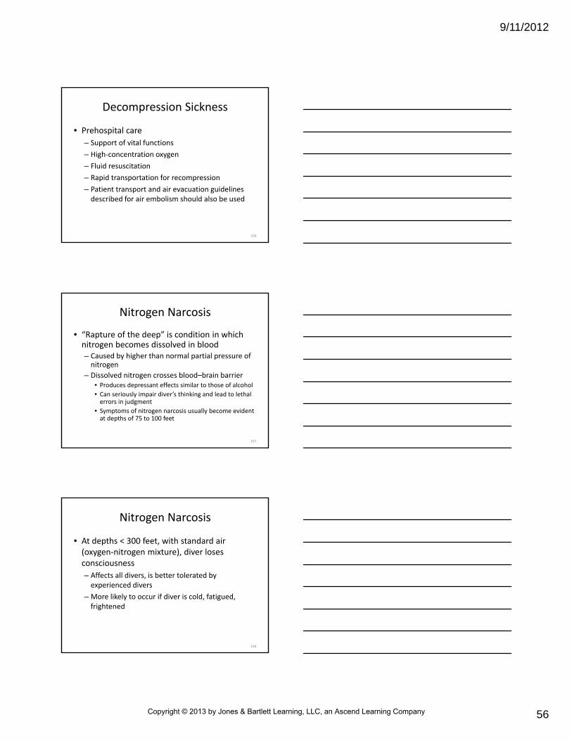
Diving Emergencies
• U.S. has over 4 million recreational scuba divers; more than 400,000 new sport divers are certified each year– Emergencies unique to pressure‐related diving include
• Those caused by mechanical effects of pressure (barotrauma)
• Air embolism
• Breathing of compressed air (decompression sickness and nitrogen narcosis)
125
Basic Properties of Gases
• Weight of atmosphere exerts a pressure of 14.7 pounds psi of force at sea level
– 1‐inch column of air as tall as atmosphere would weigh 14.7 pounds
– Weight is commonly referred to as 1 atmosphere of pressure (1 atm)
– Water weighs considerably more than air and can exert much more pressure
• Three laws of properties of gases underpin all pressure diving‐related emergencies (some high‐altitude illnesses)
– Boyle’s law
– Dalton’s law
– Henry’s law
127
Laws Pertaining to Gases
• Properties of gases can aid comprehension of these laws
– Increased pressure dissolves gases into blood
– Oxygen metabolizes
– Nitrogen dissolves
128
Boyle’s Law
• If temperature remains constant, volume of given mass of gas is inversely proportional to absolute pressure– When pressure is doubled, volume of gas is halved (compressed into smaller space), vice versa
– Expressed by equation PV = K• P is pressure
• V is volume
• K is constant
– Law explains “popping” or “squeezing” sensation in ears that person may feel when traveling by air
• Basic mechanism for all types of barotrauma trapped gases expand as pressure decreases
– Example
• When diver uses scuba tank of pressurized air, lung volumes remain constant at various depths
• If diver ascends but does not exhale, water pressure decreases and gas in lungs expands
• Greatly increases pressure in lungs
130
Dalton’s Law
• Pressure exerted by each gas in mixture of gases is same pressure that gas would exert if it alone occupied same volume
– On other hand, total pressure of a mixture of gases equals sum of partial pressures that make up mixture
– Expressed by equation Pt = Po2 + PN2 + Px
• Pt is total pressure
• Po2 is partial pressure of oxygen
• PN2 is partial pressure of nitrogen
• Px is partial pressure of remaining gases in mixture
131
Dalton’s Law
• To simplify, air we breathe is about 80 percent nitrogen and 20 percent oxygen– About 80 percent of pressure of air (i.e., gas mixture) is exerted by nitrogen in mixture
– About 20 percent of pressure is exerted by oxygen in mixture
– Means that at sea level, pressure exerted on us by nitrogen in air is 80 percent of 14.7, or 11.76 psi
– Pressure from oxygen is 20 percent of 14.7, or 2.94 psi
– Together, account for 14.7 psi of pressure at surface
• Even though gas mixtures remain with normal percentages of nitrogen and oxygen, partial pressures of these gases change at different altitudes above sea level or at depths below sea level– Principles of this law explain problems that can arise from breathing of compressed air
• Gas expansion causes partial pressure of oxygen to drop as gas molecules move farther apart, reducing available oxygen
133
Henry’s Law
• At constant temperature, solubility of gas in liquid solution is proportionate to partial pressure of gas– Means that more gas can be dissolved into liquid at higher pressure
– Less gas can be dissolved into liquid when that pressure is released
– Example• When container of carbonated beverage (pressurized with dissolved carbon dioxide gas) is opened, “pop” is heard, and bubbles form on liquid
• Occurs because pressure in container is no longer great enough to hold dissolved gas inside
134
Henry’s Law
• Expressed by equation %X = Px/Pt X 100
– %X is amount of gas dissolved in liquid
– Px is partial pressure of gas
– Pt is total atmospheric pressure
– Law explains why more nitrogen, which makes up almost 80 percent of air, dissolves in diver’s body as ambient pressure increases with descent
• Dissolved nitrogen is released from tissues on ascent as pressure decreases
• Tissue injury caused by change in pressure, which compresses or expands gas contained in various body structures
– Type depends on whether diver is in descent or ascent
– Most common injury of scuba divers
136
Barotrauma of Descent
• Barotrauma of descent (also known as squeeze)
– Results from compression of gas in enclosed spaces as ambient pressure increases with descent under water
– Air trapped in noncollapsible chambers is compressed
– Leads to vacuum‐type effect that results in
• Severe, sharp pain caused by distortion
• Vascular engorgement
• Edema
• Hemorrhage of exposed tissue
137
Barotrauma of Descent
• As a rule, squeeze usually results from blocked Eustachian tube or from failure of diver to clear (open) Eustachian tube with exhalation during descent– Ears and paranasal sinuses most likely to be affected
– Occurs in• Ears
• Sinuses
• Lungs and airways
• GI tract
• Thorax
• Teeth (pulp decay, recent extraction sockets or fillings)
• Occurs through reverse process of descent (“reverse squeeze”)
– Assuming that air‐filled cavities of body have equalized pressure during diver’s descent, volume of air trapped in this pressurized space expands as ambient pressure decreases with ascent (Boyle’s law)
142
Barotrauma of Ascent
• If air is not allowed to escape because of obstruction (e.g., breath‐holding, bronchospasm, or mucus plug), expanding gases distend tissues surrounding them
• Most common cause breath‐holding during ascent
143
Barotrauma of Ascent
• Problems from reverse squeeze are rare
– Pulmonary overpressurization syndrome (POPS) can occur as result of expansion of trapped air in lungs
• Can lead to alveolar rupture
• Can lead to leakage of air into areas outside alveoli
• Should be suspected if diver suddenly loses consciousness immediately after surfacing
– Begin BLS and ALS measures
– Rapidly transport for recompression treatment
– Thoroughly evaluate for signs of POPS, such as a pneumothorax
151
Air Embolism
• Patient suspected of having air embolism should be transported in horizontal, neutral position
– Helps to avoid aggravating cerebral edema that may develop
152
Air Embolism
• If air transport is used, should be transported by aircraft that is pressurized to sea level– Can be transported by rotary wing aircraft that flies at low altitude
– Prevents existing intra‐arterial air bubbles from expanding further
– Flight altitude must be as low as possible if internal cabin pressure cannot be maintained at sea level
• Use of elevated pressure (including hyperbaric oxygen therapy) to treat conditions within body caused by a rapid decrease in pressure (e.g., air embolism)
– Takes place in hyperbaric oxygen chamber
• Allow for delivery of oxygen at higher than normal atmospheric pressure
• Process is used to overcome natural limit of oxygen solubility in blood
154
Recompression
• Reduces intravascular bubble volume and restores tissue perfusion– Slow decompression helps to prevent bubbles from reforming
– Paramedics should know location of nearest hyperbaric treatment facility
– Follow protocol established by medical direction
– Ground transportation to hyperbaric facility is preferred over air transportation
• Increase in altitude lowers the ambient pressure and allows microbubbles to expand
• Heat illness results from one of two basic causes
– Normal temperature‐regulating functions can be overwhelmed by conditions in environment
• Conditions can include heat stress
• More often involve excessive exercise in moderate to extreme environmental conditions
– Failure of body’s thermoregulatory mechanism
• May occur in older adults or ill or debilitated individuals
• Heat cramps are brief, intermittent, and often severe, and are muscular cramps that occur in muscles fatigued by heavy work or exercise
187
Summary
• Heat exhaustion is characterized by minor aberrations in mental status, dizziness, nausea, headache, and a mild to moderate rise in the core body temperature (CBT) (up to < 103°F [39°C])
• Heat stroke occurs when the temperature‐regulating functions break down entirely
‒ Results in body temperature rises to 105.8°F (41°C) or higher
• Damage all tissues and lead to collapse
188
Summary
• Hypothermia (CBT lower than 95°F [35°C]) can result from decrease in heat production, an increase in heat loss, or a combination of these factors– Progression of clinical signs and symptoms of hypothermia is divided into three classes based on the CBT: mild (CBT between 93.2°F and 96.8°F [34°C and 36°C]), moderate (CBT between 86°F and 93°F [30°C and 34°C]), and severe (CBT below 86°F [30°C])
– Severely hypothermic patients have no vital signs, including respiratory effort, pulse, and blood pressure
– Results from environmentally induced freezing of body tissues
• Leads to damage to blood vessels
– Ischemia often produces the most damaging effects of frostbite
• In deep frostbite, this can include mummification and sloughing of nonviable skin and deep structures
190
Summary
• Drowning is a process that results in primary respiratory impairment from submersion/immersion in a liquid medium that presents the person from breathing air
– Regardless of the type of water aspirated, pathophysiology of drowning is characterized by hypoxia, hypercapnia, and acidosis, which result in cardiac arrest
191
Summary
• Three laws pertaining to the basic properties of gases that are involved in all pressure‐related diving emergencies are Boyle’s law, Dalton’s law, and Henry’s law
– Increased pressure dissolves gases into blood; oxygen metabolizes, and nitrogen dissolves