30
The Cardiovascular System and Its Control
| Date post: | 30-Dec-2015 |
| Category: |
Documents |
| Upload: | emerson-nieves |
| View: | 30 times |
| Download: | 0 times |

The Cardiovascular System and Its
Control

The Cardiovascular System:The Cardiovascular System:Major FunctionsMajor Functions
• Delivers O2, nutrients
• Removes CO2, other waste
• Transports hormones, other molecules
• Temperature balance and fluid regulation
• Acid-base balance
• Immune function
The Cardiovascular SystemThe Cardiovascular System
• Three major circulatory elements1. A pump (heart)
2. Channels or tubes (blood vessels)
3. A fluid medium (blood)
• Heart generates pressure to drive blood through vessels
• Blood flow must meet metabolic demands
The HeartThe Heart
• Four chambers– Right and left atria (RA, LA): top, receiving
chambers– Right and left ventricles (RV, LV): bottom, pumping
chambers
• Pericardium
• Pericardial cavity
• Pericardial fluid

Figure 6.1Figure 6.1
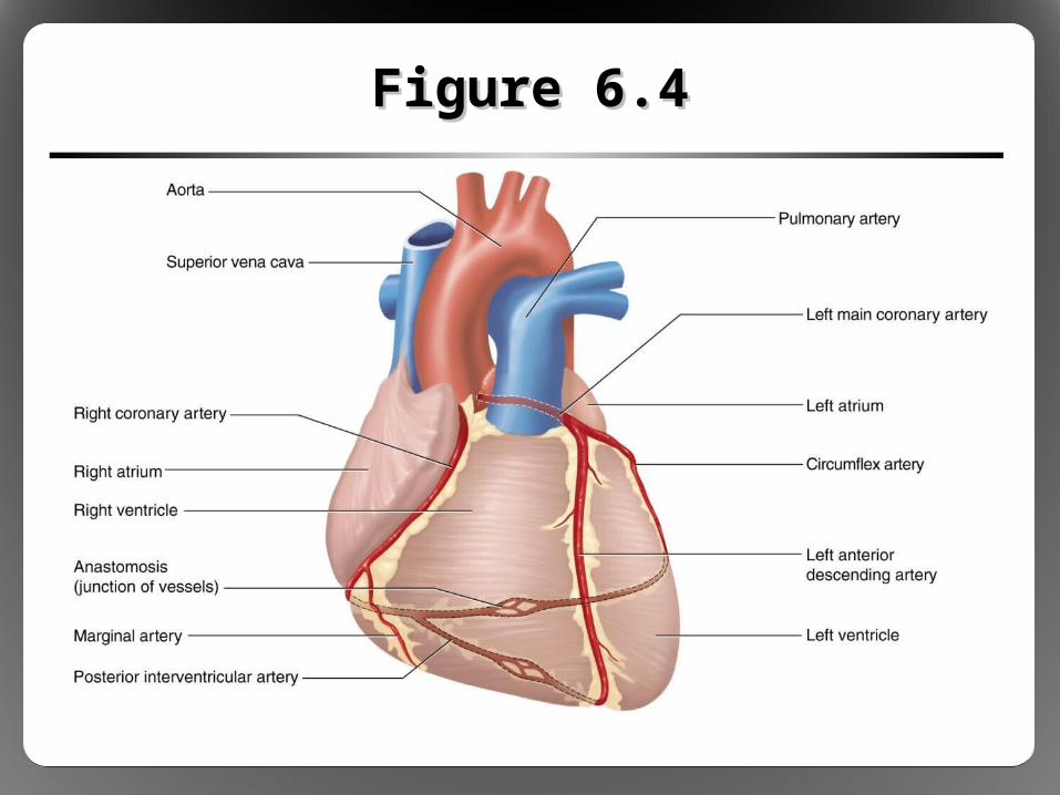
Figure 6.4Figure 6.4

Intrinsic Control of Heart Activity: Intrinsic Control of Heart Activity: Cardiac Conduction SystemCardiac Conduction System
• Spontaneous rhythmicity: special heart cells generate and spread electrical signal– Sinoatrial (SA) node– Atrioventricular (AV) node– AV bundle (bundle of His)– Purkinje fibers
• Electrical signal spreads via gap junctions– Intrinsic heart rate (HR): 100 beats/min– Observed in heart transplant patients (no neural
innervation)
Intrinsic Control of Heart Activity: Intrinsic Control of Heart Activity: Cardiac Conduction SystemCardiac Conduction System
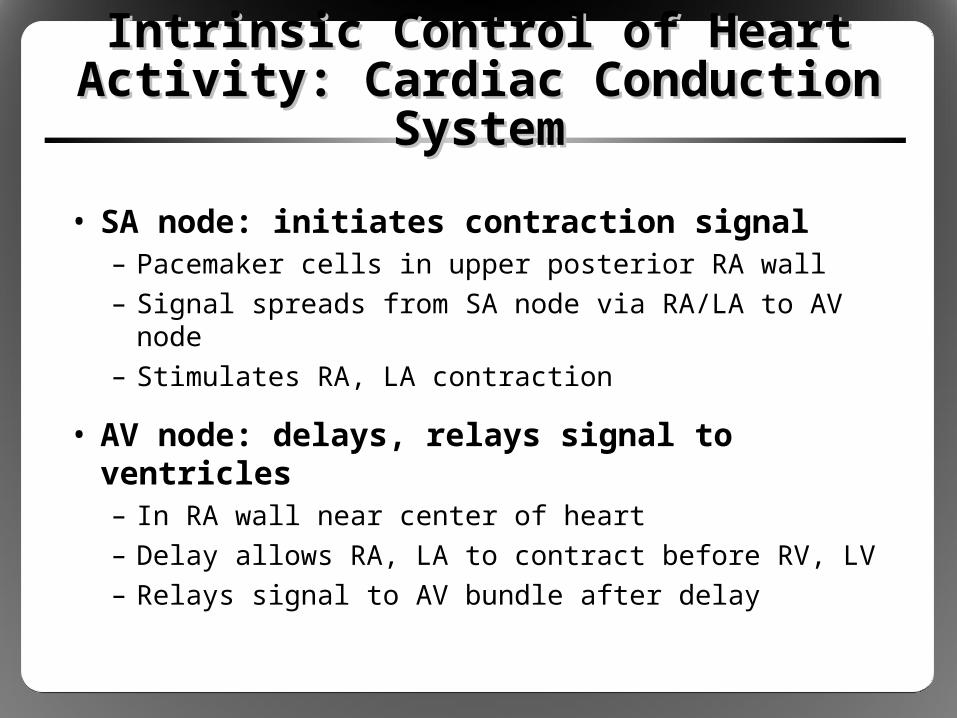
• SA node: initiates contraction signal– Pacemaker cells in upper posterior RA wall– Signal spreads from SA node via RA/LA to AV node– Stimulates RA, LA contraction
• AV node: delays, relays signal to ventricles– In RA wall near center of heart– Delay allows RA, LA to contract before RV, LV– Relays signal to AV bundle after delay
Intrinsic Control of Heart Activity: Intrinsic Control of Heart Activity: Cardiac Conduction SystemCardiac Conduction System
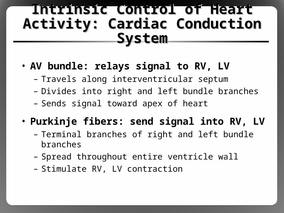
• AV bundle: relays signal to RV, LV– Travels along interventricular septum– Divides into right and left bundle branches– Sends signal toward apex of heart
• Purkinje fibers: send signal into RV, LV– Terminal branches of right and left bundle branches– Spread throughout entire ventricle wall– Stimulate RV, LV contraction
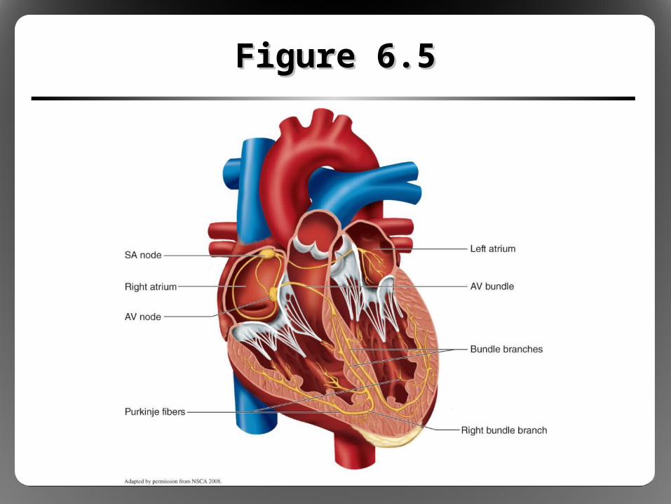
Figure 6.5Figure 6.5

Figure 3.1Figure 3.1
Figure 6.8Figure 6.8

Cardiac TerminologyCardiac Terminology
• Cardiac cycle
• Stroke volume
• Ejection fraction
• Cardiac output (Q)

Cardiac CycleCardiac Cycle
• All mechanical and electrical events that occur during one heartbeat
• Diastole: relaxation phase– Chambers fill with blood– Twice as long as systole
• Systole: contraction phase

Figure 6.9Figure 6.9

Stroke Volume, Ejection FractionStroke Volume, Ejection Fraction
• Stroke volume (SV): volume of blood pumped in one heartbeat– During systole, most (not all) blood ejected– EDV – ESV = SV– 100 mL – 40 mL = 60 mL
• Ejection fraction (EF): percent of EDV pumped– SV / EDV = EF– 60 mL/100 mL = 0.6 = 60%– Clinical index of heart contractile function

Cardiac Output (Q)Cardiac Output (Q)
• Total volume of blood pumped per minute
• Q = HR x SV– RHR ~70 beats/min, standing SV ~70 mL/beat– 70 beats/min x 70 mL/beat = 4,900 mL/min– Use L/min (4.9 L/min)
• Resting cardiac output ~4.2 to 5.6 L/min– Average total blood volume ~5 L– Total blood volume circulates once every minute

The Vascular SystemThe Vascular System
• Arteries: carry blood away from heart• Arterioles: control blood flow, feed
capillaries• Capillaries: site of nutrient and waste
exchange• Venules: collect blood from capillaries• Veins: carry blood from venules back to
heart

Blood PressureBlood Pressure
• Systolic pressure (SBP) – Highest pressure in artery (during systole) – Top number, ~110 to 120 mmHg
• Diastolic pressure (DBP)– Lowest pressure in artery (during diastole)– Bottom number, ~70 to 80 mmHg
• Mean arterial pressure (MAP)– Average pressure over entire cardiac cycle– MAP ≈ 2/3 DPB + 1/3 SBP
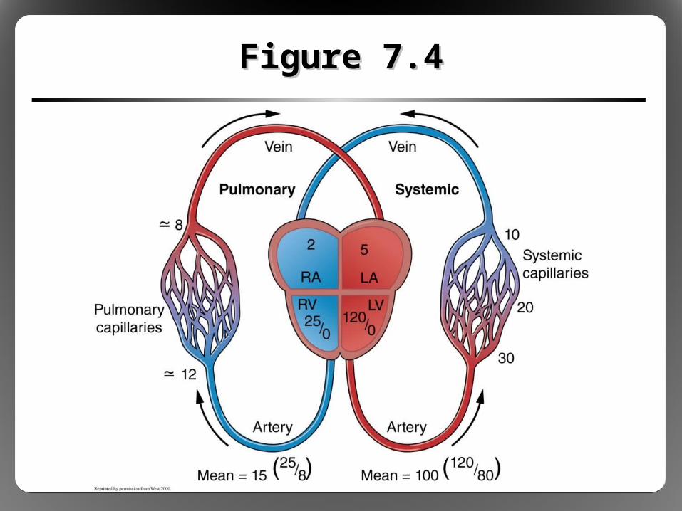
Figure 7.4Figure 7.4

General HemodynamicsGeneral Hemodynamics
• Blood flow: required by all tissues• Pressure: force that drives flow
– Provided by heart contraction– Blood flows from region of high pressure (LV, arteries)
to region of low pressure (veins, RA)– Pressure gradient = 100 mmHg – 0 mmHg
= 100 mmHg
• Resistance: force that opposes flow– Provided by physical properties of vessels– R = [L/r4] radius most important factor

Figure 6.11Figure 6.11
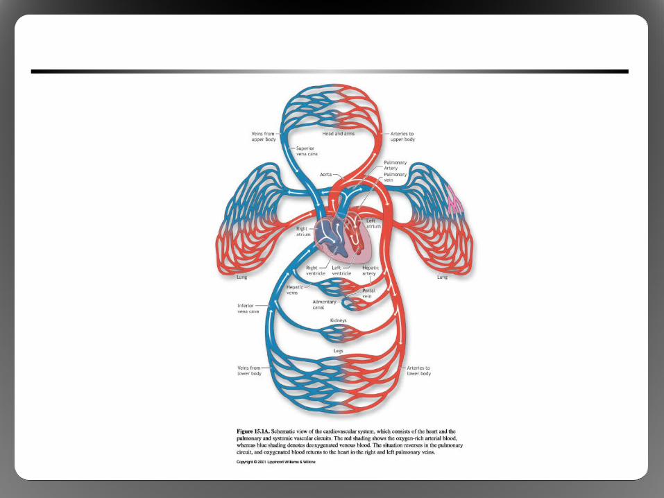
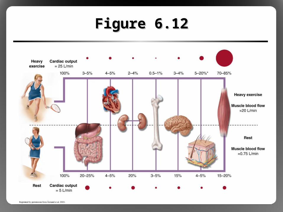
Figure 6.12Figure 6.12

Distribution of Venous BloodDistribution of Venous Blood
• At rest, veins contain 2/3 blood volume– High capacity to hold blood volume– Elastic, balloonlike vessel walls– Serve as blood reservoir
• Venous reservoir can be liberated, sent back to heart and into arteries– Sympathetic stimulation– Venoconstriction

Figure 6.14Figure 6.14

Return of Blood to the HeartReturn of Blood to the Heart
• Upright posture makes venous return to heart more difficult
• Three mechanisms assist venous return– One-way venous valves– Muscle pump– Respiratory pump
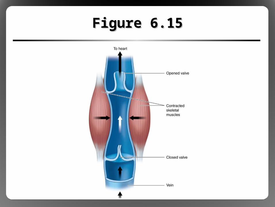
Figure 6.15Figure 6.15

BloodBlood
• Plasma (55-60% of blood volume)– Can decrease by 10% with dehydration in the heat– Can increase by 10% with training, heat acclimation– 90% water, 7% protein, 3% nutrients/ions/etc.
• Formed elements (40-45% of blood volume)– Red blood cells (erythrocytes: 99%)– White blood cells (leukocytes: <1%)– Platelets (<1%)
• Hematocrit = total percent of volume composed of formed elements


