11
11

11

10

9

8
Developmental Aspects of the Muscular System (page 221) • Increasing muscular control reflects the myelination maturation of the nervous system. Muscle
control is achieved in a cephalic/caudal and proximal/distal direction • To remain healthy, muscles must be regularly exercised. Without exercise, they atrophy (a
reduction in size or wasting away of an organ or cell resulting from disease or lack of use); with exercise, they hypertrophy (an increase in the size of a tissue or organ independent of the body’s general growth).
• As we age, muscle mass decreases, and the muscles become more sinewy. Exercise helps to retain muscle mass and strength.
• Homeostatic Imbalances: o Muscular dystrophy: a progressive disorder marked by atrophy and stiffness of the
muscles o Duchenne’s muscular dystrophy: the most common and serious form of muscular
dystrophy; expressed almost exclusively in boys and usually diagnosed between the ages of 2 and 7 years; active, normal-appearing children become clumsy and fall frequently as their muscles weaken; the disease progresses from the extremities upward, finally affecting the head and chest muscles; children with this disease rarely live beyond their twenties and generally die of respiratory failure; the diseased muscle fibers lack a protein (called dystrophin) that helps maintain the sarcolemma.
o Myasthenia gravis: a rare disease that can affect muscles during adulthood; a disease characterized by drooping of the upper eyelids, difficulty in swallowing and talking, and general muscle weakness and fatigability; involves a shortage of acetylcholine receptors at neuromuscular junctions; the muscle cells are not stimulated properly and get progressively weaker. Death usually occurs because of respiratory failure, the inability of the respiratory muscles to function.

7
Muscle Movements, Types, and Names • Muscles and Body Movements
o Movement is attained due to a muscle moving an attached bone o Muscles are attached at least 2 points:
Origin = attachment to an immovable bone Insertion = attachment to a moveable bone
o Results of increased muscle use Increase in muscle size Increase in muscle strength Increase in muscle efficiency Muscle becomes more fatigue resistant
• Types of Ordinary Body Movements o Flexion = bending; the movement that decreases the angle between bones o Extension = movement that increases the angle of a joint, e.g., straightening a flexed knee o Rotation = movement of a bone around its longitudinal axis o Abduction = movement of a line away from the midline of the body o Adduction = movement of a limb toward the body midline o Circumduction = circular movement of a body part; a combination of flexion, extension,
abduction, and adduction • Special Movements
o Dorsiflexion = up and down movement that includes lifting the foot so that its superior surface approaches the shin (standing on your heels)
o Plantar flexion = depressing the foot (pointing your toes) o Inversion = turn inward; turn the sole medially o Eversion = special movement of the foot achieved by turning the sole laterally o Supination = the outward rotation of the forearm causing palms to face anteriorly; turning
backward o Pronation = the inward rotation of the forearm causing the radius to cross diagonally over
the ulna – palms face posteriorly; turning forward o Opposition = the action by which the thumb is used to touch the tips of the other fingers on
the same hand. This unique action makes the human hand such a fine tool for grasping and manipulating tools.
• Types of Muscles o Prime mover = muscle with the major responsibility for a certain movement o Antagonist = muscle that opposes or reverses a prime mover o Synergist = muscle that aids prime mover in a movement and helps prevent rotation o Fixator = stabilizes the origin of a prime mover
• Naming Skeletal Muscles o Direction of muscle fibers o Relative size of the muscle o Location of the muscle o Number of origins o Location of the muscles origin and insertion o Shape of the muscle o Action of the muscle
• Muscles have several fascicle arrangements that influence their force and degree of shortening

6
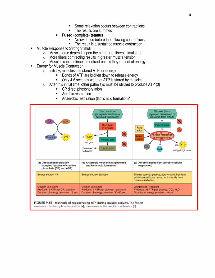
o Direct phosphorylation Muscle cells contain creatine phosphate (CP)
• CP is a high-energy molecule After ATP is depleted, ADP is left CP transfers energy to ADP, to regenerate ATP CP supplies are exhausted in about 20 seconds
o Aerobic (“with oxygen”) respiration Series of metabolic pathways that occur in the mitochondria Glucose is broken down to carbon dioxide and water, releasing energy This is a slower reaction that requires continuous oxygen
o Anaerobic (“without oxygen”) glycolysis Reaction that breaks down glucose without oxygen Glucose is broken down to pyruvic acid to produce some ATP Pyruvic acid is converted to lactic acid This reaction is not as efficient, but it is fast
• Huge amounts of glucose are needed • Lactic acid produces muscle fatigue
• Muscle Fatigue and Oxygen Deficit o When a muscle is fatigued, it is unable to contract o The common reason for muscle fatigue is oxygen deficit
Oxygen must be “repaid” to tissue to remove oxygen deficit Always occurs to some extent during vigorous muscle activity, whether fatigue
occurs or not • During the recovery period after activity, the individual breathes rapidly and
deeply. • This continues until the muscles have received the amount of oxygen that is
required to get rid of accumulated lactic acid and make ATP and creatine phosphate reserves
Increasing acidity (from lactic acid) and lack of ATP causes the muscle to contract less
• Types of Muscle Contractions o Isotonic contractions
Myofilaments are able to slide past each other during contractions The muscle shortens
o Isometric contractions Tensions in the muscles increases The muscle is unable to shorten
• Muscle Tone o Some fibers are contracted even in a relaxed muscle o Different fibers contract at different times to provide muscle tone o The process of stimulating various fibers is under involuntary control
• Effect of Exercise on Muscles o Aerobic exercises, or endurance: Muscles subjected to regular aerobic exercise become
more efficient and stronger and can work longer without tiring. Aerobic exercise also benefits other body systems
Examples: participating in aerobics class, jogging, biking o Isometric exercises, or resistance: Muscles challenged by resistance exercise to respond
(almost) beyond their ability increase in size and strength. Examples: weight training
5
• Some relaxation occurs between contractions • The results are summed
Fused (complete) tetanus • No evidence before the following contractions • The result is a sustained muscle contraction
• Muscle Response to Strong Stimuli o Muscle force depends upon the number of fibers stimulated o More fibers contracting results in greater muscle tension o Muscles can continue to contract unless they run out of energy
• Energy for Muscle Contraction o Initially, muscles use stored ATP for energy
Bonds of ATP are broken down to release energy Only 4-6 seconds worth of ATP is stored by muscles
o After this initial time, other pathways must be utilized to produce ATP (3) CP direct phosphorylation Aerobic respiration Anaerobic respiration (lactic acid formation)*

4
• Myosin filaments have heads (extensions, or cross bridges) • Myosin and actin overlap somewhat • At rest, there is a bare zone that lacks actin filaments • Sarcroplasmic reticulum (SR) – for storage of calcium
Skeletal Muscle Activity • Properties of:
o Irritability = ability to receive and respond to a stimulus o Contractility = ability to shorten when an adequate stimulus is received
• Nerve Stimulus to Muscles o Skeletal muscles must be stimulated by a nerve to contract o Motor unit
One neuron Muscle cells stimulated by that neuron
o Neuromuscular Junctions = association site of nerve and muscle o Synaptic cleft – gap between nerve and muscle
Nerve and muscle do not make contact Area between the nerve and muscle filled with interstitial fluid
• Transmission of Nerve Impulse to Muscle o Neurotransmitter = chemical released by nerve upon arrival of nerve impulse
The neurotransmitter for skeletal muscle is acetylcholine o Neurotransmitter attaches to receptors on the sarcolemma o Sarcolemma becomes permeable to sodium (Na+) o Sodium rushing into the cell generates an action potential (electrically charged gradient) o Once started, muscle contraction can not be stopped
• Sliding Filament Theory of Muscle Contraction (Myosin binding is prevented, Ca+ modifies shape, myosin binding occurs)
o Activation by nerve causes myosin heads (crossbridges) to attach to binding sites on the thin filament
Ca+ ions have to be released to bind to actin regulatory proteins (changing the shape) so myosin heads can bind to the actin
o Myosin heads then bind to the next site of the thin filament o This continued action causes a sliding of the myosin along the actin o The result is that the muscle is shortened (contracted)
• Contraction of a Skeletal Muscle o Muscle fiber contraction is “all or none” o Within a skeletal muscle, not all fibers may be stimulated during the same interval o Different combinations of muscle fiber contractions may give differing responses o Graded responses = differing degrees of skeletal muscle shortening o Graded muscle contraction can occur in 2 ways:
By changing the frequency of muscle stimulation By changing the number of muscle cells being stimulated
o Types of Graded Response Twitch
• Single, brief contraction • Not a normal muscle function
Tetanus (summing of contractions) • One contraction is immediately followed by another • The muscle does not completely return to a resting state • The effects are added
Unfused (incomplete) tetanus

3
Smooth Muscle Characteristics • Has no striations • Spindle-shaped cells • Single nucleus • Involuntary – no conscious control • Found mainly in the walls of hollow visceral organs (other than heart)
Cardiac Muscle Characteristics • Has striations • Usually has a single nucleus • Joined to another muscle cell at an intercalated disc • Involuntary • Only found in the heart
Functions of Muscles • Produce movement • Maintain posture • Stabilize joints • Generate heat
Microscopic Anatomy of Skeletal Muscle • Cells are multinucleate • Nuclei are just beneath the sarcolemma • Sarcolemma = specialized plasma membrane • Sarcoplasmic reticulum = specialized smooth endoplasmic reticulum • Myofibril (many in a muscle fiber)
o Bundles of myofilaments
o Myofibrils are aligned to give distinct bands
I band = light band
A band = dark band
• Sarcomere o Contractile unit of a
muscle fiber (from Z-disc to Z-disc)
Organization of the sarcomere • Thick filaments = MYOSIN
filaments o Composed of the
protein myosin o Has ATPase
enzymes • Thin filaments = ACTIN
filaments o Composed of the
protein actin

2
o Sites of muscle attachment Bones Cartilages Connective tissue coverings

1
Anatomy Study Guide
Test: Thursday 1/27/11; covers pages 182-206, & 221
The Muscular System • Muscles are responsible for all types of body movement • Muscle Types (SEE COMPARISON CHART AT END)
o Skeletal o Cardiac o Smooth
Characteristics of Muscles • Muscle cells are elongated (muscle cell = muscle fiber) • Contraction of muscles is due to the movement of microfilaments • All muscles share some terminology
o Prefix myo- refers to “muscle” o Prefix mys- refers to “muscle” o Prefix sarco- refers to “flesh”
Skeletal Muscle Characteristics • Most are attached by tendons to bones • Cells are multinucleate • Striated – have visible banding • Voluntary – subject to conscious control • Cells are surrounded and bundled by connective tissue • Connective Tissue Wrappings of Skeletal Muscle
o Endomysium – around single muscle fiber o Perimysium – around a fascicle (bundle) of fibers o Epimysium – covers the entire skeletal muscle (to tendon) o Fascia – on the outside of the epimysium
• Skeletal Muscle Attachments o Epimysium blends into a connective tissue attachment
Tendon – cord-like structure (mostly collagen) Aponeuroses – sheet-like structure


